Forum Replies Created
-
AuthorPosts
-
Hi Sylvia, Thanks for your reply. I see the Register button has reappeared now; I’m sure that wasn’t there a week or so ago when a couple of local lichenologists wanted to join the forum. Now that there is a button to press, I will encourage these people to try to register, and to be persistent if it doesn’t work on their first attempt.
In addition to the
Rinodina calcarea, we also added Strigula tayloriiand Porina byssophilafrom the trunk of an ash tree in Riseley churchyard, a tree which has received a lot of attention over the past decade or so. It seems that these species continue to get more common in my area, after being rare or absent until relatively recently. We also added a lichenicolous fungus, Pyrenidium actinellums. lat. on Caloplaca teicholyta. Even in such a well-worked churchyard we continue to add things to the list on almost every visit. For people travelling by public transport, Riseley is rather difficult to get to (except by taxi) because the timing of the buses would only allow a short day. We are also conducting an ongoing survey of Foster Hill Road Cemetery in Bedford which is a superb site with excellent examples of very many common saxicolous species along with many recherche and obscure ones. This cemetery is within easy walking distance of the train and bus stations. I will let people know when we have another day planned for either site. If you get impatient feel free to suggest a day to suit you and I can often set aside a day and then invite others.
I can’t offer any additional insights or advice but, from my limited observations, individual colonies of A. ciliarison gravestones are also experiencing some sort of decline. The BLS account states that scattered occurrences on limestone rock are surviving better than on trees, which I can’t contradict, but something seems to be causing decline on calcareous gravestones. Significant portions of the colonies seem to be disappearing and the remaining portions look rather unhealthy in their mature parts, a sort of brownish discolouration. The only slight hope for their future is that the growing tips and youngest parts of the lobes still look healthy and give the appearance of active growth. The first side-by-side below is from a Bedfordshire cemetery, the photos taken in 2012 and 2022. 
[attachment=1]Screenshot 2026-03-27 10.27.39 AM.png[/attachment] The next side-by-side is from a Northamptonshire churchyard, the photos taken in 2018 and 2022.

[attachment=0]Screenshot 2026-03-27 10.28.43 AM.png[/attachment] In both cases you can see where significant portions of the colonies have disappeared. It is possible that the missing portions were removed by some physical process, either human or animal activity, but my suspicion is that these are unhealthy portions which just fell away.
My observations which are from just two sites don’t really prove anything but I provide them for general interest.
I recently came across some well-developed material of what I would normally record as Buellia griseovirens on a wooden bench seat in Bedford Cemetery. This gives the following reactions, K+ yellow, C+ faint yellow and Pd+ faint yellow. In a previous post I speculated that this sort of material might be a chemotype of B. griseovirens which produces stictic rather than norstictic acid. However, the faint Pd+ yellow reaction seems to suggest otherwise, the presence of stictic acid ought to produce a much more intense Pd+ yellow-orange reaction. When the K spot dried and UV was shone at it, an intense K/UV+ yellow reaction was seen, especially on the thallus. This might indicate the presence of atranorin which would be right for B. arborea. I dug out an old specimen of K+ yellow turning red B. griseovirens from ash bark in Cambridgeshire and that didn’t produce an intense K/UV+ yellow reaction, just a very faint yellowish colour. This perhaps adds weight to Paul”s suggestion to consider B. arborea. If the sort of material that Di and I have recently collected from wooden seats really is B. arborea then we would need to have a complete rethink of the distribution. I suspect that many records of ‘B. griseovirens’ are made on sight, without testing with K. Most specimens I have encountered in the Midlands and Home Counties have been K+ yellow.
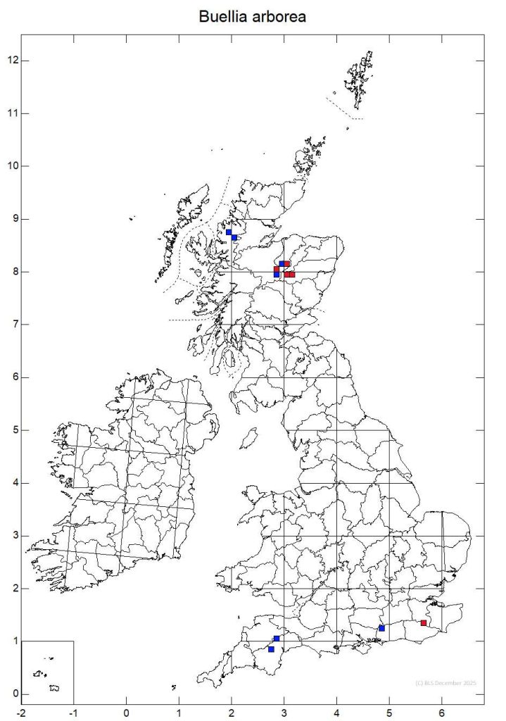
[attachment=0]Buellia arborea.jpg[/attachment] I was sent an interesting specimen of Phaeographis dendritica by James Rowland, collected from a young Fraxinus in Aversley Wood in Huntingdonshire. Since this occurrence was so far out of its known geographical range James wanted to be sure of the identity. Everything seemed to fit, thallus K+ yellow turning red, ascospores turning pale brown when young-mature and dark excipular layer continuous beneath the hymenium. The one thing that disconcerted both of us was the septation of the spores, neither of us could find any with more than seven septa, while the published accounts state that the septation is (5-) 7- to 9- (10-)-septate. Brian has kindly confirmed the specimen as P. dendritica and provided the following note: “The majority of P. dendritica specimens I’ve seen have mainly 7-septate spores, so no worries there”. I think you are almost certainly on the right lines with Petractis clausa. The overall appearance is spot on and I have only seen that distinctive perispore in P. clausa (but that doesn’t rule out it occurring in related species I am not familiar with). I suspect that P. clausa has a layer of redeposited calcite covering its thallus and thalline margins, or at least that is the impression that it gives. When dry, and the apothecia darker and more immersed than when swollen with moisture, P. clausa can be overlooked as one of the Verrucaria/Baggliettoa species that have a lithocortex of redeposited calcite (e.g. B. calciseda). This appearance, as if it has a lithocortex, isn’t really stressed in most published accounts and the closest hint we have is that the thallus is described as ‘immersed’ in the calcareous stone. By contrast, the Gyalecta species you are likely to come across in that habitat, such as G. jenensis, has pinky orange-brown thallus and thalline margins without a white covering. I don’t think the distinctive perispore in P. clausa is described in most accounts, it looks as though the gel of the perispore is drawn out into radiating spines, somewhat giving the effect of rays of light streaming out of the spore in conical columns. For your own satisfaction you will want to try to observe and identify the photobiont. I wonder if you can find any photobiont cells by investigating the thallus immediately beneath the apothecia, or close to the apothecia deep in the thallus? Look for patches which look bluish green (rather than grass green) within thallus that looks pale and ‘clean’. When I was working on the identity of Watsoniomyces obsoletus (Lecidea lichenicola as was) nobody really knew what the photobiont was and at first I suspected Trentepohlia due to the yellow-orange staining that the chalk often has where this chalk pebble lichen occurs. I found that the upper parts of the thallus of W. obsoletus has a mixture of algae and cyanobacteria, perhaps a bit like the various bits and bobs you found in your suspected P. clausa. Eventually, I found the consistent presence of Scytonema, especially when my investigations moved from the ‘contaminated’ upper thallus and I started to look deeper down, beneath and close to the apothecia. I think the same may be the case in P. clausa. I don’t have a micrograph of the Scytonema photobiont in Petractis to hand but from memory it is rather similar to that in W. obsoletus. The following image is of lichenised Scytonema in W. obsoletus, stained with ink-vinegar. The sheaths of Scytonema are orange-brown and I think it is staining from these sheaths that often gives the chalk in the vicinity of W. obsoletus an orange tinge and hence my initial suspicion that Trentepohlia might be involved.
I’m grateful to Paul for reintroducing the possibility of B. arborea being much more widespread, a species that I think I convinced myself of once on an Islay fencepost and should still have a specimen to re-examine. Since then I had, in my own mind, run with the idea that the relatively common lignicolous and corticolous lichen with bluish delimited soredia in the Midlands and Home Counties were different from the Islay material and that they seemed like chemotypes of one taxon, B. griseovirens, most K+ yellow and a few identical looking specimens being K+ yellow turning red as they should be according to the literature. I am just stating the way I had thought about the situation up to now, not holding on to any personal cherished hypothesis. I do know that I observed the apparently ‘unexplained’ C+ yellow reaction in some of my K+ yellow ‘Buellia griseovirens’, perhaps in all or most of those that I bothered to test with C. As the C+ yellow reaction is described as ‘unexplained’, including in LGBI3, I am not sure what importance we should attribute to it. I am still erring towards Di recording this as a K+ yellow form of Buellia griseovirens unless or until someone can prove that it is something different. In the meantime I will look back at some of my specimens of B. griseovirens and hopefully find that I also have a snippet of B. arborea and see if any of this changes my opinion.
Mark
As for look-alikes of Buellia griseovirens, I could add some others to the short list on the BLS species description page. It is not impossible that Porpidia tuberculosa could turn up on a churchyard bench but yours isn’t that because you would have seen a very distinct K/UV+ blue-mauve due to confluentic acid. Lecanora pannonica is somewhat similar to Buellia griseovirens, especially thin and neat material growing on tops of sandstone headstones; it is not impossible that this would turn up on lignum though admittedly I have never seen it on other than stone. The holy grail of such things on lignum is Thelomma ocellatum which I have found but once, on a very old weathered oak fence post at Toot Baldon churchyard in Oxfordshire. T. ocellatum is perhaps best described as P. tuberculosa on steroids and, despite never having seen it before in the flesh, I was fairly sure I had found it at Toot Baldon at my first glance of it and before it was confirmed. I think the most likely identity of your lichen on the wooden bench is one of the K+ yellow forms of Buellia griseovirens but I thought I would provide my list of somewhat look-alikes for your general interest.
I don’t think you should give up on the possibility of Buellia griseovirens because in my experience most specimens of this species in the Midlands, East Anglia and Home Counties give a K+ yellow reaction, rather than a K+ yellow turning red. Even when mounted microscopically in strong K solution I have failed to produce any red crystals with these K+ yellow forms of B. griseovirens. The published information suggests that the reaction may be weak but I suspect that some populations, perhaps a lowland race, produce stictic acid rather than norstictic acid. There is a similar phenomenon in an unrelated species, Aspicilia grisea. Although the published information states that this contains norstictic acid, the lowland material, including the rare occurrences on brickwork, and notably the specimens on the sarsen stones such as at Fyfield Down, are consistently K+ yellow. Brian has speculated that, in a similar way to B. griseovirens, these K+ yellow forms of A. grisea contain stictic rather than norstictic acid.
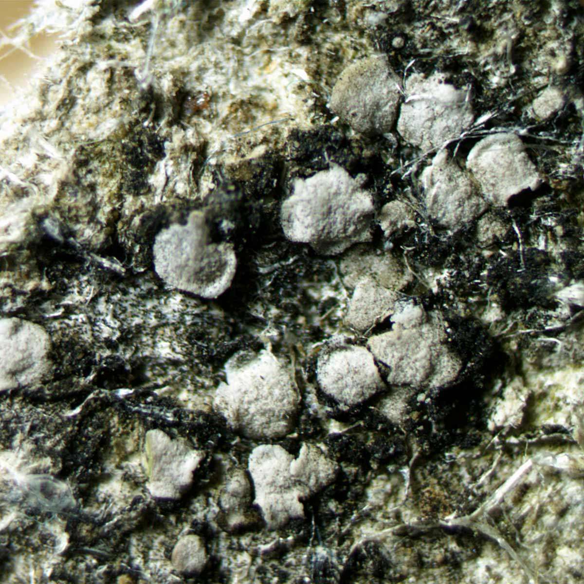
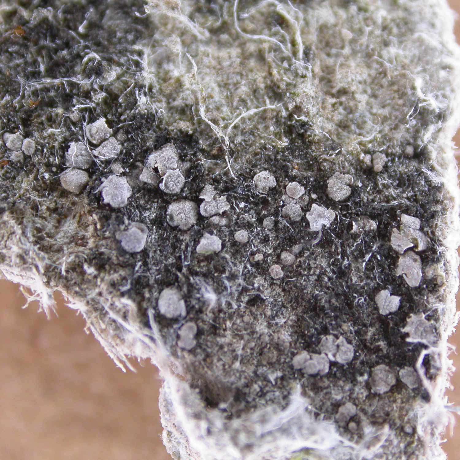
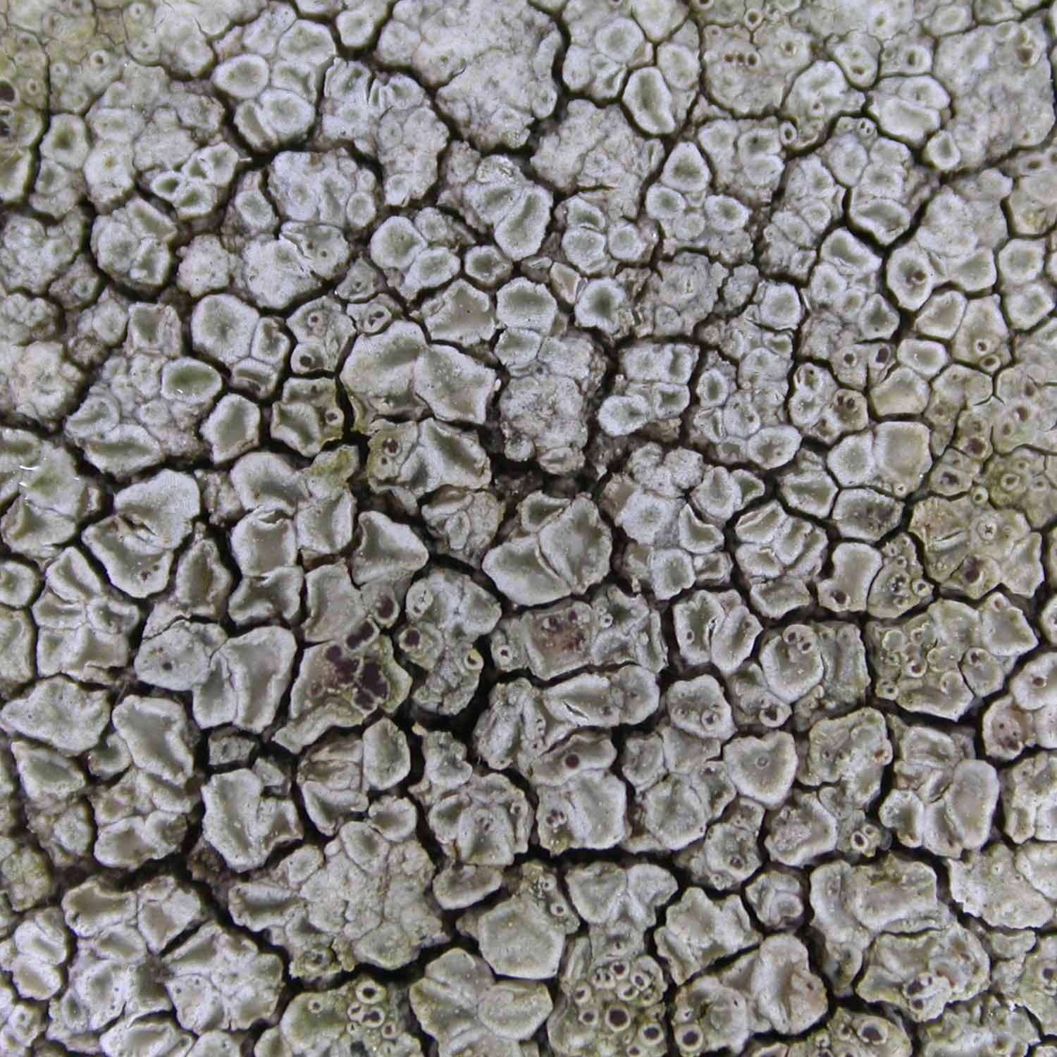
Many thanks for taking the trouble to go back and find this and for the great set of photographs, perhaps some of the best I have seen of this species (for there can be no real doubt that this is A. moenium). Your oblique photographs really capture the shape of the squamules, and including a finger for scale shows how tiny these squamules are. A synonym of Acarospora moenium is Aspicilia excavata and that name is more evocative of the lichen even though it has been superseded. The white-pruinose areoles/squamules are reminiscent of some ‘Aspicilia’ species such as contorta and ‘excavate’ describes the way the edges of the squamules become undercut as soredia are formed. Having looked at your photo again, I am very close to confirming it as A. moenium, though if it would be a new Vice-county record then perhaps best if it checked. It would be an interesting little excursion for anyone who lives close enough. Here are a couple of my own images and an annotated drawing. 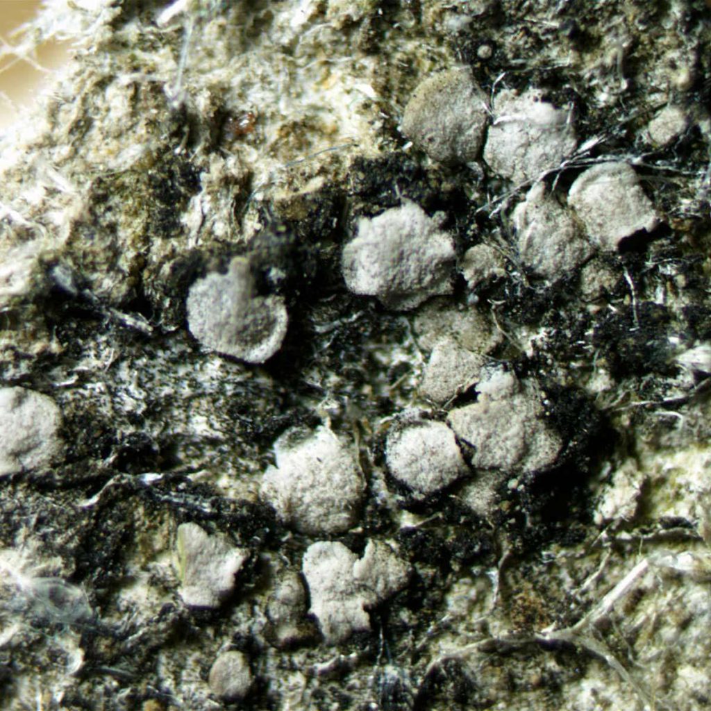
[attachment=2]moenium3.jpeg[/attachment] 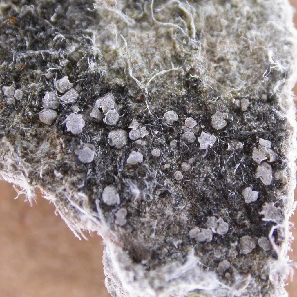
[attachment=1]moenium2.jpeg[/attachment] 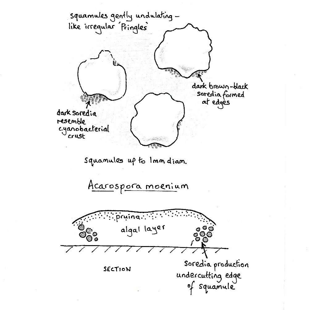
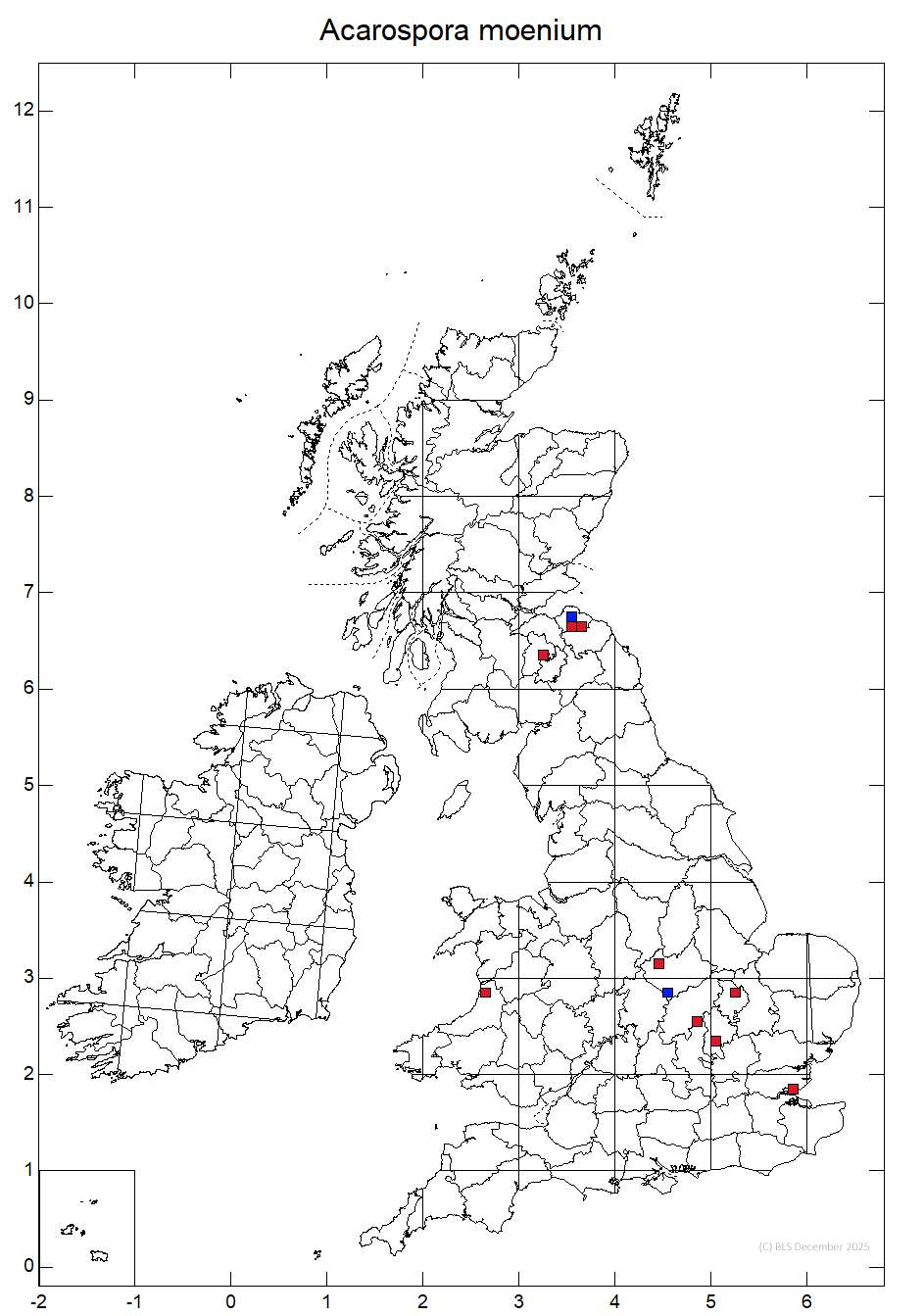
[attachment=0]moenium1.jpeg[/attachment] The lichen in your photo looks remarkably good for A. moenium, a species which I have quite a lot of experience of. I encountered it in the first year of looking at lichens and thought it looked like something interesting though I was given all sorts of different suggestions for its potential identity. I collected lots of specimens which I took from the broken asbestos cement roof of a collapsed Nissen hut and eventually worked out its identity, subsequently confirmed by Brian C. It could potentially be confused with Aspicilia contorta, which has a similar ecology and similar size and colour of areoles but the areoles of A. moenium are more squamulose, they are a bit like little Pringles and I think I can see in your photo the right sort of arched voids under the edges of some of the areoles. If there is anyone else in the area interested in following this up I do think it would be worth it. It is certainly not common, perhaps surprising because when it does turn up it is, for example, on the rough concrete of manhole covers, decaying concrete on multi-story car-parks, all sorts of mundane situations but even when you know the species you rarely find it.
The distribution map, a cluster in East Lothian and a few in ‘my’ region, hint that it might be overlooked.

[attachment=0]Acarospora moenium.jpg[/attachment] Thanks for letting us all know; it is an interesting phenomenon and useful for people to know what such heavily modified Lecanora campestris and L. horiza can look like. The separation of these two species is a bit tricky because there don’t seem to be any absolute diagnostic differences between them. They both lack epithecial granules and both are K+ yellow. Deciding on one or the other depends on an appraisal of a small suite of morphological differences and slight differences of ecology but all the features used to distinguish them are somewhat overlapping. Now that I am familiar with them, I think I can fairly confidently assign a name to half or perhaps up to two thirds of specimens while the other specimens leave me in some doubt and I tend not to record these more intermediate specimens. I have no doubt at all that you can reliably recognise good specimens of each species, especially those specimens which are towards the more typical ends of the spectrum. Lecanora horiza tends to lack a fimbriate margin (though it sometimes has a suggestion of a fimbriate margin on smooth surfaces such as aspen bark), the thallus of L. horiza tends to be whiter and lumpy compared to the pale grey warted thallus of L. campestris. The apothecia of L. horiza tend to be larger, often exuberant, with flexuose margins and often giving the impression that they might start to fall off the substratum. When I was working on how to separate these two, I became briefly very excited when I found several occurrences of typical L. campestris infected with Muellerella lichenicola and several occurrences of typical L. horiza infected with Vouauxiella verrucosa. I was excited because I hoped these lichenicolous fungi might prove to be host specific but further work showed that either LF can occur on either host.
I think the conidia being strongly hooked at least at one end is quite diagnostic for Bacidina modesta or at least that is what we used to think when we called it B. sulphurella. It would be interesting to hear from anyone who can confirm or dispute my assertion that a lichen such as yours, with a thallus composed of small goniocysts and with pale pycnidia containing filiform, strongly hooked conidia, can be reasonably named as B. modesta even without apothecia. There was a time, perhaps between ten and fifteen years ago, when I seemed to find B. ‘sulphurella’ quite commonly on shaded bark but it seems to have become rather scarce now in my region. This follows the pattern of so many lichens, which seem to have a sort of heyday at certain periods during our changing atmospheric pollution regimes. I can well imagine B. modesta growing on the remains of an old bracket fungus so the substratum doesn’t cause me any concern.
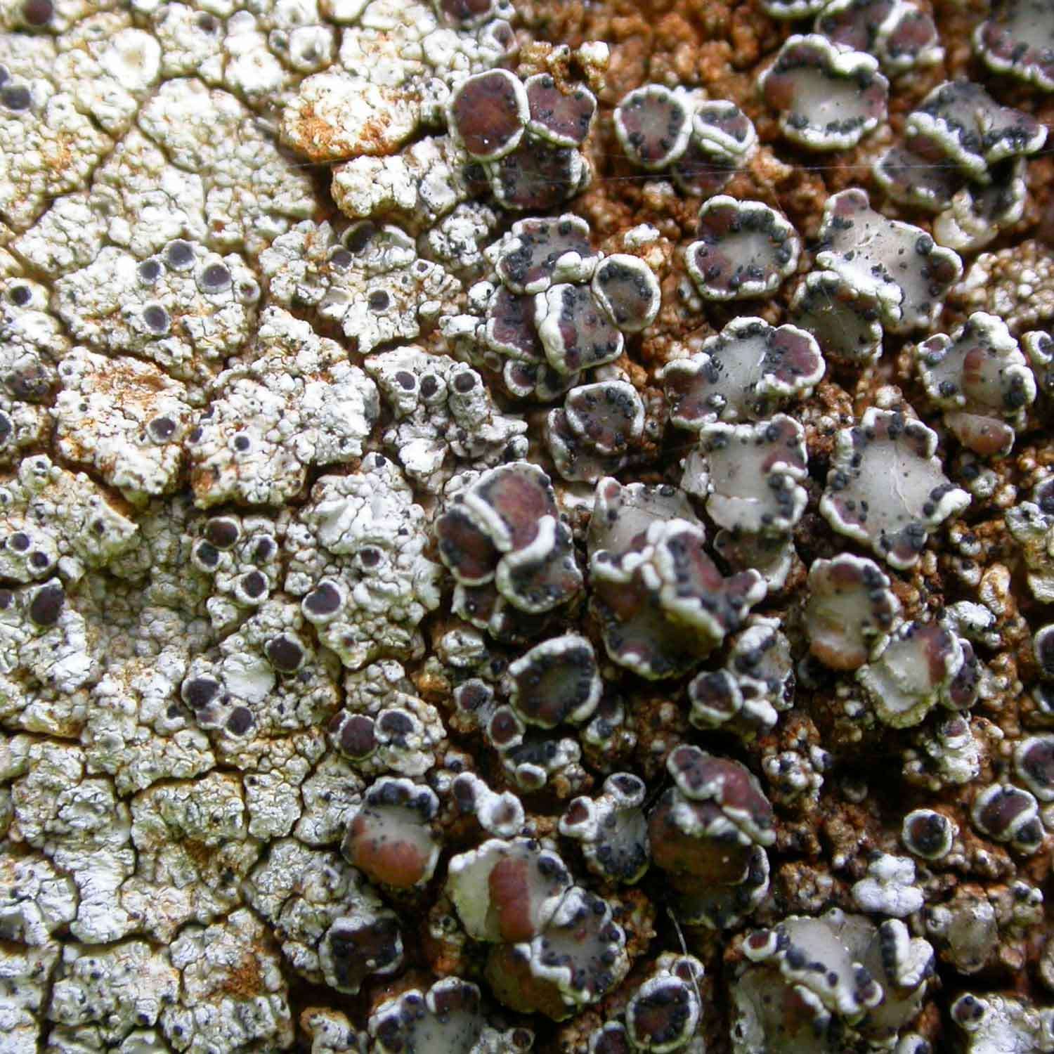
If you can’t find any truly squamulose lichens which seem to fit then I will tentatively suggest an alternative idea. I have sometimes come across colonies of various Lecanora species which have had the hymenium browsed out of their apothecia leaving only the empty margins. These then sometimes make an attempt to produce some small secondary discs from the hollowed-out original apothecia. The following photo is an example, which I identified as L. campestris at the time. 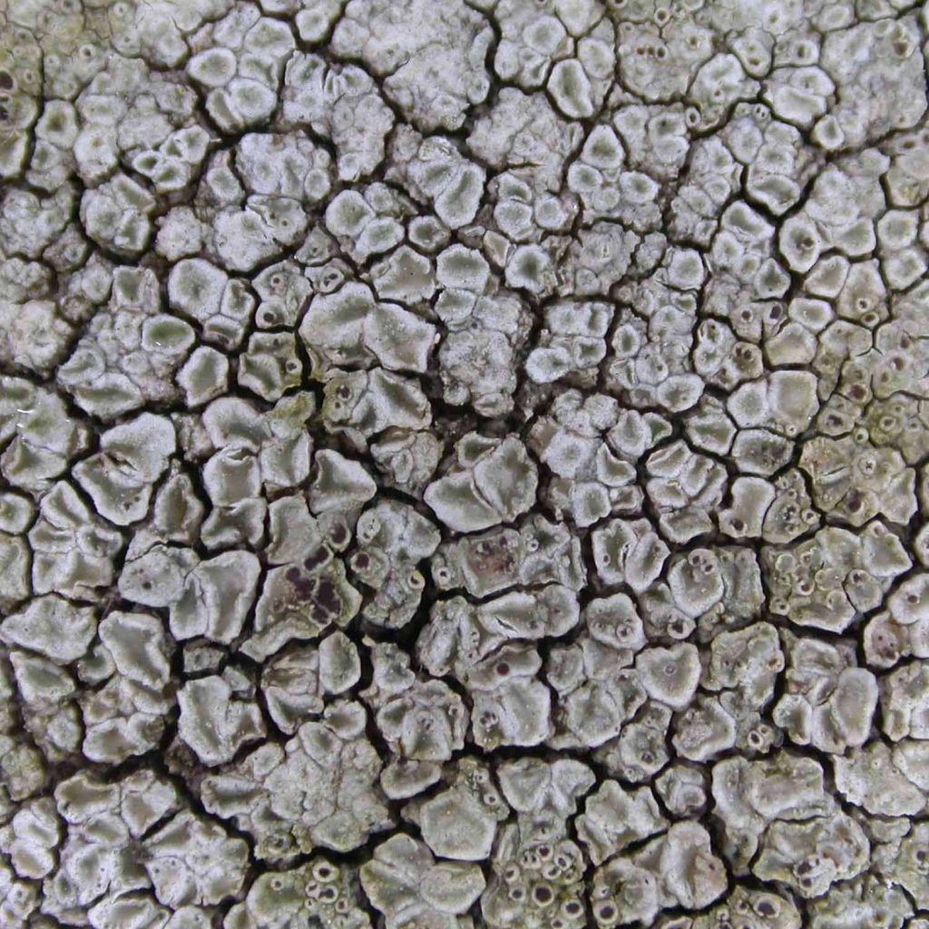
[attachment=1]campbrows.jpeg[/attachment]
On a calcareous gravestone, and especially in the company of Physconia grisea, you might have L. horiza which might produce even more of a squamulose appearance if its apothecia were hollowed out, its apothecia tending to be rather large, with flexuous margins and appearing almost as if falling off the stone. In the following photo we have L. campestris on the left side of the image, L. horiza to the right (heavily infected with Vouaxiella verrucosa), neither of them very much hollowed out by browsing in this instance.
[attachment=0]horiza.jpeg[/attachment]
My suggestion is merely that, a possibility to consider rather than anything definite.Pachnolepia pruinata, along with Cresponea premnea, occurs near the base of one of the large upright stones of Stonehenge. 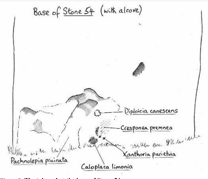
[attachment=0]stonehenge.png[/attachment] -
AuthorPosts