Forum Replies Created
-
AuthorPosts
-
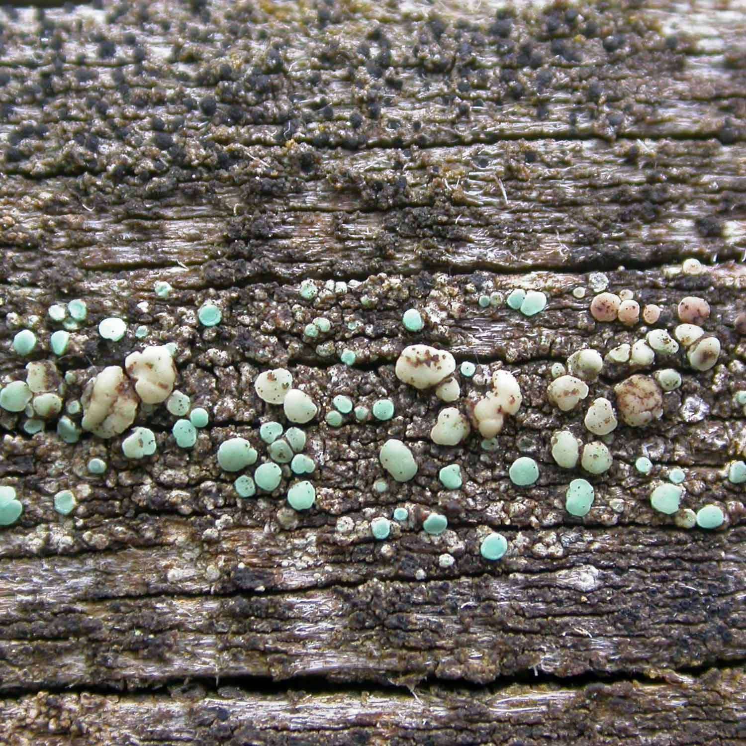
I can’t provide a definite identification but I will speculate about what might be going on here, always with the possibility that others may provide different and perhaps more convincing suggestions. I wonder whether you have a bark fungus with perithecia which are protruding through the thallus of a thinly developed lichen. If I had been in the field I would have scanned across the bark to get an impression of whether the perithecia always seemed to be associated with the ‘sorediate’ thallus or whether there were places where the perithecia grew in ‘bare’ bark. The following is not as much as a suggestion of an identity for your potential bark fungus but gives you an idea of the sort of thing I have in mind, Navicella pileata:
https://www.irishlichens.ie/pages-fungi/f-98.html As for the lichen thallus in your photo, it appears suppressed in the way that crustose lichens sometimes are on smooth bark, with the presumed soralia not having properly developed powdery soredia. Two common species which spring to mind which might look like that on smooth ash bark are Lecanora expallens and Caloplaca obscurella. The former ought to exhibit a C+ orange reaction but this is often difficult to demonstrate on suppressed or shaded material. The K/UV+ green (wet) test is much more sensitive for detecting xanthones in L. expallens. The soralia of C. obscurella sometimes fail to develop into their distinctive crater-like forms when suppressed.
Bear in mind that I might be leading you astray with my suggestions! Whatever the identity of your crust, you raise an interesting point, that “pyrenocarps don’t seem to go in for soredia”. Offhand I can’t think of a single instance and it will be interesting to see if anyone can suggest any sorediate pyrenocarps whether or not relevant to your specimen. Some taxa in Verrucaria form blastidia, such as V. macrostoma f. furfuracea and V. nigrescens f. tectorum. Some Verrucaria species have ‘goniocyst-like units but these are usually embedded in the thallus, such as the apparently undescribed thing like V. murina but with speckled green thallus, which crops up on pebbles. Some Porina species are isidiate. Are there any pyrenocarps which have true soralia?
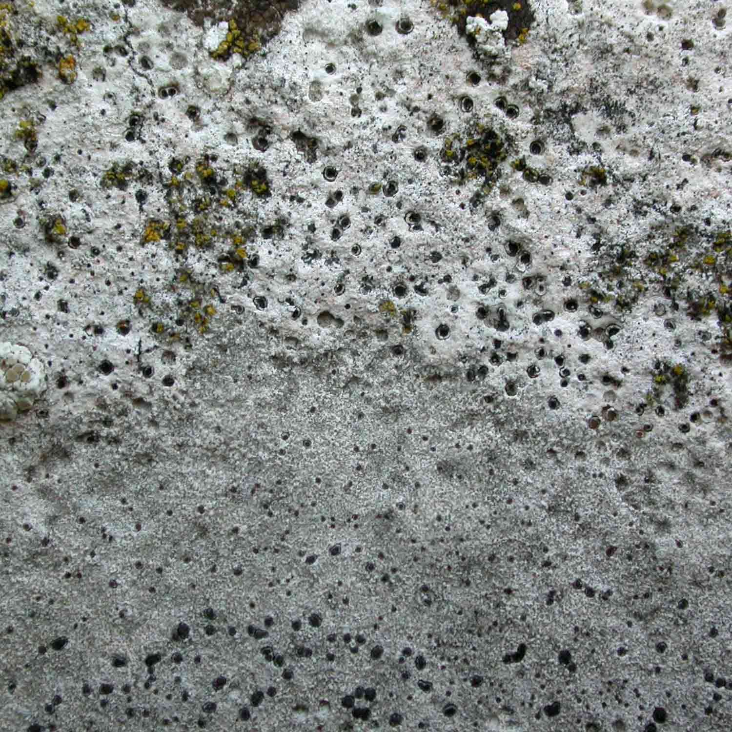
Yes, Verrucaria and its allies are a challenge but rather an important one in churchyard recording. They constitute well over 10% of the list for most churchyards, certainly churchyards in limestone areas. As a guide, in the northern Home Counties that I know best, I would expect to find the following in most churchyards: Thelidium incavatum, T. pyrenophorum, Verrucaria calciseda, V.elaeina, V. fuscella, V. hochstetteri, V. macrostoma f. furfuracea, V. ochrostoma, V. muralis, V. nigrescens f. nigrescens, V. nigrescens f. tectorum and V. viridula.
Less commonly, but not very rare, I find: Polyblastia albida, P. dermatodes, Thelidium decipiens, Verrucaria baldensis, V. obfuscans, V. polysticta, V. macrostoma f. macrostoma and V. romeana.
Of course some of my Verrucaria species are now placed in genera such as Bagliettoa and Placopyrenium but I have clumped them in Verrucaria for this rough and ready analysis.
We have come quite a long way in our understanding of churchyard Verrucariaceae in the past two decades. Thelidium pyrenophorum was completely overlooked or misidentified in the past. Verrucaria obfuscans was added to the British list in 2015 despite it being so common and often present in abundance on church buildings. Up until about 2010 or so, Verrucaria calciseda was poorly known by churchyard recorders and often recorded as V. baldensis. It is quite common for me to come across Verrucaria specimens which I can’t name. I feel that there is still quite a lot of unrecognised diversity out there with this and related genera. If we have only just got to grips with such common and distinctive species as T. pyrenophorum, V. calciseda, V. obfuscans and V. ochrostoma, it seems likely that there are many other less common and less distinctive taxa that we are overlooking. How many lichenologist’s eyes have scanned over sheets of V. obfuscans in the past and not registered anything more than vague thoughts such as ‘a dull brown Verrucaria, perhaps something like V. nigrescens that has gone wrong with some metal contamination’.
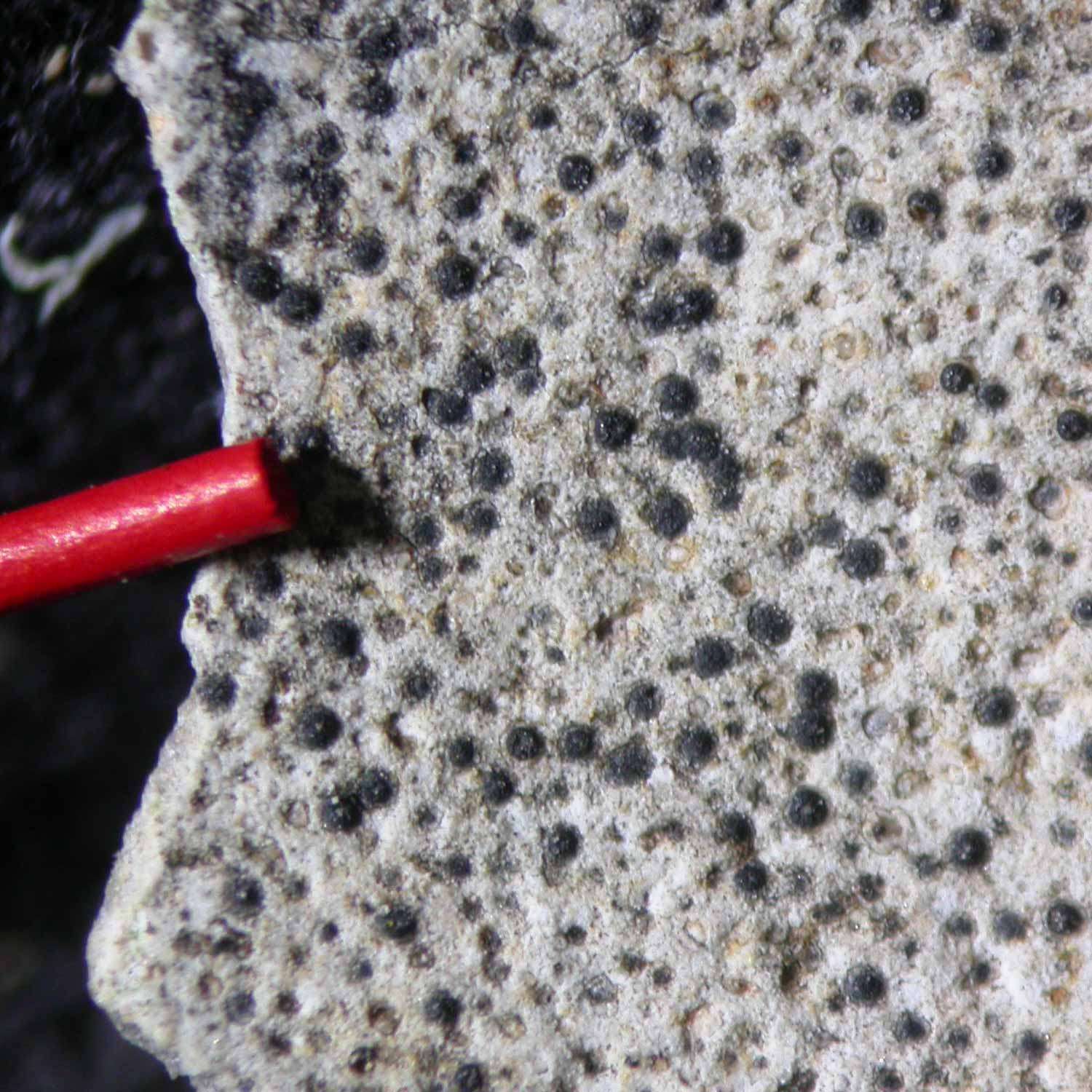
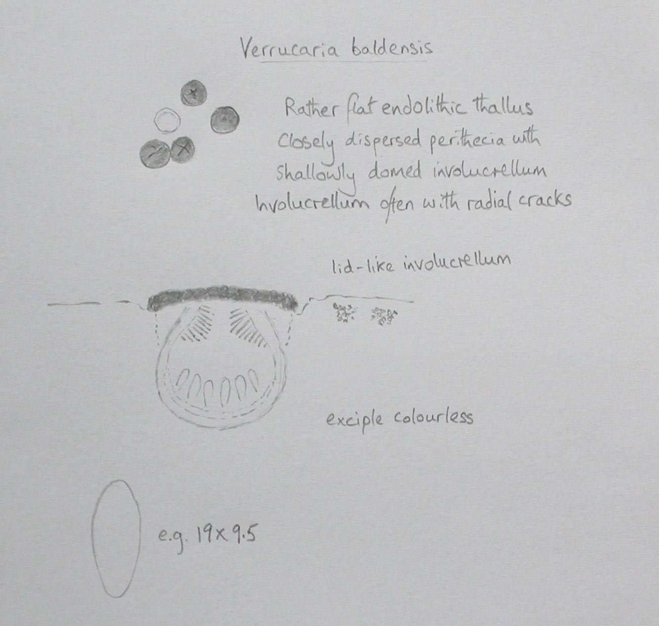
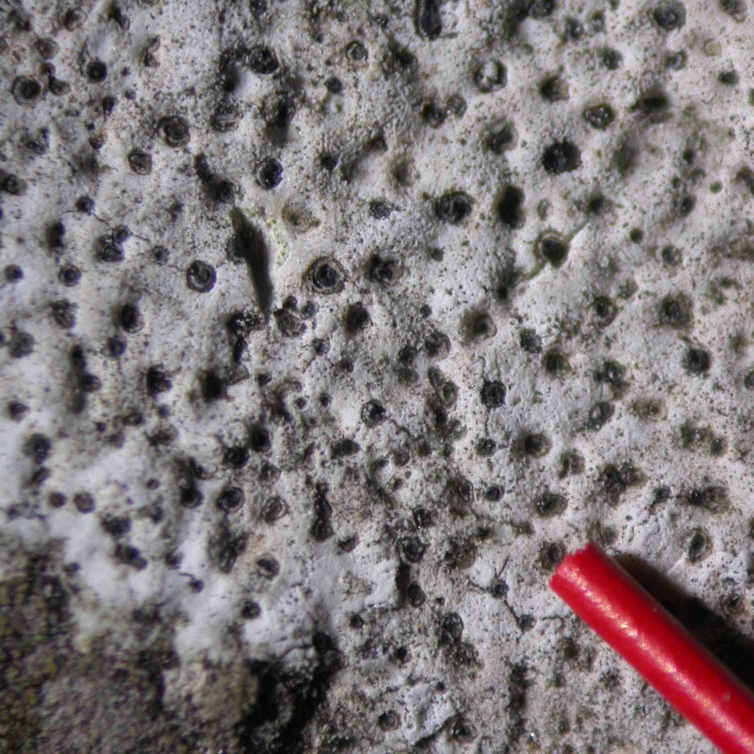
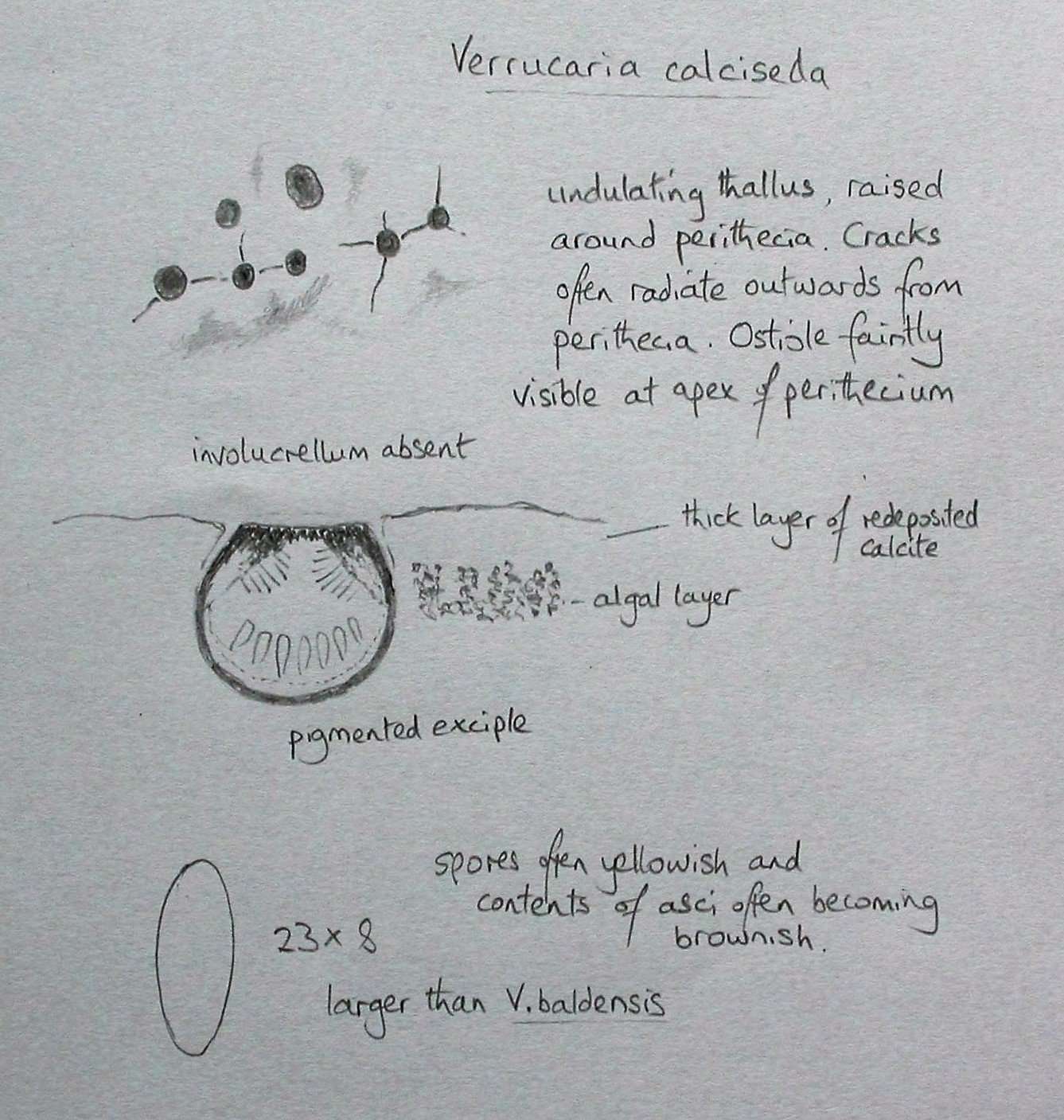
Here are some images and details of V. baldensis s. lat. and V. calciseda (both now placed in Bagliettoa). These are the two churchyard immersed pyrenocarps with the thickest lithocortex, especially V. calciseda which looks like a thick coat of masonry paint. Thelidium incavatum and its three look-alikes have a thinner cortex than this pair but thicker than V. hochstetteri.

[attachment=3]bald1.jpeg[/attachment] 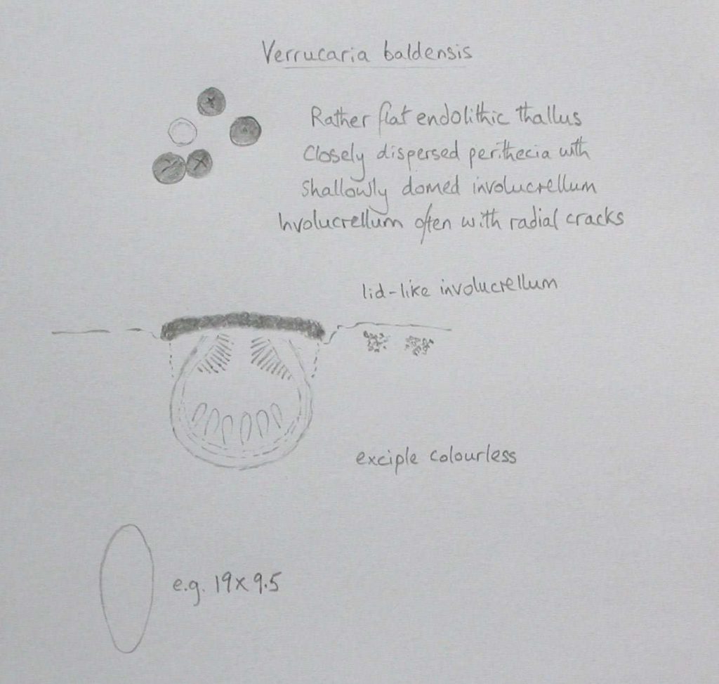
[attachment=2]bald2.jpeg[/attachment] 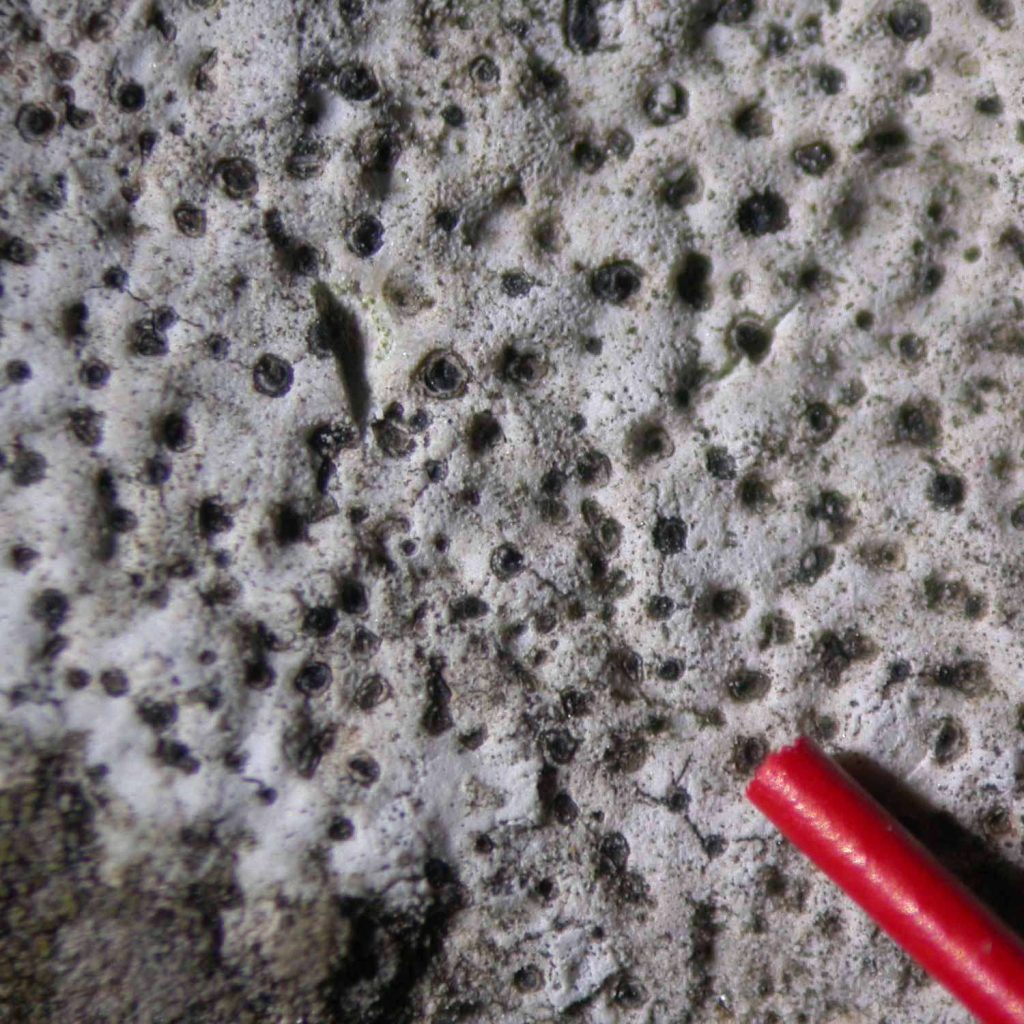
[attachment=1]calc1.jpeg[/attachment] 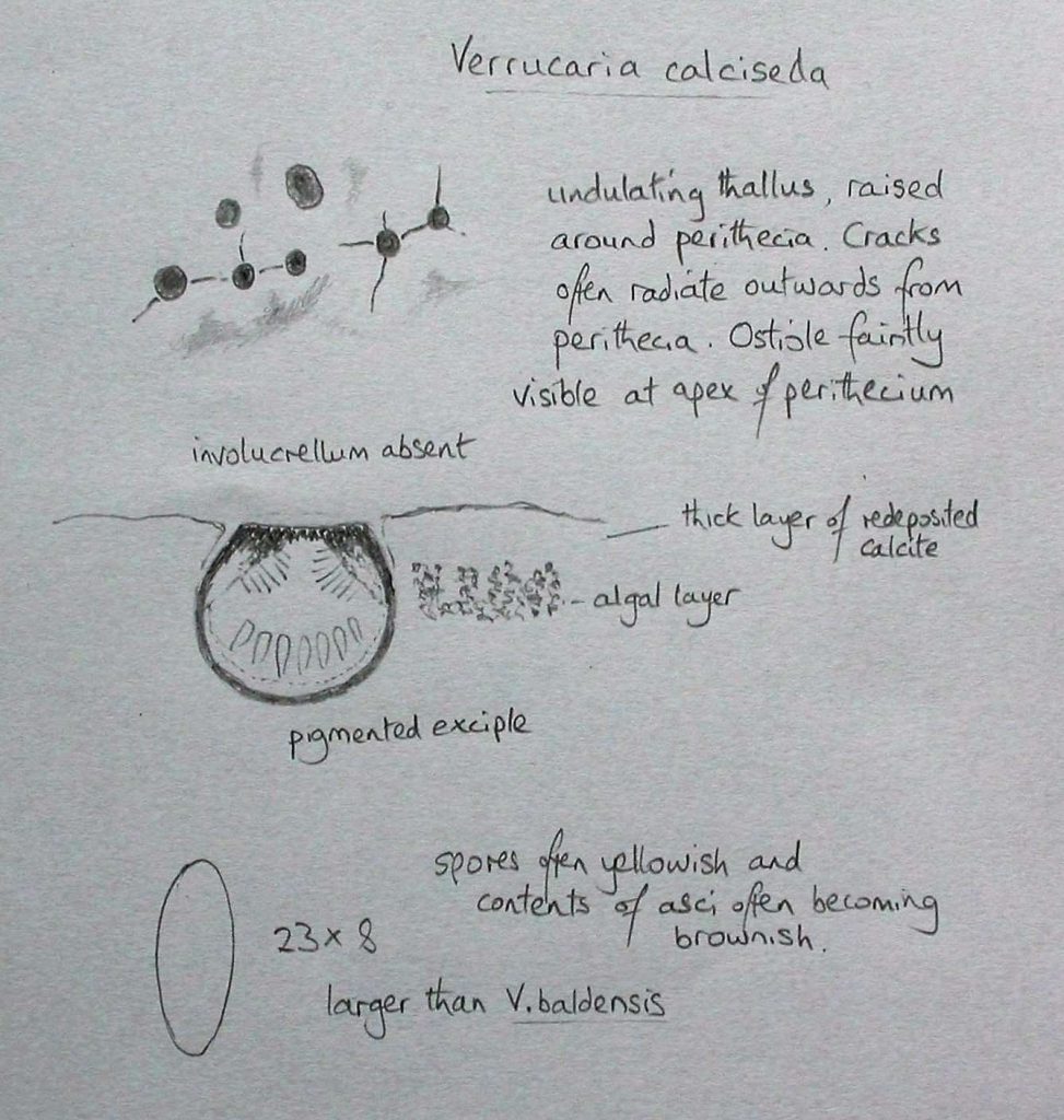
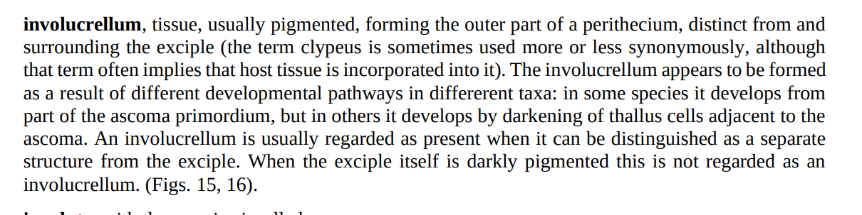
[attachment=0]calc2.jpeg[/attachment] Recording three of the four from a churchyard is really good going. In the past there were many good churchyard recorders who hadn’t developed appropriate collecting techniques, and who didn’t trouble themselves too much with microscopy, who almost completely overlooked Thelidium incavatum and its look-alikes. In my experience T. incavatum is the most common by some margin, followed by T. decipiens (which I have a slight suspicion has a slight preference for slightly shady situations such as the chamfered plinth on the north side of a church). Polyblatia albida and P. dermatodes both seem to turn up quite a lot less commonly and I usually find them when I take a bit of potential T. incavatum for checking. I am often a bit disconcerted by P. dermatodes but if the spores are submuriform I feel that P. dermatodes is the best name. The decision of whether an involucrellum is present or not is often not as straightforward as it sounds. An involucrellum is not a single evolutionary homologous structure; in different groups it arises from different developmental pathways. Really, it is probably accurate to say that an involucrellum is any structure that a taxonomist has decided is sufficiently distinct to consider as being a separate structure to the exciple itself.
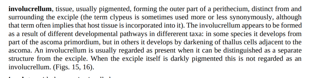
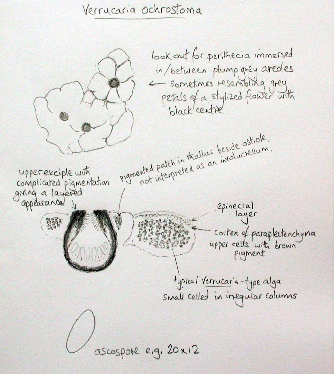
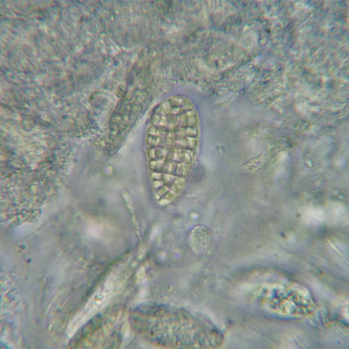
[attachment=1]Screenshot 2025-12-15 4.12.23 PM.png[/attachment] When I was working on the identity of Verrucaria ochrostoma, a lichen that was virtually unknown to British lichenologists before 2013, despite being so common and widespread, I agonised over the issue of whether an involucrellum was present or not. The upper part of the exciple is multilayered and there is often a little patch of pigmented thallus near the apex of the perithecia but neither of these features is considered to be an involucrellum.
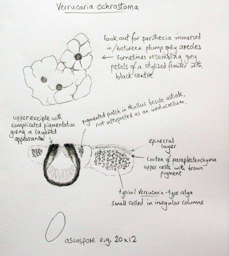
[attachment=0]ochrostoma.jpeg[/attachment]
Similarly, when I found the first occurrence of Verrucaria sphaerospora in Britain, I had the difficulty of deciding whether its perithecia had an involucrellum or not. In this case it does have an involucrellum but it is fused with the exciple, forming a complete globose covering hardly distinguishable from the exciple.I can’t think what else it is likely to be other than Polyblastia albida and, as you say, it seems to fit. Rather than rushing into a confirmation straight away, I will explain my thinking when looking at your photos. One of the first things I checked was that there weren’t any paraphyses which would help confirm that we are looking at a member of the Verrucariaceae. Your stained section of a whole perithecium shows that the periphyses are nicely stained but there don’t seem to be any distinct interascal filaments/paraphyses. In Verrucaria and related genera the hymenium often has a suggestion of linear structures, like trails of inclusions or lines separated elongated holes in the hymenial gel but no actual hyphae. I think I am convinced by your micrographs that no interascal filaments are present. When one comes across a member of the Verrucariaceae with muriform spores then another genus to briefly consider is Staurothele but this genus has algal cells within the hymenium and these are usually quite numerous and rather obvious and I can’t see any suggestion of them in your photos. I have very rarely encountered Staurothele in churchyards or elsewhere on old stonework but it is not unknown.
Assuming we are dealing with Polyblastia the two next important things to note are that the perithecia are immersed and that no involucrellum is present. The latter is sometimes difficult to interpret. I would say that your perithecia have an exciple (perithecial wall) which is thickened in the upper part but that there is no distinct separate structure which would be interpreted as an involucrellum.
By this stage, assuming we have interpreted things correctly, we have narrowed things down and we end up at P. albida.
Here is Polyblastia albida in the upper part of my photo and Verrucaria hochstetteri below.
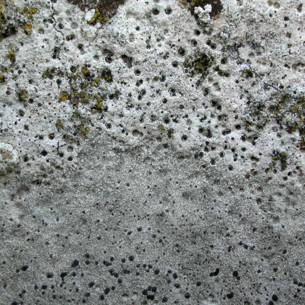
[attachment=6]albid1.jpeg[/attachment]
Polyblastia albida and its look-alikes have a lithothallus of redeposited calcite which make them seem like a thick layer of (woodwormed) white paint, contrasting with the much less developed lithocortex of V. hochstetteri resulting in very slight depressions in the stone. As an aside Thelidium pyrenophorum produces even more pronounced depressions in the stone (due to repeated browsing not having the protection of a lithocortex). Thelidium decipiens, T. incavatum, Polyblastia albida and P. dermatodes form a quad of species which are indistinguishable in the field. Verrucaria baldensis and Verrucaria calciseda have a thicker lithocortex than the quad. These subtleties help to distinguish the various immersed pyrenocarps on limestone in churchyards which seem impossible at first.Here are ascospores from my collections of P. albida and I think they ring true when set against yours.

[attachment=5]albid2.jpeg[/attachment] 
[attachment=4]albid3.jpeg[/attachment]
A few more notes and images of P. albida and related species.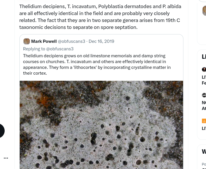
[attachment=3]Screenshot 2025-12-15 9.08.24 AM.png[/attachment] 
[attachment=2]Screenshot 2025-12-15 9.09.41 AM.png[/attachment] 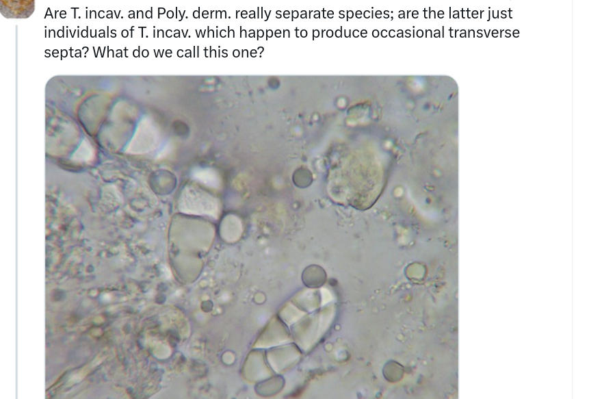
[attachment=1]Screenshot 2025-12-15 9.10.14 AM.png[/attachment] 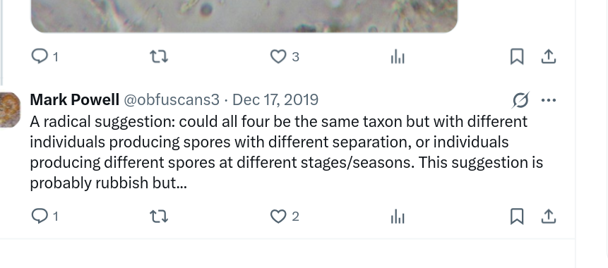
[attachment=0]Screenshot 2025-12-15 9.10.42 AM.png[/attachment] As long as you are happy that your specimen lacks distinct interascal filaments, has no distinct involucrellum, has no hymenial algae then I think you will be justified in recording it as P. albida. I usually do find P. albida and the rest of the ‘quad’ on fairly hard limestone, most commonly on Victorian/Edwardian limestone memorials (headstones and coped/coffin tombs) so I note your hesitation about the substratum. There is no problem with finding it on a vertical surface. As long as the particular stone was at least somewhat calcareous, as probably indicated by the presence of Verrucaria species nearby, then I wouldn’t worry too much.
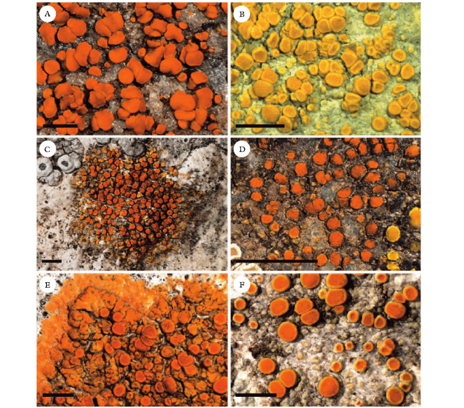
I think your attractive collection of ‘Caloplaca’ species are just as you suggested based on appearance and what is likely. So we have what looks to me like cerinella to the left, pyracea in the middle and ‘cerina’ to the right. I am not experienced with the cerina group so I won’t comment further on that one (C. ‘cerina’ is rare or absent still in my region). A. cerinella has, at least, something distinctive and diagnostic microscopically (12- to 16-spored asci) which is more than can be said for many ‘Caloplaca’ species. I am not suggesting you spoil your specimen as cerinella is now frequent, especially on Sambucus and Fraxinus twigs, but it is easy to confirm if necessary. As for the suspected A. pyracea, I think I can see some hint of grey thalline margins on the young apothecia. If that is so then I think you can reasonably consider your lichen to be A. pyracea. Although LGBI3 suggests that A. holocarpa has a (poorly developed) grey thalline margin, I think this assertion is lifted from LGBI2 in which ‘C. holocarpa’ was treated as a group rather than a single taxon. In my studies, I find that the thalline margin of C. holocarpa s. str. is yellowish-orange and scarcely distinguishable from the similarly coloured proper margin. The description of C. holocarpa s. str. in Arup’s 2009 paper seems to align with my own careful observations on the margins of C. holocarpa s. str. The actual thallus of C. holocarpa s. str. is usually difficult to see because of the closed-packed apothecia but when apparent, it is seen to consist of tiny orange warts.
Here is one of the illustrations from Arup’s paper, C. holocarpa s. str. A and B, and C. pyracea F.
[attachment=1]Screenshot 2025-12-14 11.45.36 AM.png[/attachment]
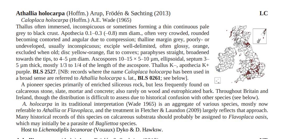
Here is the LGBI3 account of A. holocarpa and if I am right about the thalline margin of this taxon, then I think the suggestion here that it has a grey thalline margin may cause confusion. If anyone wants to find some A. holocarpa to study then these days it is probably easiest to find it on chemically treated fence rails where it often grows with Candelariella vitellina and Lecanora stenotropa.
[attachment=0]Screenshot 2025-12-14 11.27.46 AM.png[/attachment]
If one wants to find it on stone then it is more profitable to look at relatively recent gravestones rather than old ones and it is important to look for it on siliceous/acid stone (fine grained pure sandstone or ‘granite’) rather than on calcareous stone (on which C. oasis is common).Back to the A. pyracea and when I surveyed a site in Derbyshire some years ago, I found A. pyracea fairly frequently on smooth Fraxinus bark so ash trees are a likely host even though in my region it is much more commonly found on Populus. As so often in lichenology, it is very instructive once you have specimens of the various options side by side for examination.
Your lichen looks fine for Athallia pyracea to me. I think you have found a slight problem with couplet 59 in LGBI3 because, as you suggest, the apothecia of A. pyracea do have have a thalline margin and it is this feature which helps to distinguish it from species such as corticolous A. holocarpa. I suppose the key was trying to separate those species such as Caloplaca cerina which have a thick and persistent thalline margin and the wording didn’t quite take into account that A. pyracea also has a thalline margin, albeit a less conspicous one. 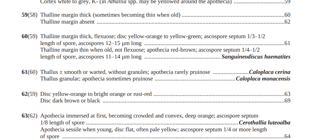
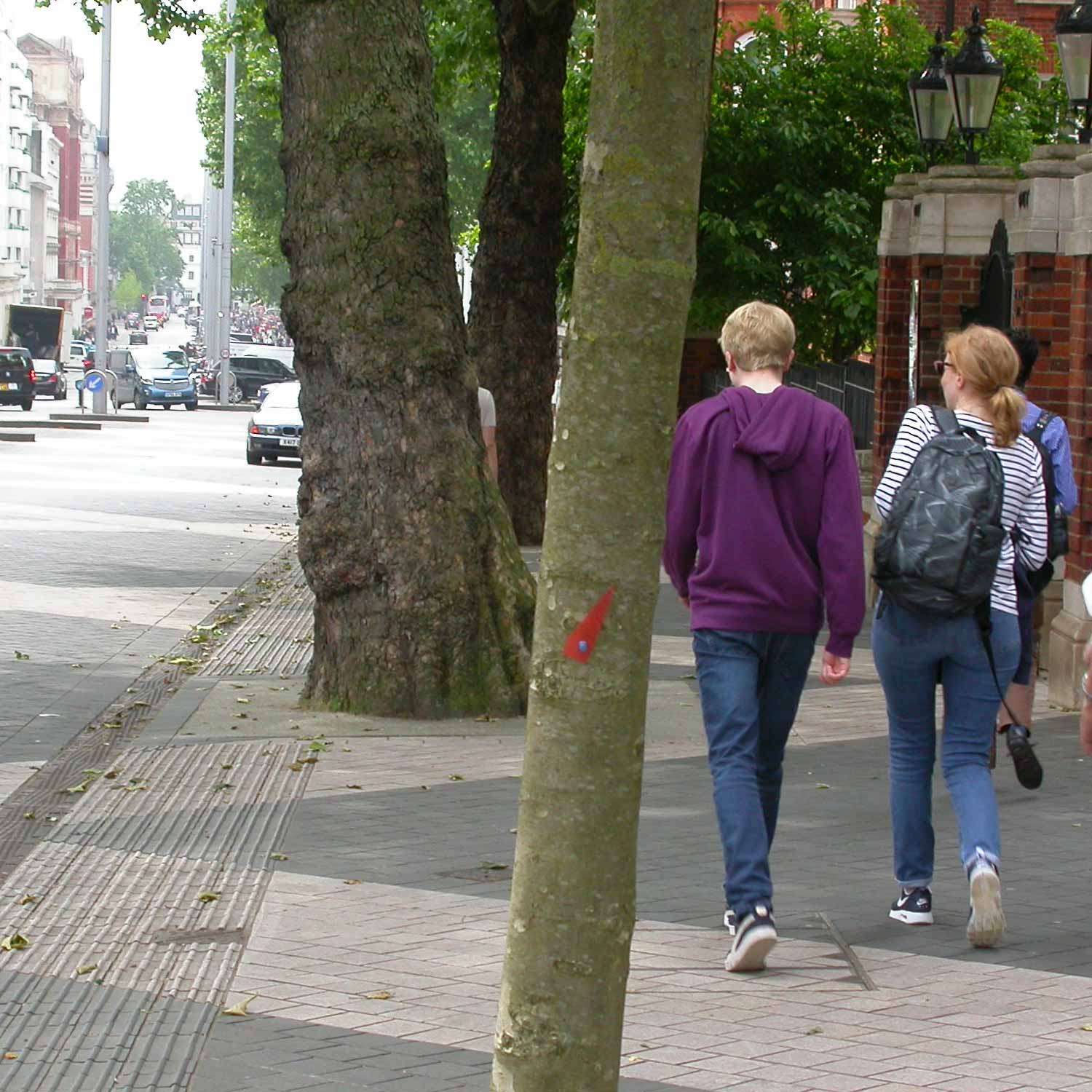
[attachment=3]Screenshot 2025-12-10 6.47.49 PM.png[/attachment] I find A. pyracea to be fairly common now on large twigs of Populus species, especially P. tremula in the wild and P. alba in parks and cemeteries. It is much less common in my experience on other species of tree but your finding of it on cherry doesn’t worry me too much because it does crop up in surprising places and on surprising hosts. The red marker on this young London Plane tree points to a colony of A. pyracea in Exhibition Road in London.

[attachment=2]pyracea exhib.jpeg[/attachment] As you have described, the thalline margin of A. pyracea tend to be conspicuous on young apothecia but then become rather excluded as the apothecia mature. I tend to identify A. pyracea on sight now that I am familiar with it. If I found something in this group of holocarpa type things on a damaged tree trunk I might allow myself to get excited by the prospect of C. luteoalba and look for the somewhat distinctive spores that species is supposed to have but so far I haven’t come across C. luteoalba. When growing on young bark, especially twigs and small branches, I don’t often bother looking at the spores because there is not much diagnostic to distinguish between A. pyracea and A. holocarpa. More important seem to be the larger apothecia of A. pyracea with a thalline margin (visible at least on young apothecia).
Here are some images of specimens which I have named A. pyracea.

[attachment=1]pyracea1.jpeg[/attachment] 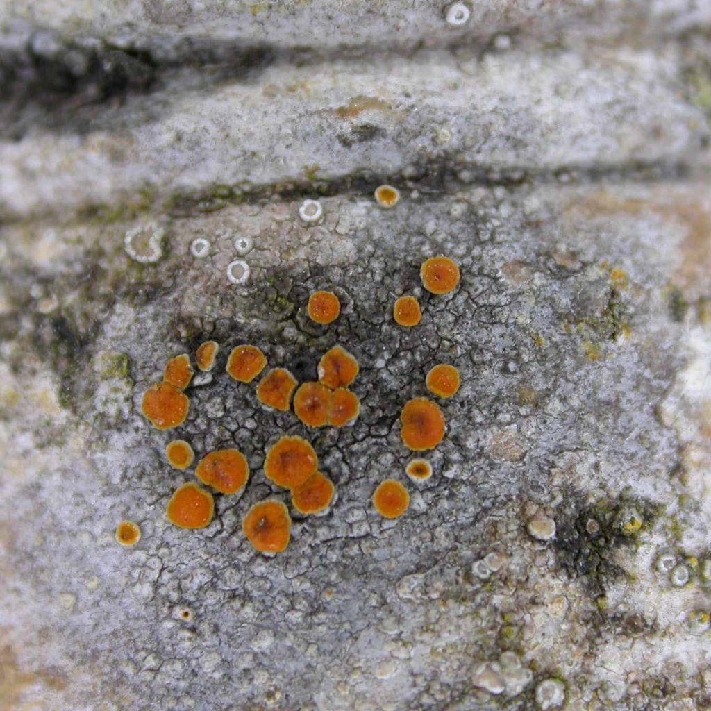
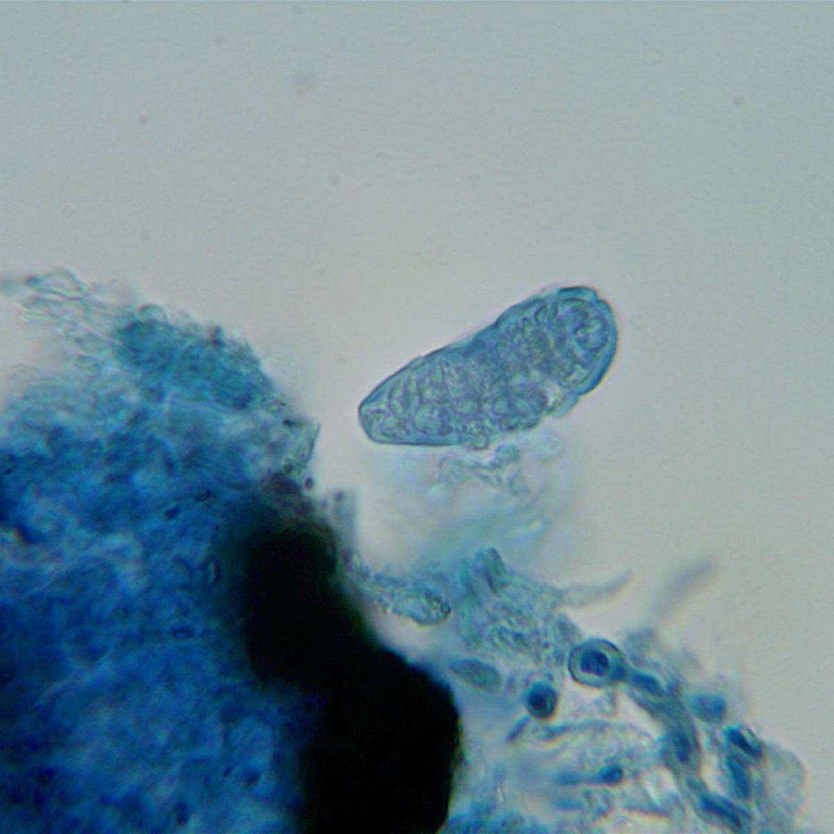
[attachment=0]pyracea2.jpeg[/attachment] I’m not quite sure what to say about the algal cells. You are probably right but the ones that are most unambiguously dividing by binary fission don’t have any associated hyphae attached to them. Any such hyphae have either come loose on squashing or one also has to be careful because many lichen thalli have some unassociated algae growing on them, often Chlorella type which divides by binary fission. I think this lichen will have to remain unnamed for the moment since we don’t have any apothecia, no C reaction has been detected and I can’t be certain about the photobiont. There have been many instances where I have found mysteries in churchyards which I couldn’t name when I first encountered them. A fair proportion of them eventually became resolved one way or another. I will keep your lichen in mind just in case I have a flash of inspiration with a different/better suggestion.
Since nobody else has made any suggestions, I am going to give a very tentative gut reaction. The habitat and the appearance of the convex areoles put me in mind of Trapelia glebulosa s. lat. This ought to be ruled out by your report of a lack of C reaction but the C+ red of Trapelia species is often missed, especially if performed in the field. With moist material in the field the reaction is often completely inapparent. As with all spot reactions, the only truly reliable way is frugal application of strong fresh reagent to dry material under a dissecting ‘microscope. There is something rather distinctive about Trapelia and a few other genera and that is the way the photobiont cells divide. In Fuscidea, Trapelia, Trapeliopsis (and a special case in Micarea) the algal cells divide by binary fission rather than forming a cluster of daughter cells before division. Here is a drawing of the algal cells and host hyphae in Trapelia; the binary fission producing a paired or quad appearance.

[attachment=2]EvfC6YKXAAQ_Pqf.jpeg[/attachment]
Here is a section of Fuscidea lightfootii thallus.
[attachment=1]DoWX2p2XkAETQRu (1).jpeg[/attachment]
For comparison here is the formation of a cluster of daughter cells as seen in Caloplaca obscurella.
[attachment=0]GFGvWsKWMAU2bLK.jpeg[/attachment] So, if you want to pursue this, you might want to look at the cell division of the photobiont to strengthen or disprove my suggestion. Of course I have taken photos of particularly convincing examples of binary fission and of division into clusters of daughter cells and it isn’t always so easy to decide which you have but a careful examination of a portion of thallus, mounted in K to relax the structure, usually allows one to decide between these two types of division.
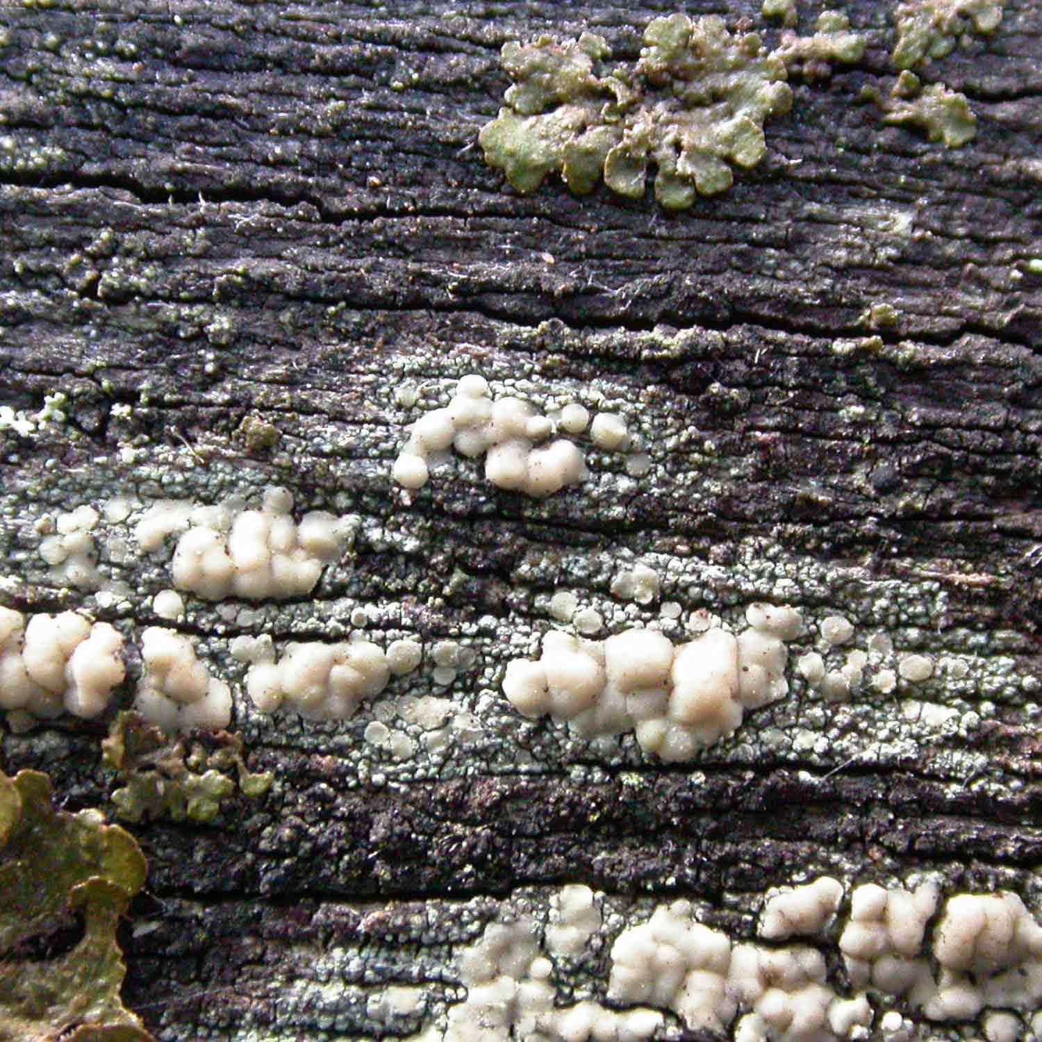
Here is L. symmicta. 
[attachment=2]symmicta.jpeg[/attachment]
Here is L. stenotropa on sandstone, showing the slight blue tinge.
[attachment=1]stenotropa2.jpeg[/attachment]
Here is some L. stenotropa on chemically treated lignum, the bright bluish colour presumably due to accumulated copper compounds.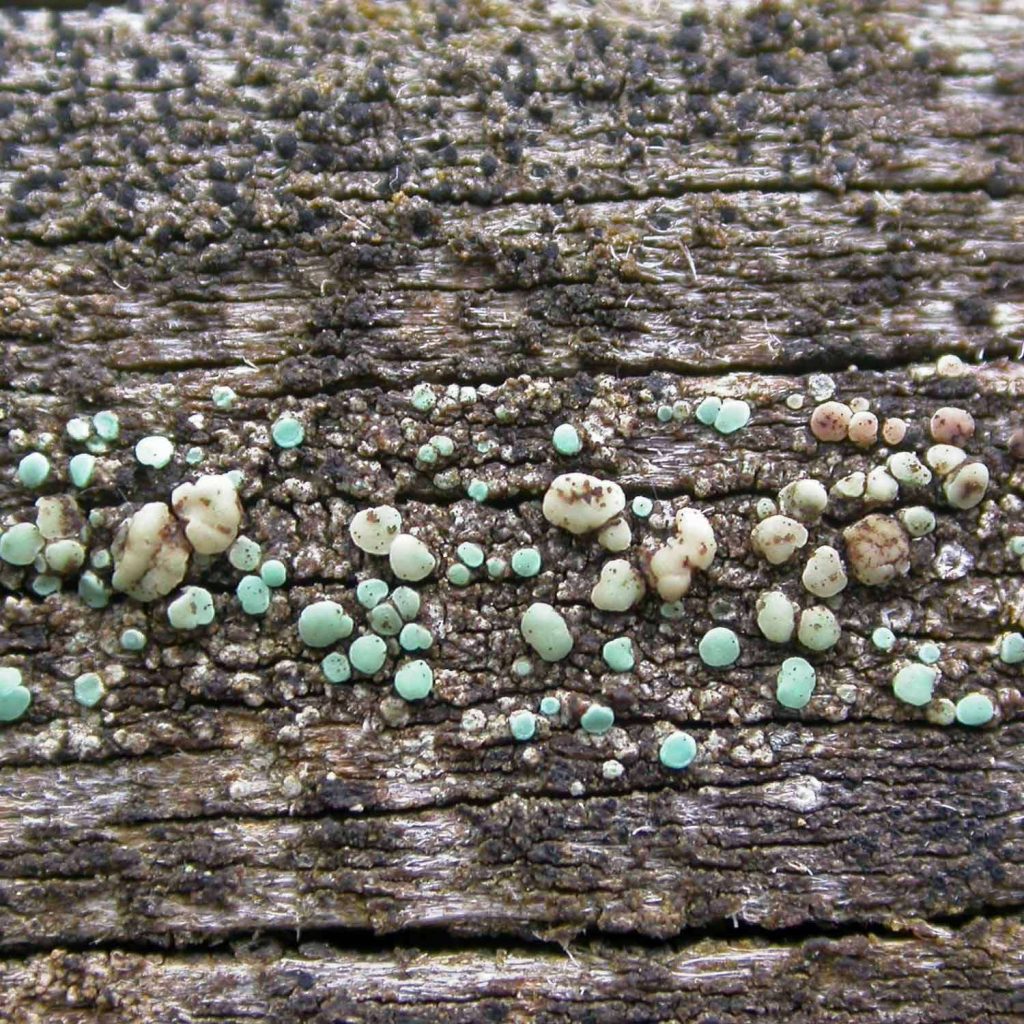
[attachment=0]stenotropa1.jpeg[/attachment]
When I am uncertain about which species I have, I often compare sections of known specimens of the main possibilities. There are a lot of subtle differences which defy easy description and hence aren’t often found in published accounts. For example there are almost certainly differences in the epithecial granules between L. symmicta and L. stenotropa/polytropa but if I have ever made detailed comparisons of these I don’t have the notes to hand. This has often helped my way towards an identification even if I eventually have to use published characters to formally decide on the name I am going to apply. In the case of the lichen in this topic/post I think I am happy to confirm it as L. stenotropa unless anyone has any doubts.I am going to suggest Lecanora stenotropa. This was my gut reaction on first glance at the photos and seems to be backed up by your observations. I think there is a bit too much margin showing for L. symmicta in which the apothecial margins get excluded at a very early stage. The discs of L. symmicta are usually distinctly (pale) brownish (or sometimes darkened) whereas the apothecia in your material are mostly a cleaner paler colour, much of the colony having the slight blue-rinse appearance typical of L. stenotropa. L. symmicta contains arthothelin and perhaps other xanthones and your chemical tests have not detected any hint of xanthones (no C+ orange, no K/UV+ green [wet]). I think you have demonstrated the presence of usnic acid with your KC+ yellow reaction of the apothecial disc and the absence of xanthones. I also carefully add K then C (with cocktail sticks) to apothecial discs when I suspect L. stenotropa or L. polytropa and want to make sure it is one of the usnic acid containing Lecanora species. Assuming we have ruled out L. symmicta we now have the two most likely usnic acid-containing contenders on such lignum to consider, L. polytropa and L. stenotropa. Out of these two, your spores have the narrowly ellipsoid shape of L. stenotropa. LGBI2 states that the spores of L. stenotropa are 3 to 4 microns wide but I usually find some are wider, up to 4.5. Rightly or wrongly, I am sort of ignoring the one that is just over 5 microns wide, thinking it might look wider due to an optical effect apparently caught up in the hymenium. The spores of L. polytropa are broadly ellipsoid, more like hens eggs. In the East Midlands I come across L. stenotropa much more commonly on lignum (chemically treated fence and gate rails) than on its more ‘typical’ habitat of sandstone. I do find it occasionally on old sandstone headstones and I pick out candidates for it by its less yellowish colour compared to L. polytropa and it often having a slight blue tinge. On lignum L. stenotropa sometimes becomes more spectacularly bluish coloured due not only to its inherent appearance but also by apparently concentrating copper compounds in its structure.
I think its appearance strongly suggests Lecidella scabra. I base this on the presence of a distinct lumpy/warted grey thallus and a hint of bluish green in some of the exposed soredia. In contrast, Lecanora expallens tends to have nothing more than a thin pale film-like thallus and soredia generally in a more even colour (pale green without bluish green tinges) and a more even and thin cover of powdery soredia. It appears to be growing next to Candelariella vitellina, both of which grow on exposed lignum and the C. vitellina indicates that the surface is also suitable for L. scabra. With its bluish tinged soralia, your lichen also somewhat resembles Buellia griseovirens but that species is ruled out because it is UV- (doesn’t contain xanthones which your lichen certainly does). If you ever get the chance, if you compare the fluorescence of L. expallens and L. scabra, I think you will find that the latter has noticeably more intense UV+ orange like that shown in your photos.
I must admit that I never got very far with investigating C/UV reactions but that doesn’t mean that they have no potential so I was pleased to see your C/UV+ yellow even though it didn’t inform my opinion of your specimen.
As an example of how difficult it is to use morphology to distinguish these brown Acarospora/ Myriosporaspecies we need only to look at A. fuscatawhich has a convenient C+ red reaction so we can recognise it. I will post a couple images which show that it can vary from an intricate dark blackish-brown angular mosaic sort of appearance to a pale brown semi-squamulose appearance. The dark pigmentation is concentrated in the apothecia and hence slow-growing, highly fertile material is dark and intricate while rapidly growing vegetative portions tend to be pale and exuberant. 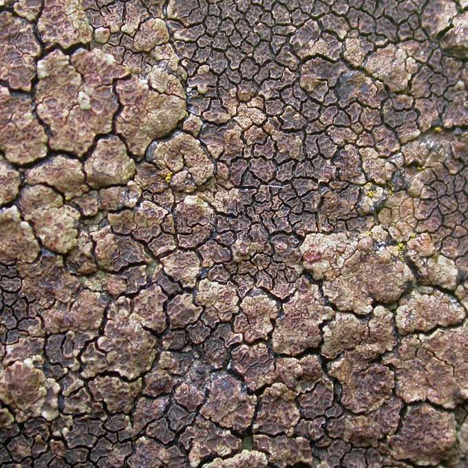
[attachment=4]FMmlk-XWYAUurW0.jpeg[/attachment] 
[attachment=0]FMmk8TrXwAIjsU_.jpeg[/attachment]
Here is a rather thinly developed colony ofM. rufescensshowing the orange glow on wetting (but as mentioned in an earlier post, this may be characteristic but not diagnostic). 
[attachment=5]DVsTIuUX4AAJw7F.jpeg[/attachment]
I mentioned the mis-identification ofPolysporina simplexas A. nitrophila( muddii) and here is an image of it in the lower two thirds of the photo with A. fuscataabove. Note that P. simplexhas a habit of seeming to pluck out grains from the substratum (more aggressively than other species) and incorporating into the thallus. This is also seen when it has a thinner thallus growing on slate where it seems to pluck at the slate. 
[attachment=3]EPzDedPUwAEP53h.jpeg[/attachment]
Here we have one of the few images I have of a specimen which I recorded asA. nitrophila( muddii). Admittedly it has lots of apothecia which seems to slightly contradict my earlier comment about ‘lots of thallus compared to apothecia’. 
[attachment=2]EPzANEiUYAA8kQR.jpeg[/attachment]
Here is what I record asP. simplexin the left hand side and the right hand side was C+ red ( A. fuscata). 
[attachment=1]EPy_V2mXkAIQIWG.jpeg[/attachment]
I looked up Knudsen’s comments aboutA. versicolorin which he highlights the problem of a lack of distinctive microscopic characters in these brown Acarosporaspecies: “The hymenium height, ascospore size and cortex thickness of A. versicolorare rather common in the genus. The regular occurrence of pruina and negative C reaction are the main diagnostic characters”. While I am happy to give my two penn’orth, these brown Acarosporas. lat. things are difficult to identify when they lack positive spot reactions and also lack other distinctive characters. When I added A. versicolorto the British list, with Paul’s help, I was only confident enough to do so because the presence of pruina combined with negative reactions seems to be reasonably diagnostic for this species. Unfortunately microscopic characters seem to be little help in separating many of the species. When I was working on the identity of British A. versicolorI was met with comments such as “ A.versicolorbelongs to the morphological group of brown Acarosporaspecies lacking secondary metabolites. There are many names and many taxonomic problems in this group”. I am not sure whether the thallus of the Trotton specimen is nodulose; I would err towards the interpretation that the nodulose overall appearance results from the abundance of apothecia, including many immature ones developing within the thallus. Even if the thallus is nodulose, this character may not be particularly diagnostic for
A. muddii. I will include some screenshots from a paper that suggests that Myriospora rufescens can also have a nodulose thallus. 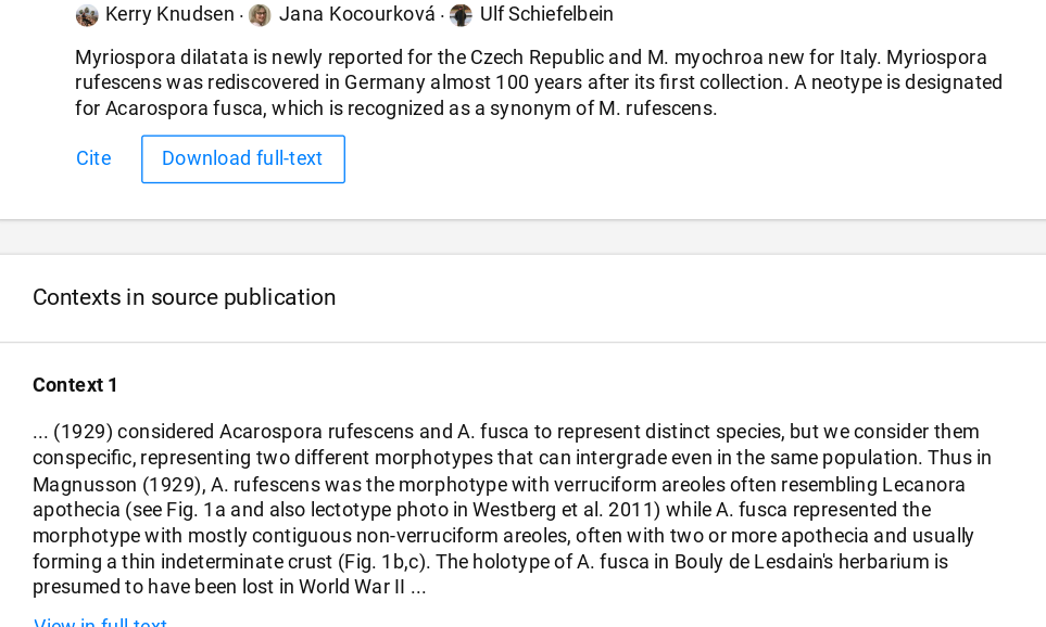
[attachment=1]Screenshot 2025-11-27 2.50.13 PM.png[/attachment] 
[attachment=0]Screenshot 2025-11-27 2.51.56 PM.png[/attachment] As Peter has noted, the taxon which we record as BLS number 18 is still listed as
A. nitrophilain the Taxon Dictionary and the Species Maps. The distribution map shows a scatter of records for A. nitrophilaacross England but there is an additional problem about the reliability of these records. When I started attending meetings of the Churchyard Working Group, I noticed that A. nitrophilawas used as a bit of a dump category name for brown crusts on acid stone which lacked spot reactions. I subsequently realised that some or many of these crusts were actually Polysporina simplexin its pycnidiate form with a well-developed, dark brown thallus, proved to my satisfaction by some such material occasionally having typical P. simplexapothecia. Back to the Trotton specimen and my gut reaction leans towards a rather atypical form of
M. rufescens, perhaps modified in morphology by the suspected metal influence. The apothecia glow orange when wet like those of M. rufescensbut I this may not be a particularly diagnostic character. My impression of the few times I have (perhaps tentatively) recorded A. muddii/ nitrophilais that there tends to be quite a lot of thallus compared to the number of apothecia but that again may not be too reliable. I do have a few collections of
Acarosporawhich I encountered as one-offs; they looked different and I was unable to name them and they remain unresolved in their packets. It was only when I got to know A. versicoloras a distinct entity through repeated encounters that I felt motivated to work on its identity. If I found the Trotton colony and I really wanted to pursue it, I think I would try making comparative sections, comparing it microscopically with known material of
M. rufescens, A. muddii(if such a specimen was available) and any other brown Acarosporaspecies I could obtain. There are many subtle features that are difficult to describe and often not present in published descriptions but which can strengthen or weaken a hunch. Although I found this method useful when working out the identity of various lichen species, it is not scientifically rigorous enough to use for naming but certainly helps in getting to know a group and sometimes leads to the discovery of useful new characters. I apologise for such an inconclusive reply!
It may be useful if I remind people of the two main ways of using Pd. The best and preferred method is to drop a couple of drops of alcohol (meths, vodka or other strong ethanol you have to hand) on to a very few crystals of Pd (much less than a pinch of salt). This makes a very strong solution of Pd which can be transferred to the specimen using a cocktail stick, corner of a razor blade or fine glass rod. The traditional container in which to make the solution is a laboratory watch glass but I often use an old bottle top which can then be wrapped and discarded when finished with. You don’t have to trash the tiny amount of Pd in the ‘watch glass’ after the first use. It will soon dry and can then be kept covered for several more uses. Pd oxidises rapidly and becomes useless when in solution but oxidises slowly when dry. You will know when the crystals in the watch glass are getting too oxidised because they go dark (blackish) in colour. I suggest keeping the watch glass covered in between uses because the powdery (recrystallised) residue can disperse in air currents and, as well as the potential health risk with Pd dust in the air, any escapee dust can cause permanent dark stains on anything it comes into contact with. If you are at all house proud it is probably best to use Pd in a garage or shed.
All spot tests (K, C and Pd) are best made on dry material, using frugal spots of strong reagent and observed under a dissecting microscope. Of course we often want to conduct spot tests in the field and this is fine as long as we realise that the reactions may be weak or even missed altogether, especially if the specimen tested is at all damp or we clumsily dump an excessively large drop of reagent on it. The traditional way of using Pd in the field is Steiner’s solution, an aqueous solution of Pd made stable by the addition of sodium sulphite. Steiner’s solution has a deserved reputation for producing weak reactions. This is because Pd is sparingly soluble in water. I think I can claim to have made the simple innovation of a strong and stable solution, Powell’s alcoholic Steiner’s solution. Pd is readily soluble in alcohol, sodium sulphite readily soluble in water. I use something like vodka as a happy mixture of alcohol and water in which to create a strong, perhaps even saturated solution of both sodium sulphite and Pd. This creates a strong solution of Pd which can be in used in the field and usually lasts for several months. I only make up about 5 mils at a time, plenty for numerous tests.
I agree that this is an interesting post, investigating a source of a Pd substitute for those who can’t get hold of the real thing. As for getting hold of Pd itself, I managed to purchase a couple of kilos soon after I started to study lichens and when I realised how difficult it was to get hold of. I can supply small quantities of Pd crystals, free of charge (within UK), to anyone who has a genuine interest in lichen studies and who reassures me that they have read about and understood the potential health risks. I treat Pd with respect but I am not personally concerned about the minimal exposure to it that results from careful and frugal usage of it. I always appreciate it if anyone who receives some Pd from me sends me some stamps or other token acknowledgement of the small amount of trouble and expense of sending it out. I supply it in resealable plastic bags (triple-bagged to minimise any risk of spillage) which should be fine for long term storage but some prefer to transfer it to small glass bottles. The important thing is to try to minimise oxidation and the most important things are to keep it dry and minimise the air it is in contact with. Anyone with a genuine purpose for using Pd, including general amateur lichen recording, can private message me on this website or can probably find an email address for me on the BLS website under the ‘Contact Us’ tab (Local Contacts).
If anyone knows of other sources of Pd then it is always useful to hear of them.
-
AuthorPosts