JennyS

Forum Replies Created
-
AuthorPosts
-
Might they be the pycnidia of a bryophilous fungus? https://bryophilous.co.uk/https://bryophilous.co.uk/” class=”bbcode_url”> Yes, utter nightmare – but my apologies to both you and Janet as this is my fault for not double-checking and correcting the townland name I’d noted down when recording and, as you realised, this Tullig More is in the Barony of East Muskerry. And for the photographs in this thread:
Quote:Dromore, located near Bantry in County Cork, is in the
Barony of West Carbery (West Division). It is specifically situated within the civil parish of Caheragh.
I got thoroughly confused whether to name locations from my site grid references first by Electoral Division, Civil Parish or Barony and then townland that I’ve feeling in the end I sent her most of my Irish records by townland and then grid reference. I’ll email her and check her preferred format to avoid future headaches!
Thanks Neil – those sparse granules in the epithecium were confusing me! The location for the specimen I sent you was
R619 – Tullig, W479774, mid Cork vc H4. On a twig from a roadside Ash, collected 2012-04-19.
Other species from there included Acrocordia gemmata, Arthonia cinnabarina, Enterographa crassa, Lecania naegelii, Lecanora carpinea, Parmelina pastillifera, Phaeographis smithii, Pyrenula chlorospila and macrocarpa and Rinodina sophodes. There was a small amount of Xanthoria parietina but no Physcia.
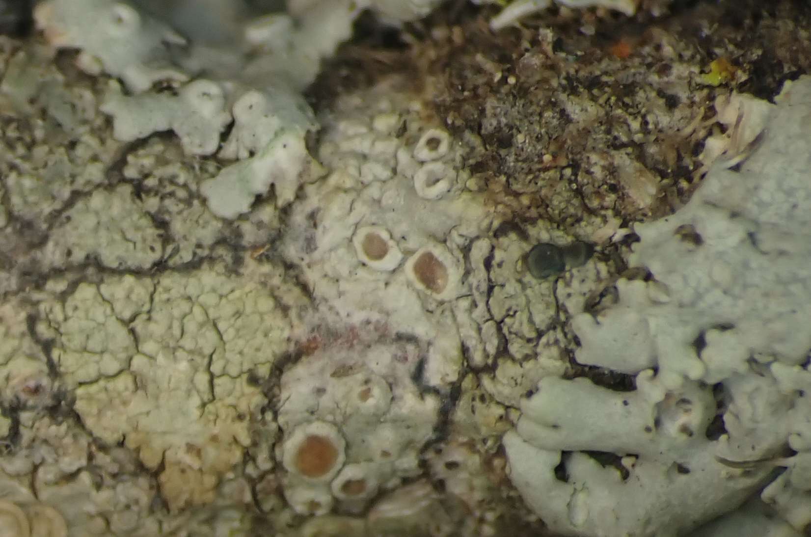
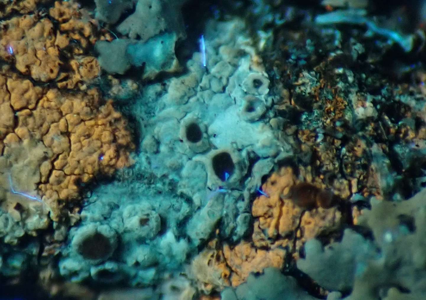
The one shown here was from Bantry – Dromore, W051463, West Cork vc H3, collected 2025-06-26.
It was on a branch rather than a twig, and probably from my daughters woodpile so either hazel, ash or possibly oak.
Species recorded there
https://www.irishlichens.ie/habitat-dromore.htmlhttps://www.irishlichens.ie/habitat-dromore.html” class=”bbcode_url”> I emailed the details and images to Paul Diederich and his reply is below:
Quote:From the anatomy of the pycnidial wall, I can confirm that it is a Didymocyrtis, and not Briancoppinsia, which is also growing on parmelioid hosts.
As D. cladoniicola and C. foliaceiphila are both known with certainty from hosts that are not related with Cladonia, e.g., Parmelia or Ramalina, confirmed by DNA sequences, an occurrence on Punctelia does not represent any problem.
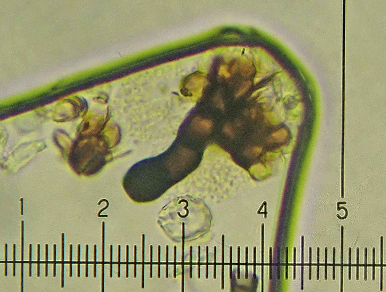
Conidia of D. cladoniicola are (3.8–)4.7–5.9(–7.3) × (2.0–)2.4–3.0(–3.5) µm, l/b (1.4–)1.7–2.2(–2.
 , while those of D. foliaceiphila are (5.0–)5.8–7.1(–7.5) × (2.0–)2.2–2.7(–3.0) μm, l/b ratio (2.0–)2.4–3.0(–3.5)
4×6 agrees with cladoniicola, while 2-2.5 is more close to foliaceiphila. In my opinion, you should measure a larger number and also calculate the lenght/width ratio statistically, as that character may be the best to distinguish both.
Nothing remained of the original specimen so it will have to remain as Didymocyrtis sp. but I was hoping someone could explain how to calculate the lenght/width ratio statistically?
Hi Neil – investigating more Irish Lecanora’s last night and I think this L. argentata (POL crystals, no granules) also seems to be showing a trace of gangaleoidin in the margins? 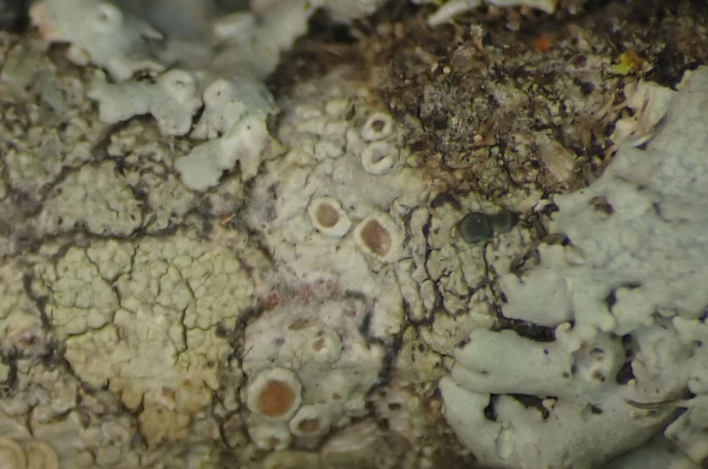
[attachment=1]Lecanora argentata 240828 013.jpg[/attachment] 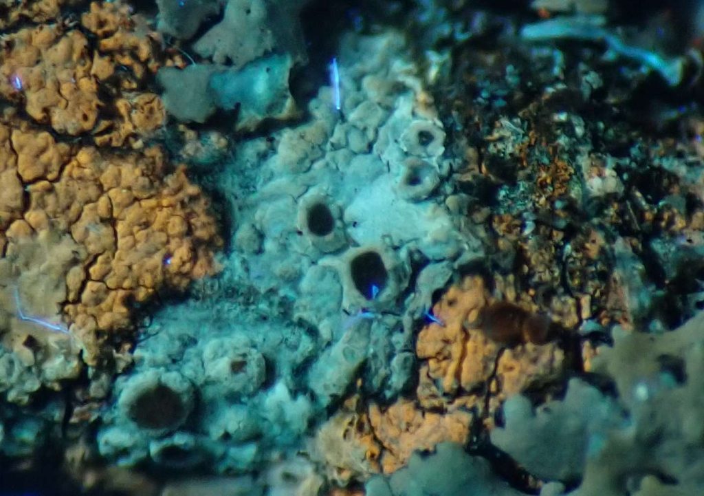
[attachment=0]Lecanora argentata 240828 019 UV.jpg[/attachment]
Lecanora argentataThanks Neil, I should have said the whiter thallus (UV photo) with smaller apothecia on the right was L. albella. I’ve a few more to check and then I’ll send you any possibles
Found an Ash twig collected 2012 from mid Cork (vcH4) and started investigating the Lecanora’s on it. Might this one be L. pseudargentata, though I’m slightly wary of the UV results I get? (Added bonus, specimen I can send!) 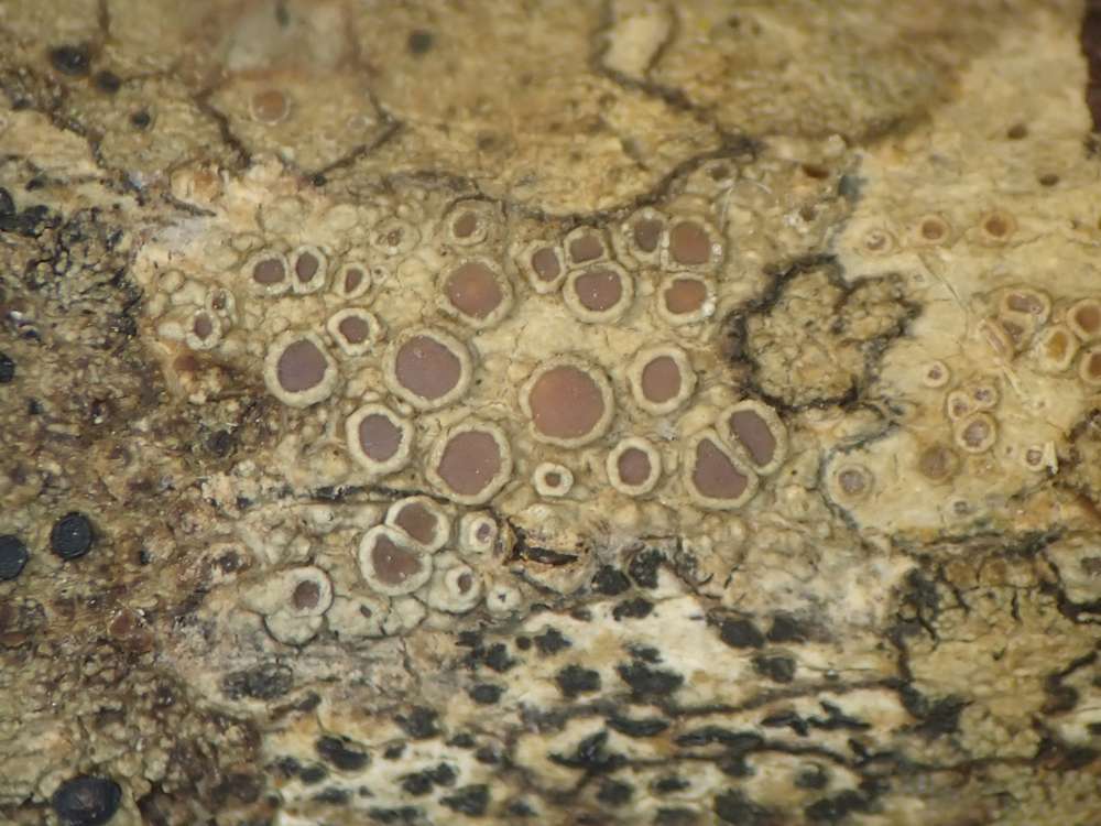
[attachment=3]w Lec 2 120419 009.jpg[/attachment] 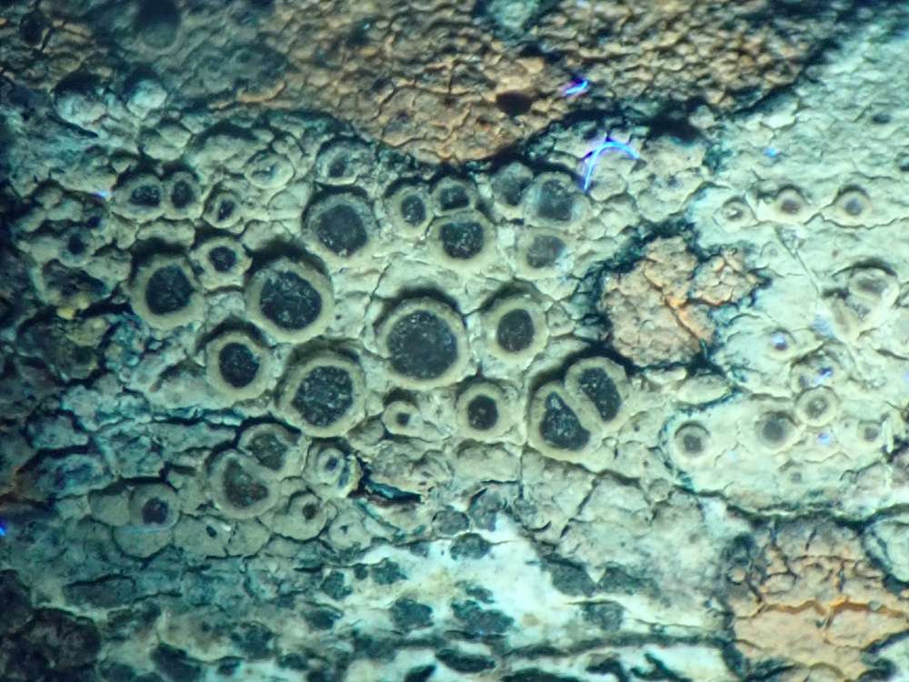
[attachment=2]w Lec 2 120419 010 UV.jpg[/attachment] 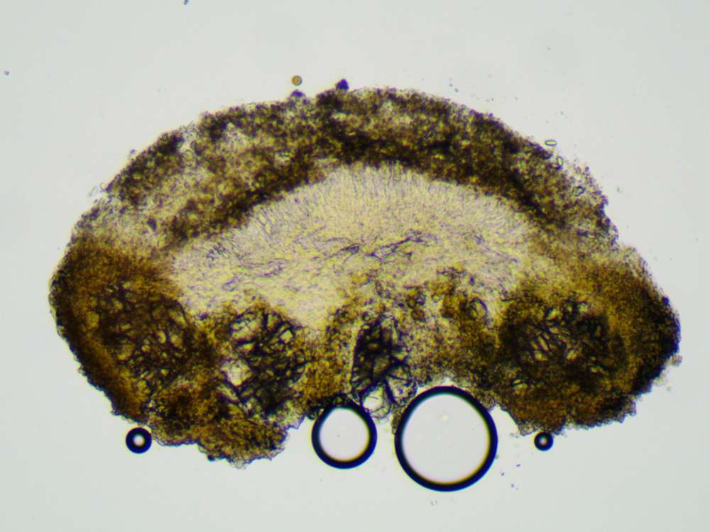
[attachment=1]w Lec 2 120419-3.jpg[/attachment] 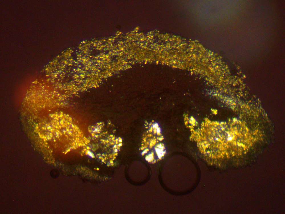
[attachment=0]w Lec 2 120419-3b.jpg[/attachment] Many thanks for further details and UV photographs Neil, and the photographs and micrographs sent – though gmail seems to have scrambled the order so checking the file names (1,1b etc) will clarify which micrograph goes with which photograph. Thanks Neil – and identification is so much more straightforward with spores! When I posted the query on the FB Lichenicolous page Wolfgang von Brackel suggested perhaps a Roselliniopsis (and Roselliniopsis tartaricola is recorded on Pertusaria leioplaca).
However what must be the “apical phialidic synanamorphs” on this one appeared slightly different and seemed to more resemble those shown in Lichenicolous Fungi vol.2 page 355 for Roselliniella stereocaulorum than the Roselliniopsis tartaricola illustration on page 356?
But I am so totally out of my depth with these!!!
? Roselliniella
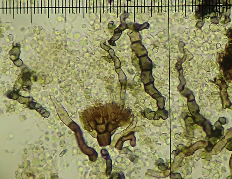
[attachment=1]zz Pert Q1 120108 064.jpg[/attachment] Roselliniopsis tartaricola (from previous post here)
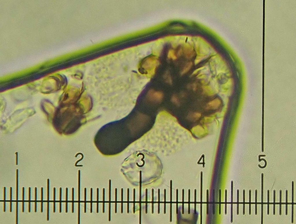
[attachment=0]zz Roselliniopsis tartaricola 100209 029.jpg[/attachment] Thanks Neil, and Cladonia cf coniocraea was my best guess but I tend to run a mile from Cladonia squamules! December 5, 2025 at 3:51 pm in reply to: Taeniolella pertusariicola on Varicellaria hemisphaerica? #46774And a slightly better image of the “apical phialidic synanamorph” (I think I need to swallow a taxonomic dictionary to make sense of the descriptions in LF1 and 2!)
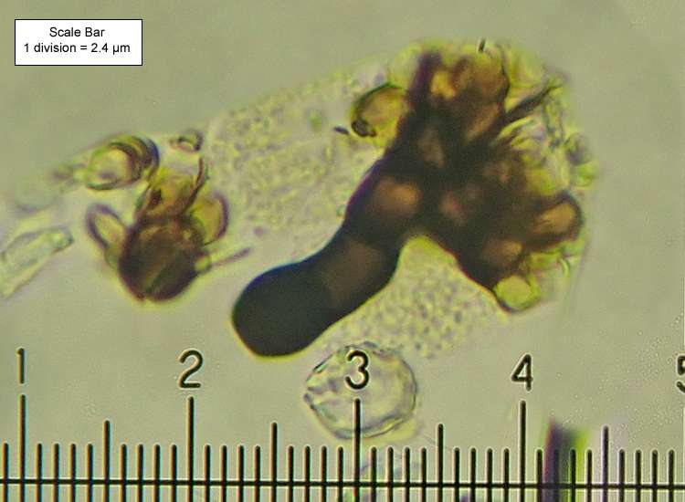
[attachment=1]z Roselliniopsis tartaricola 100209 029.jpg[/attachment] 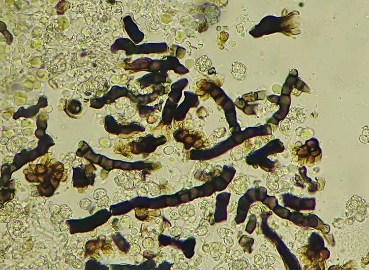
[attachment=0]z Roselliniopsis tartaricola 100209 041.jpg[/attachment] December 4, 2025 at 6:22 pm in reply to: Taeniolella pertusariicola on Varicellaria hemisphaerica? #46772Ah, thanks Neil – I checked the FLF2 Taeniolella pertusariicola description but not Roselliniopsis tartaricola. Are these below what is meant by “apical phialidic synanamorph”?
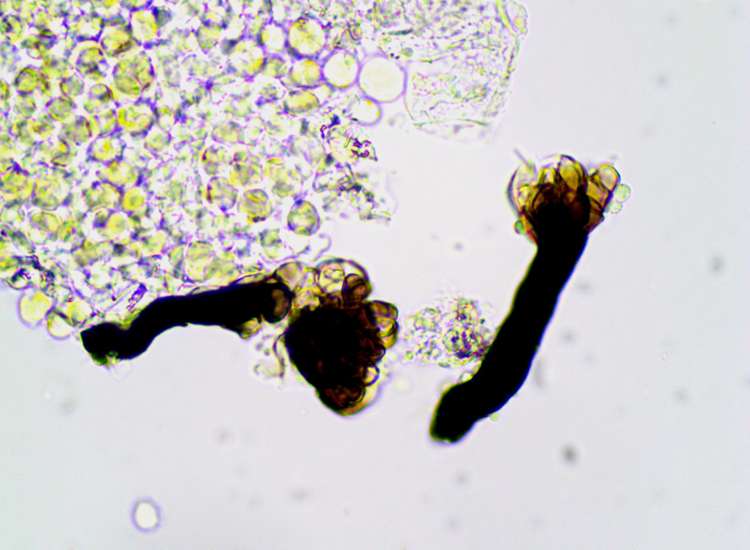
[attachment=0]z Q Roselliniopsis tartaricola 100209-3.jpg[/attachment] December 4, 2025 at 1:36 pm in reply to: Lichenopeltella pannariacearum on Parmeliella thriptophylla #46768There’s a tiny bit of the host thallus left, but as it took two attempts to find spores I’m not sure if any Lichenopeltella pannariacearum remains – will send in hope….. Glad the other specimen arrived safely!
December 4, 2025 at 12:01 pm in reply to: Lichenopeltella pannariacearum on Parmeliella thriptophylla #46766Thanks again Paul. Yes, spore measurements match with those given on the FGBI website (I got briefly lucky with the description but the images wouldn’t load)
Quote:Ascospores (14.5-) 16-17 x 5-5.5 µm (15.5-18 x 4.9-5.5 µm in GB&I material), hyaline, slightly constricted at the single ± median septum, smooth, the upper cell with three pairs of lateral flexuous hair-like setae inserted together near the septum, without a gelatinous sheath or appendages.
Thanks Paul, I hadn’t managed to find conidia measurements when I posted this but yes, mine seem much too big! There was some left with what appeared to be pycnidia in varying degrees of development so I’ll get it in the post to you. -
AuthorPosts