JonasRavn

Forum Replies Created
-
AuthorPosts
-
For what it’s worth, I have collected several specimens similar to yours from exposed faces of roadside trees and concrete in Denmark, in the hope that they might be P. sciastra and so far I have concluded that they are all forms of P. orbicularis. In more well developed specimens, it is usually possible to see orbicularis-like circular soralia with quite granular soredia, that eventually develop cortex and appear isidia-like. It is my understanding that the isidia in P. sciastra are mainly marginal, so the presence of the “isidia” in circular laminal/submarginal patches might be a way to rule out P. sciastra. I am not an expert in the genus, but I have looked at hundreds of thalli in the past year, trying to come to terms with the bewildering variability of P. orbicularis. I was lucky to have a few of my collections sequenced, one small-lobed morph with terminal capitate soralia and one with long marginal soralia and marginal rhizines, but so far everything has been orbicularis! 
I might be wrong – determining wether something is a lichenicolous fungus or the pycnidia of the “host” is a constant struggle for me. I have spent more time than I care to admit on lichenicolous fungi that turned out to be nothing more than host pycnidia The cilia/rhizines look fine for C. foliacea and I suspect that the pycnidia actually belong to the “host” as well. They can be quite numerous on the squamules of some specimens. Thank you for your detailed reply! After reading your notes I am both relieved that I am not the only one struggling with this specimen and I am now leaning heavily towards R. pityrea as the most likely ID. I have seen that species in fertile condition and much to my delight, the spores are very characteristic and makes for an easy identification in an otherwise quite difficult genus.
My main concern with identifying the current specimen as R. pityrea lies in the quite well developed (and in places sub-squamulose) thallus. But that could be a variation of the species that has yet to make it into the various descriptions – I would not be surprised if this is the case.
I will visit the site again in the hope of finding an apothecium or two. If I do I will update the thread here.
All the best,
Jonas
Hi Mark and Alan, First of all, thank you for your replies.
C. contorta ssp. hoffmanniana was actually one of the two IDs I considered (the other being C. caesiocinerea), but the habit was very different from the other examples of this taxon I have seen, so I rejected it at first, but perhaps I should reconsider.
There is no reaction with K – neither in section or from spot tests directly on the thallus.
I have tested the rock with different acids with no visible reaction. There are a few calcareous rocks in the stone wall, so perhaps runoff from these is what is causing the “shift” in substrate.
I don’t remember offhand if any of the calcareous rocks were directly above the sandstone, but I think that it was one of the rocks on top of the wall. The spore count and shape fits with C. contorta spp. hoffmanniana, so if nothing else comes of this discussion, I will tentatively name it as such
All the best,
Jonas
If it helps, I can add that the species was quite abundant on the east-facing surfaces of old Pinus sylvestrisstanding at the edge of a forest and thus exposed – about a kilometer from the nearest coast. Good luck on the search! Thank you Neil for the reply! I am convinced that you are right – the fit is very good. I am glad to put this troublesome specimen to rest!
All the best,
Jonas
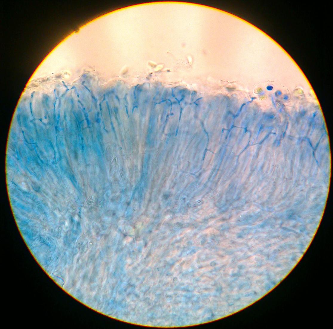
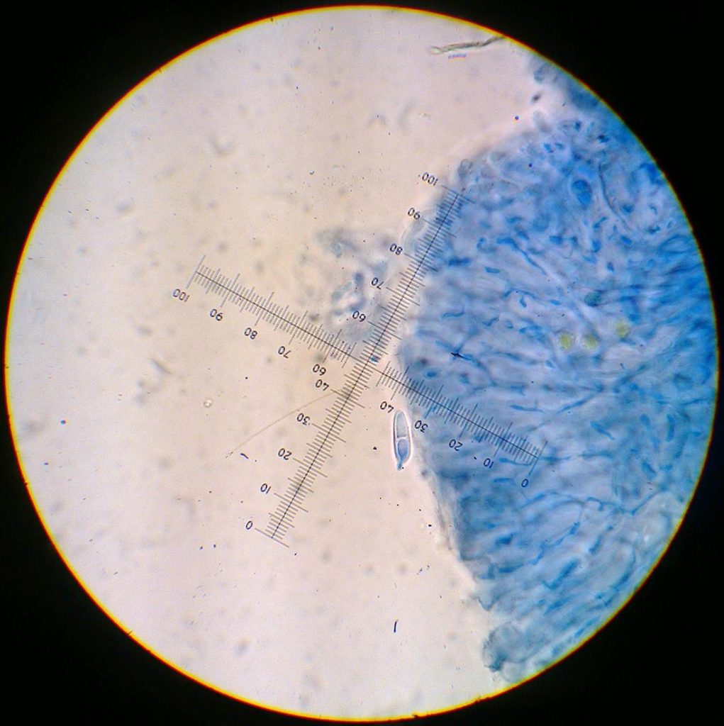
I made an k/ink/vinegar-stain and found that while some paraphyses were simple, branching and anastomosis was common: 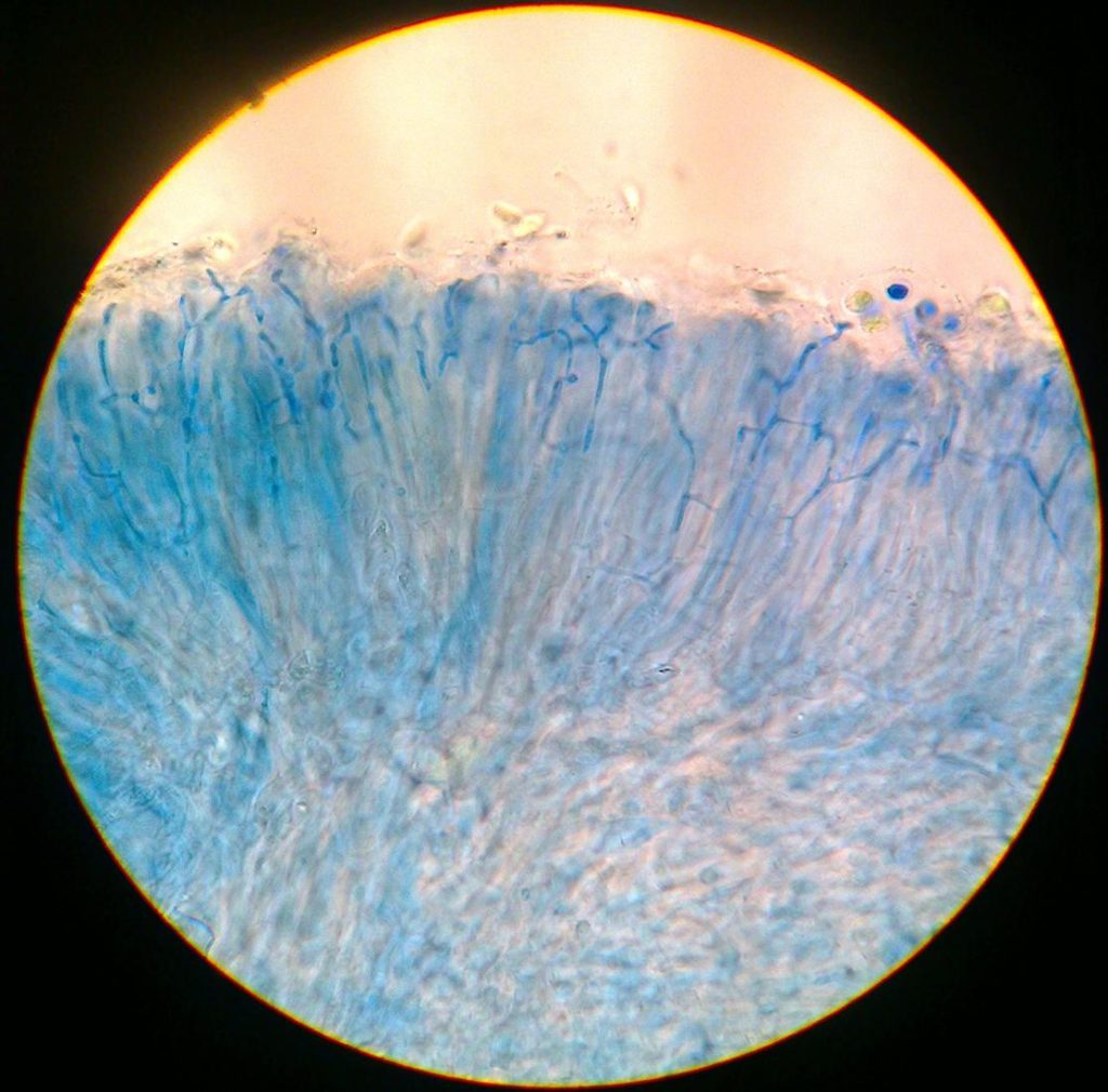
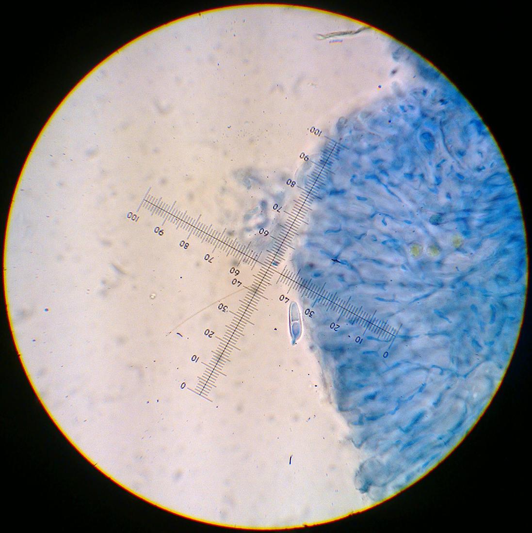
[attachment=2]Billede3080.jpg[/attachment] And much to my surprise, some spores appeared one-septate (I found three out of ~50 spores like this):
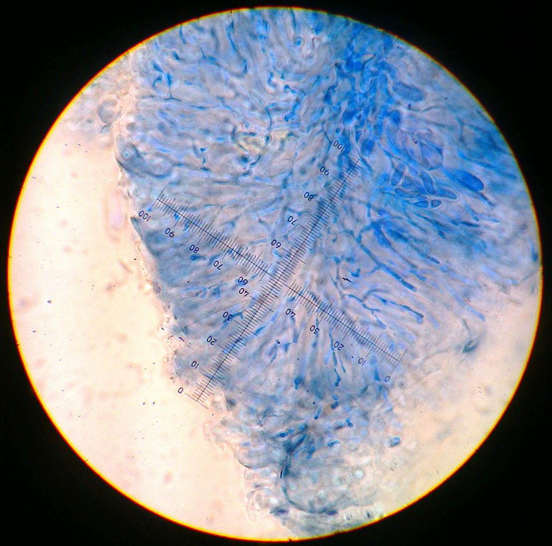
[attachment=1]Billede3081.jpg[/attachment] Lastly a picture of the excipular hyphae:
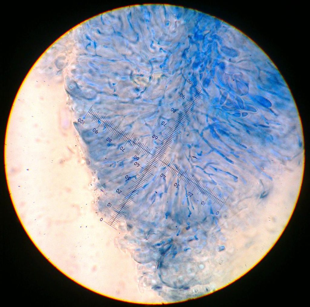
[attachment=0]Billede3083.jpg[/attachment] The thallus might have an extremely faint UV+ orange reaction (only barely visible with powerful torch in pitch black room, xanthone?), but it is too weak to capture on a photograph.
I have tried keying it as a Lecidea without much luck – it shares some characteristica with L. hypopta, but it is not a perfect fit. I cannot rule out L. exigua, but the paraphyses do not appear capitate above which causes some hesitation.
The Revision of British and Irish Lichens vol .11 (Ramalinaceae) points me in the direction of Biatora, but it does not key out to anything meaningful.
I hope these new pictures might shed some light on the true identity of my specimen.
Thank you for your replies, Alan and Mark. I have been/still am busy with field work, but tonight I had some time to follow up on your input.
I am not much closer to an ID, but your suggestions prompted me to do some more testing/staining that might help.
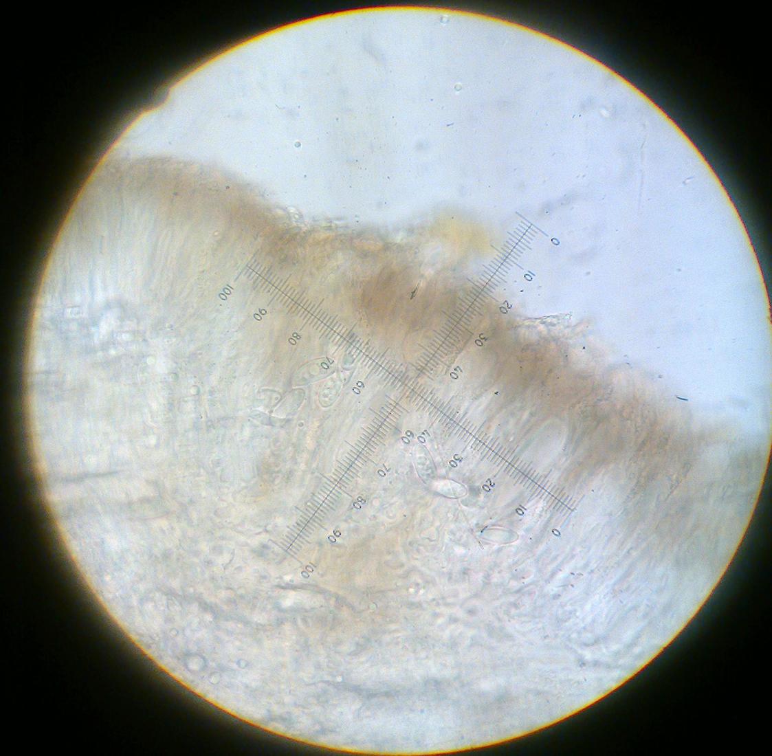
First of all, some apothecia have a bluish pigment in the excipulum (consistent with the Lecidea-hypothesis?):
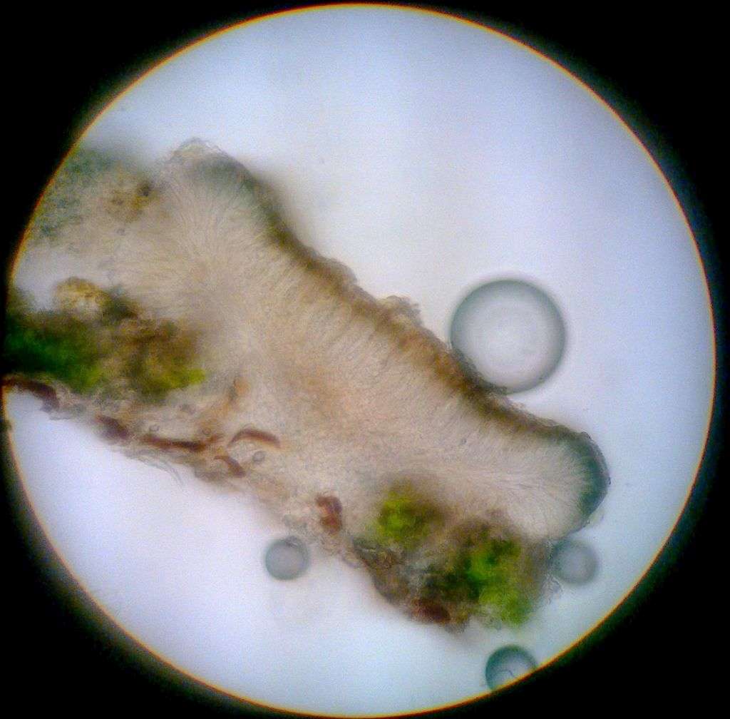
[attachment=2]Billede3070.jpg[/attachment] The brown epithecial pigment reacts N+ red and the crystals are not soluble in N:
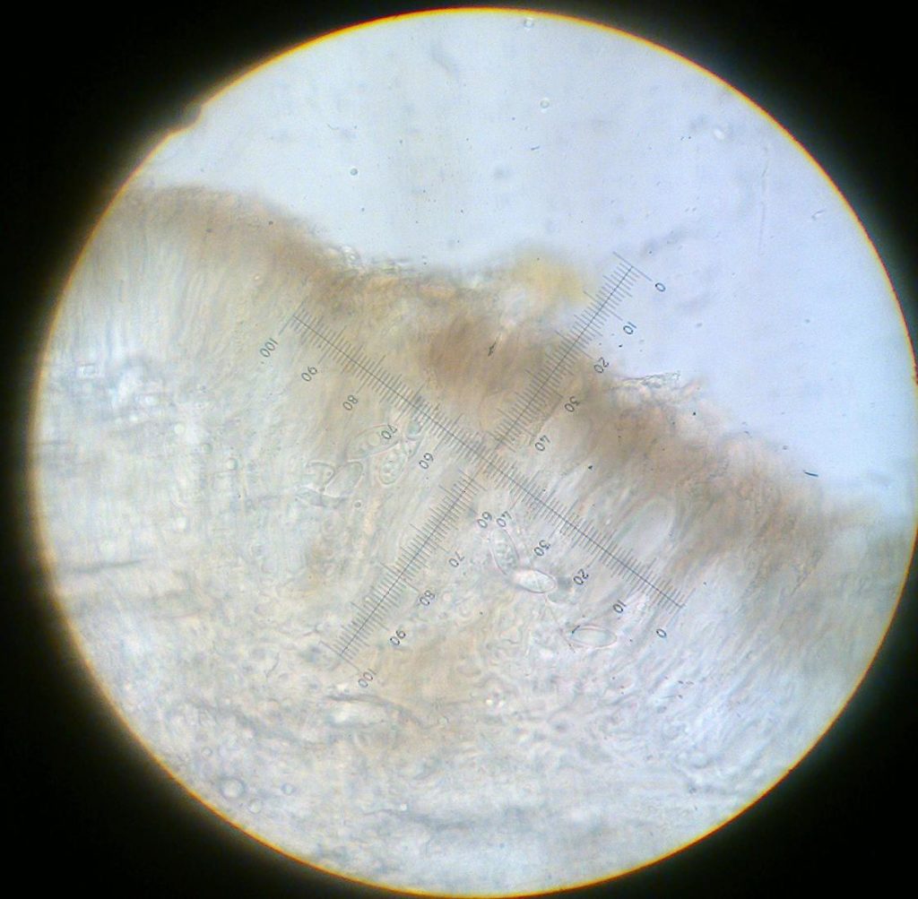
[attachment=1]Billede3072.jpg[/attachment] 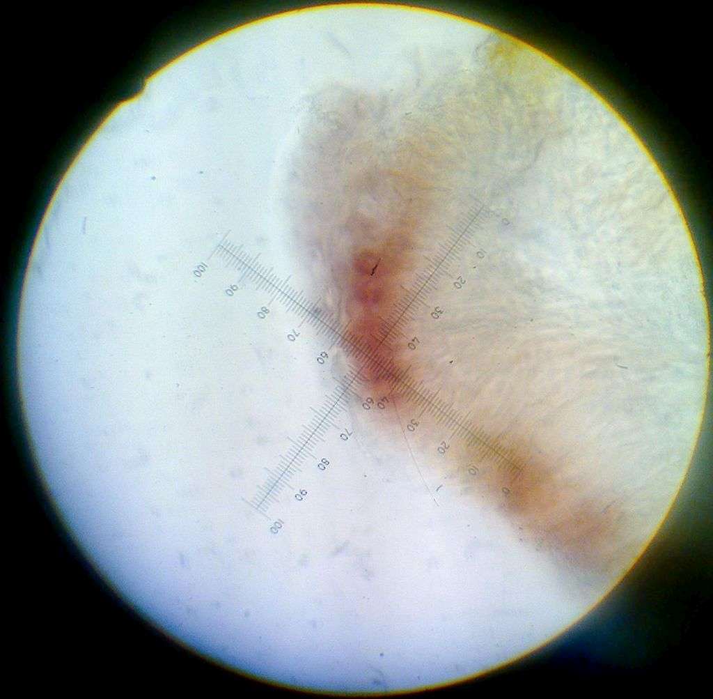
[attachment=0]Billede3074.jpg[/attachment] Here’s a photo of the K/UV-reactions 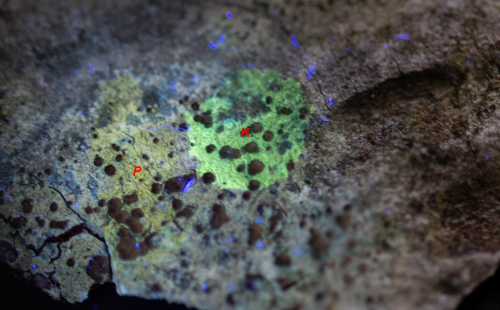
[attachment=0]cf. Lecanora sp. 1-2.jpg[/attachment] Hi again, Mark The french site “Lichens Marins” describe the spores from V. ochrostoma as “ovoid”, which fits the spores from my material, but is seemingly in contradiction to the british material. Wether this is due to taxonomical confusion or intraspecific variation in spore-shape is another matter, that would take some work to sort out. I have found that spore-shape varies quite a bit in some species, but seems conserved in others (V. bryoctona, for instance). I don’t have the largest sample size of any species, so it’s quite possible that they all vary.
I don’t have enough material to split into two, so I propose that I send you all my material for comparison. You are also more than welcome to take a closer look at the proposed LF, should that pique your interest. If at all possible, I would like at least parts of the material returned for storing in the national lichen herbarium.
Once again, I’d like to thank you for your time. You have been a great help.
Here’s a link to the “Lichens Marins” page for V. ochrostoma:
http://www.lichensmaritimes.org/index.php?task=fiche&lichen=690&lang=en Thank you for your great effort in confirming this ID! I have rechecked my material and made some follow-up photos with scale-bars attached.
From what I can see, the algae fit fine: they are rather small and generally look as in your attached photo. Furthermore, it looks like they form in vertical “rays” in thin sections.
A few photos of the algae:
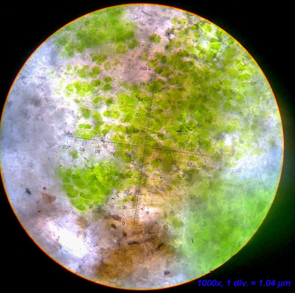
[attachment=4]Billede2990.jpg[/attachment] 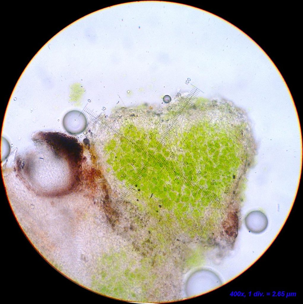
[attachment=2]Billede3000.jpg[/attachment] 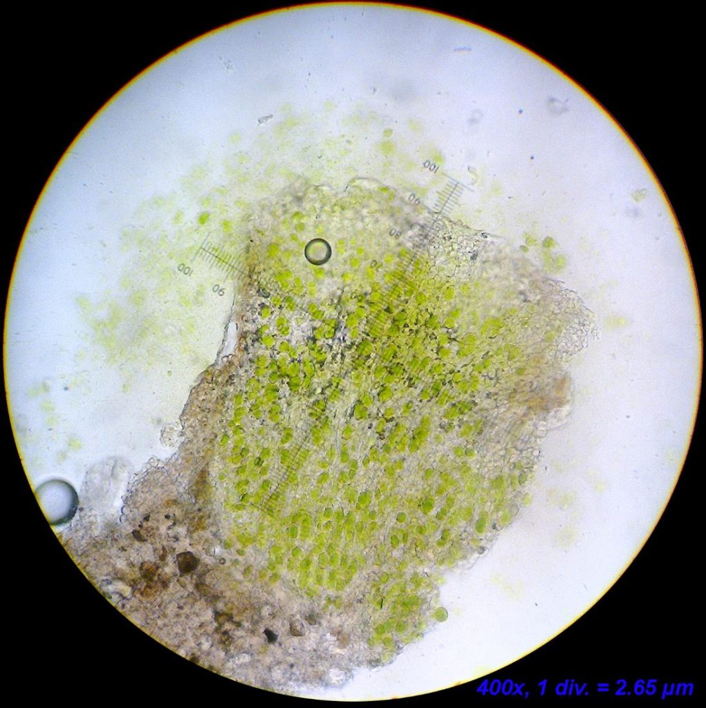
[attachment=1]Billede3001.jpg[/attachment] The algal units seem to fit in size – although a few exceed the range (~350 µm). These are very oblong units so perhaps it is ok?
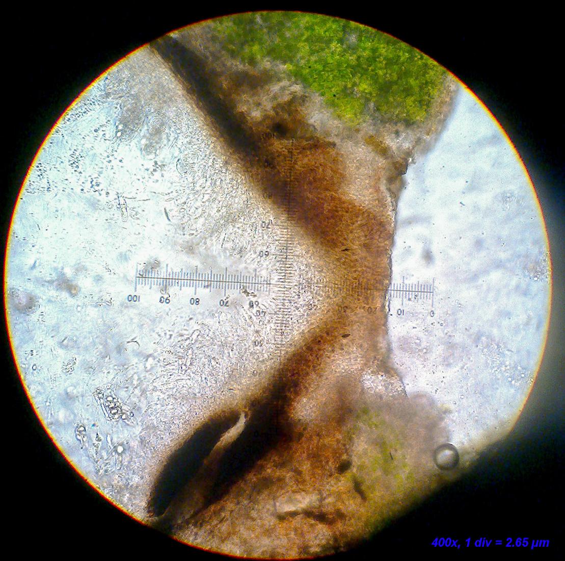
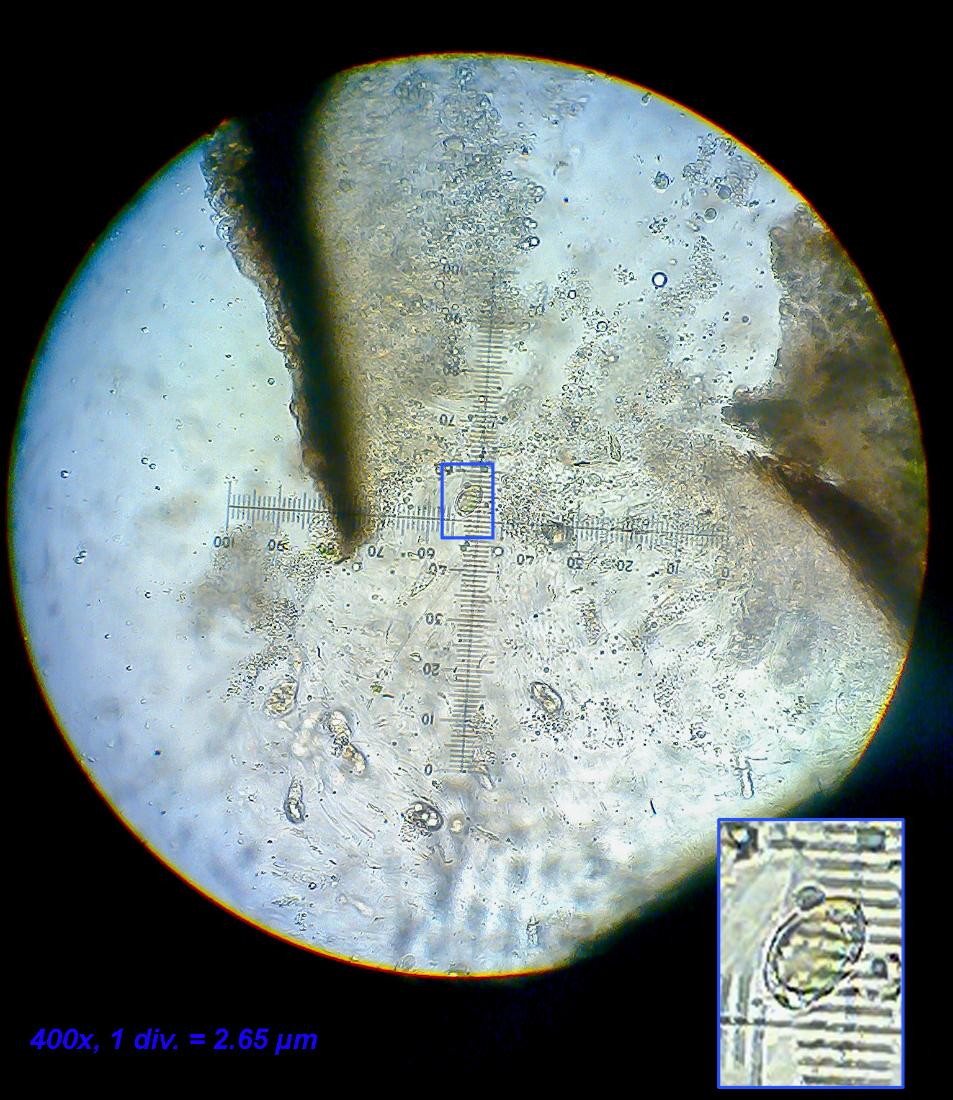
The size of the units may be judged from this photo. The perithecium in the middle is the one from which the sections in the following have been made.
Note: there’s a small black pycnidium or perithecium (no contents found so far), which I am certain must belong to a lichenicolous fungus.
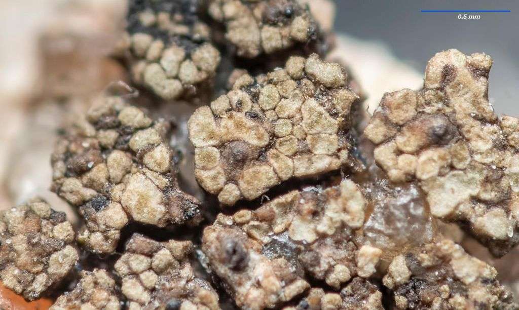
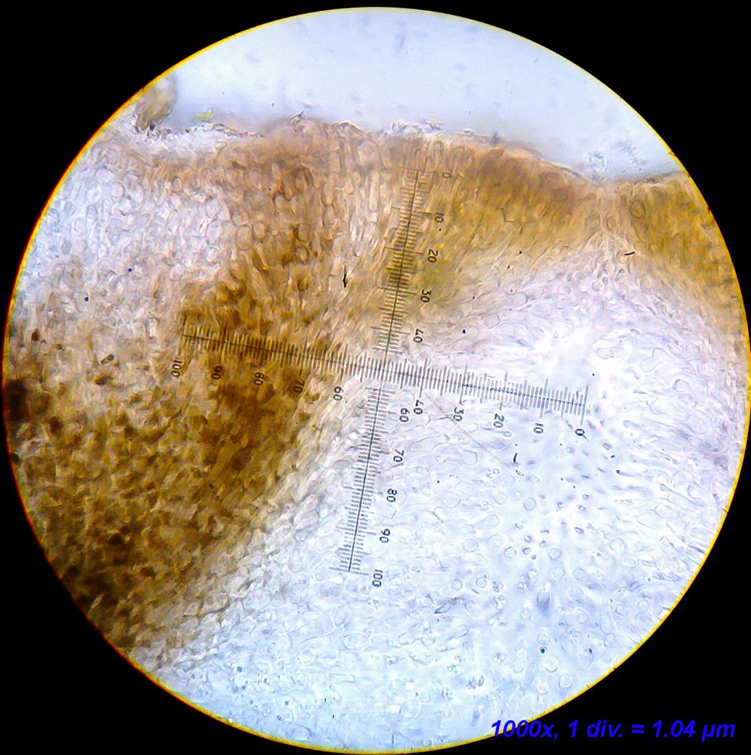
[attachment=0]Verrucaria cf. ochrostoma 1-5.jpg[/attachment] The hamathecium is without interascal filaments (although with “characteristic” Verrucaria-inclusions), but with periphyses and/or periphysoids (not entirely sure how to tell the two apart if I am being honest)
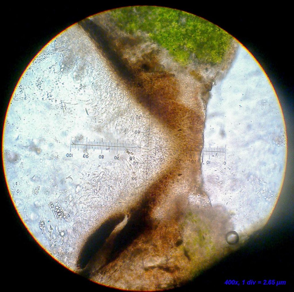
[attachment=7]Billede2987.jpg[/attachment] 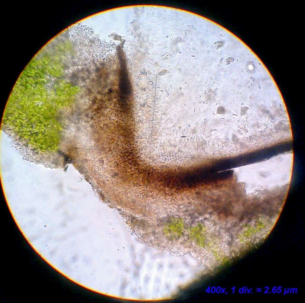
[attachment=5]Billede2989.jpg[/attachment] 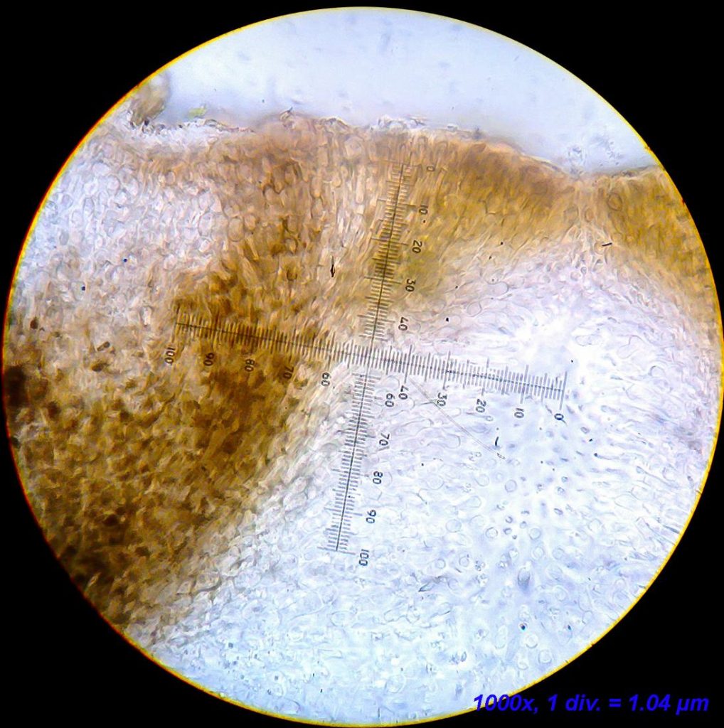
[attachment=3]Billede2992.jpg[/attachment] For whatever reason, I still struggle finding mature and well developed spores. Most asci just contain shriveled spores or simply an amorphous substance. I did find two healthy looking spores in the last section, but they are clearly underdeveloped and rather egg-shaped rather than narrowly ellipsoid. The lack of good spores is my only cause for concern regarding the correct ID, but so much of the other characters fit very well, so I am inclined to overlook this.
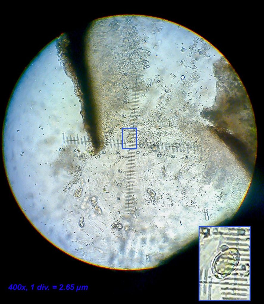
[attachment=6]Billede2988.jpg[/attachment] Hello Mark, Thank you for the swift reply! I understand and appreciate your hesitation, but I am happy that you agree with the ID even if it is tentatively. I have already studied your photos and drawings of the species – you may remember that a while ago I asked about a sterile specimen that you thought looked very much like V. ochrostoma (link to the thread:
https://www.fungi.org.uk/viewtopic.php?t=3318 ). Since then I have looked for a fertile thallus and during an excursion to the south-eastern part of Jutland, I found this one.– Jonas
Hello Francesco, To my eye, this is definitely not F. caperata. For one thing, the color is not right – although the pale yellow color produced by usnic acid that has given rise to the latin name for the genus (Flavoparmelia; flavus = yellow in latin), can be quite tricky to capture with a digital camera, I find. Did it appear disctinctly yellow in the field? Another, bigger issue, is the presence of punctiform pseudocypellae on the upper surface (the little white dots near the edge of the thallus). These point to the genus Punctelia, a hunch that can be confirmed by a spottest of the medulla (the white layer beneath the greenish upper cortex) with bleach (sodium hypochlorite = Cl). Species from the genus Punctelia react a reddish to pink color when tested this way. The reaction can be quite fleeting, so make sure to observe the reaction immidiately after applying the bleach.
I am not quite sure about the potential species of Punctelia in your area, but I would suggest looking into that genus for a good match.
Best regards,
Jonas
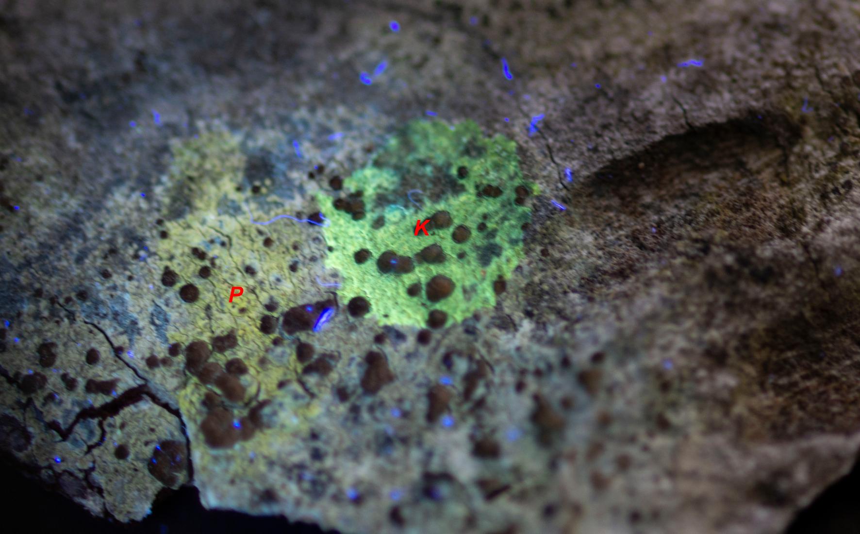
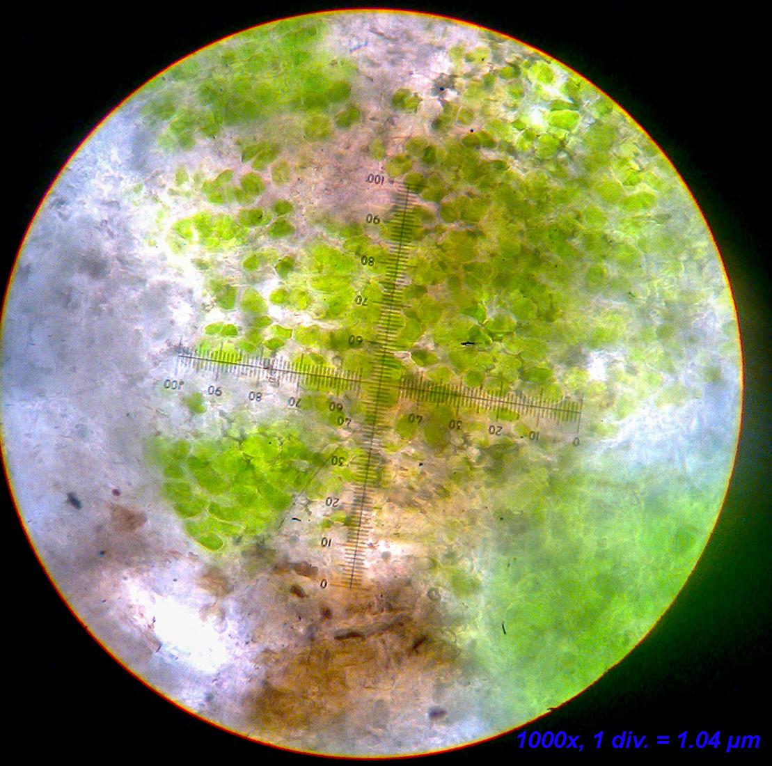
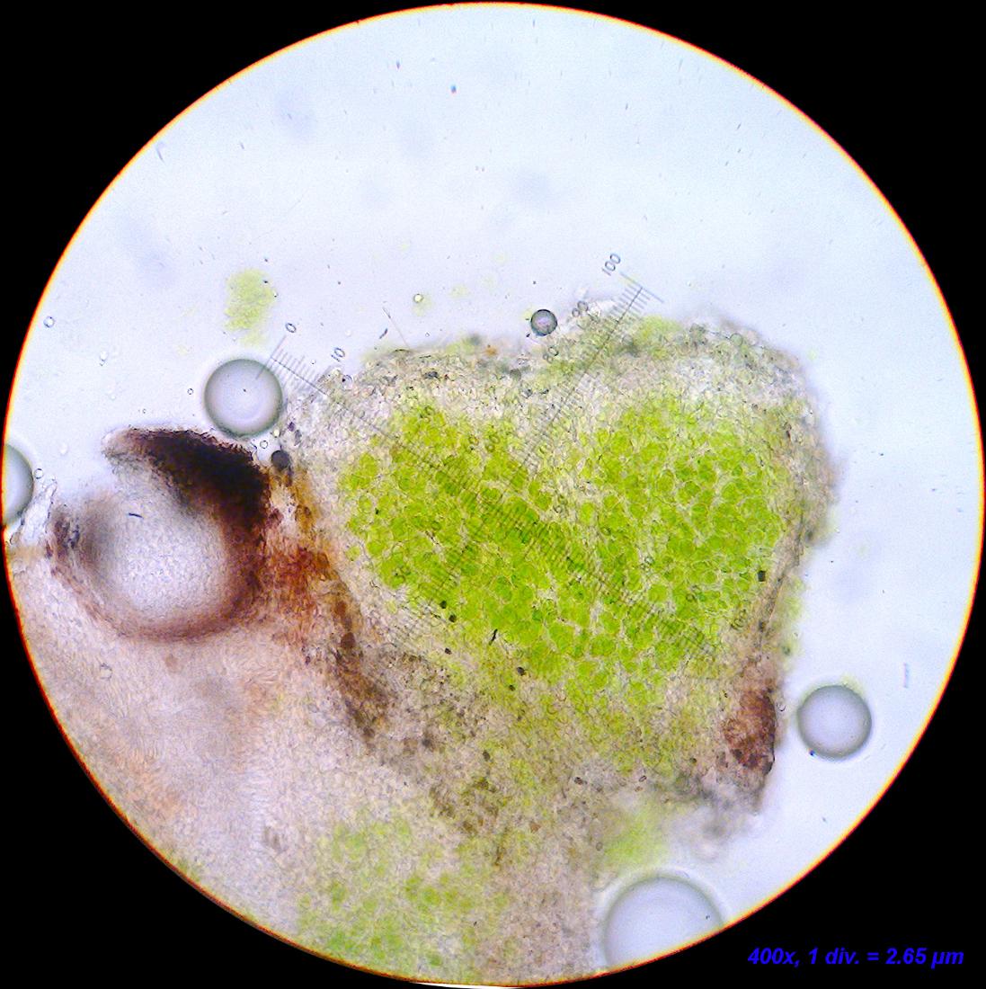
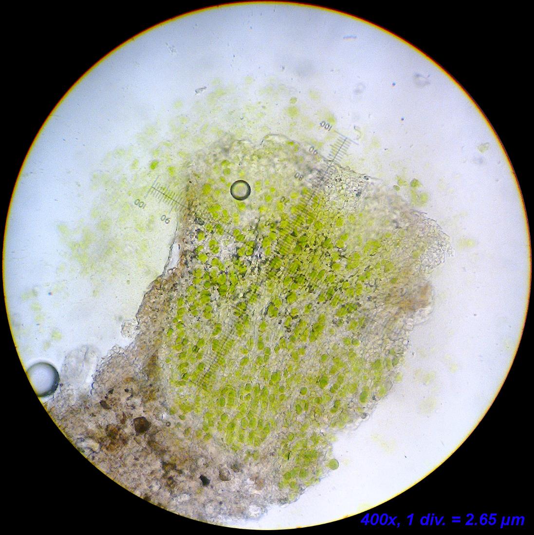
The plot thickens… I was asked in the facebook-group “Lichens Connecting People” if I was certain that there was no C+ reaction, so I redid all the chemistry (C, K and P) and to my surprise I found pretty localized K+ wine-red reactions. Not all squamules reacted in this way and I have not been able to pinpoint exactly what it is, that’s reacting even under the microscope.
The pigment that is formed reacts K/UV+ dullish orange.
– Jonas
-
AuthorPosts