
Verrucaria ochrostoma from Denmark?
- This topic is empty.
-
AuthorPosts
-
April 9, 2022 at 3:25 pm #32340
Dear all, I have been looking for Verrucaria ochrostoma in Denmark for some time now, as it is not currently known from here, but appears widespread in some of our neighbouring countries. I think that I may have finally found it, but I hope that some of you can assist me in confirming the ID.
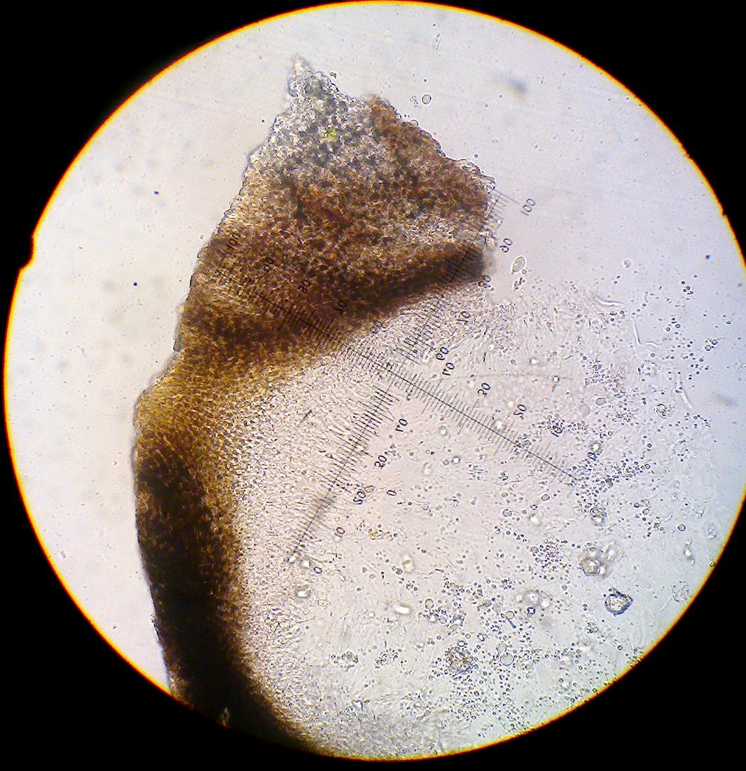
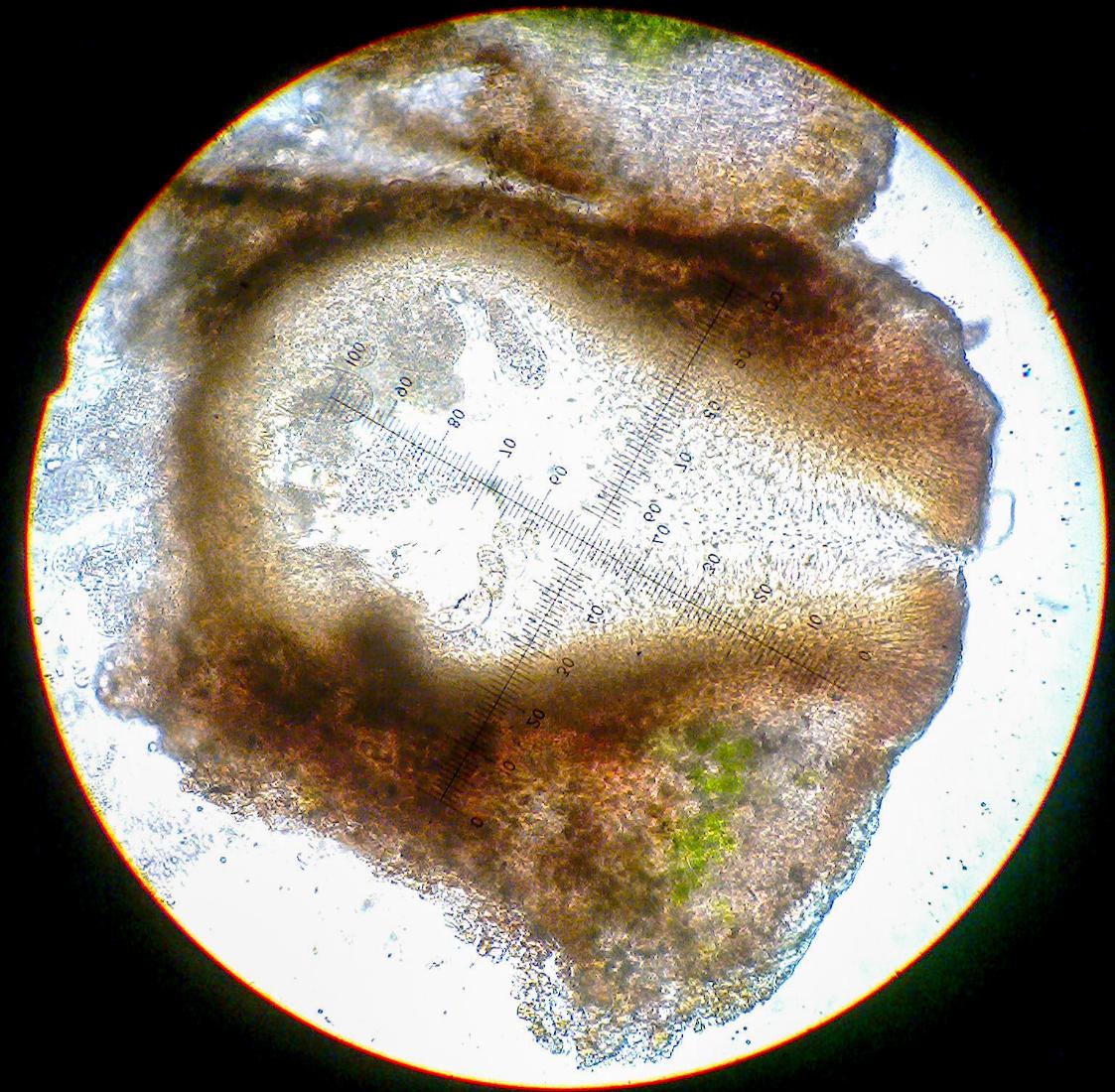
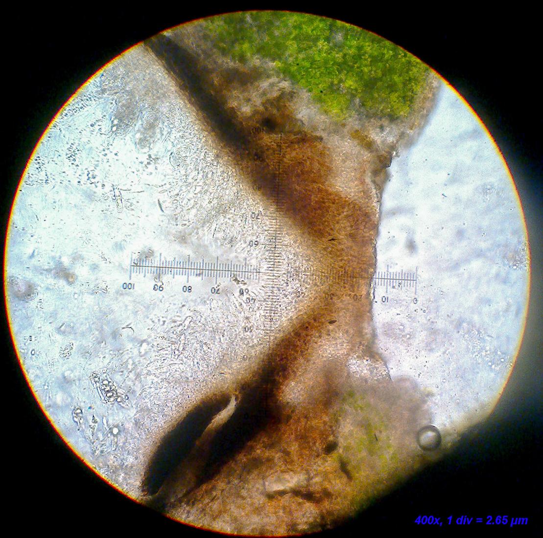
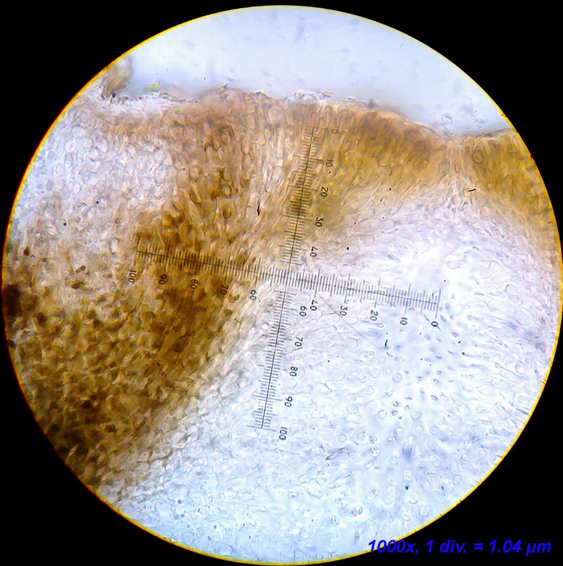
This specimen was found growing on vertical sections of coastal concrete wall. The thallus is quite pale and consists of small squamule-like areoles that are eventually compressed into a more compact thallus that cracks secondarily. Perithecia are quite immersed in the thallus and appear brownish (especially with flash photography). There is no involucrellum as far as I can see and the excipulum widens quite considerably towards the top, showing some irregular pigmentation. I have not found fully mature spores, but those I did found were much shorter than 25 µm (~18 µm).
Any and all comments regarding a verification or alternative IDs are more than welcome.
Best regards,
Jonas

[attachment=5]Verrucaria cf. ochrostoma 1-1.jpg[/attachment] 
[attachment=4]Verrucaria cf. ochrostoma 1-2.jpg[/attachment] 
[attachment=6]Verrucaria cf. ochrostoma 1-3.jpg[/attachment] 
[attachment=7]Verrucaria cf. ochrostoma 1-4.jpg[/attachment] 
[attachment=3]Billede2949c.jpg[/attachment] 
[attachment=2]Billede2950.jpg[/attachment] 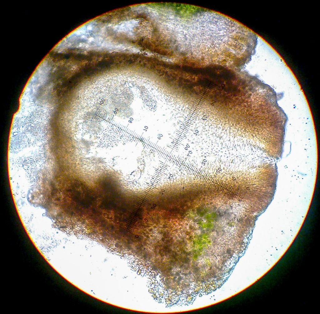
[attachment=1]Billede2952.jpg[/attachment] 
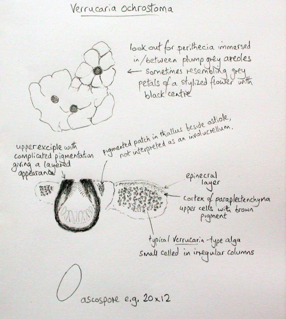
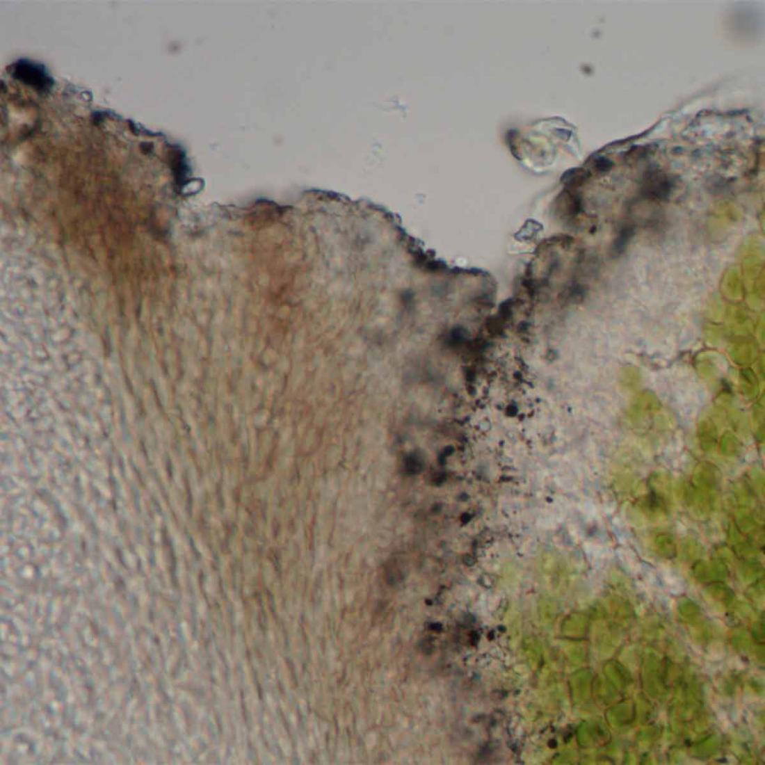
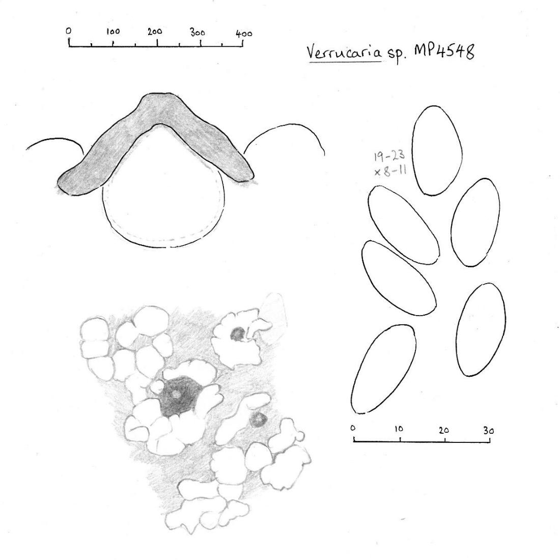
[attachment=0]Billede2954.jpg[/attachment] April 9, 2022 at 4:48 pm #44756I am very hopeful that your lichen is indeed Verrucaria ochrostoma and I am very close to confirming it as such. My only hesitation arises from the responsibility of adding a new species to a country and hence wanting to be especially certain. All of the features seem right, even down to the brownish (rather than black) colour of the apex of the perithecia. The thallus looks right, with pale primary areoles (algal units) often resembling little blobs of putty which have been pushed into place around the perithecia, these blobs sometimes rather separate, sometimes converging, and forming a thallus which is roughly level with the apices of the perithecia. As in your photographs, the primary areoles and perithecia form a crust which becomes secondarily cracked. When I was first working on the identity of V. ochrostoma, I found it difficult to decide whether an involucrellum was present because, as shown in your micrographs, the pigmented exciple widens markedly towards the apex and has a somewhat complicated, layered appearance. Sometimes there is even a separate small piece of pigmented tissue in the thallus near the apex, resembling perhaps fancifully, a poorly developed vestigial involucrellum. V. ochrostoma was very poorly known in Britain before about 2013 (despite it being very common, including in the immediate environs of the Natural History and Science Museums in London). I have the advantage of having examined Borrer’s type specimen which is housed in BM and was able to convince myself that the common colonist on concrete and limestone was the same as Borrer’s type of V. ochrostoma. I have posted some images on the FGBI website and your query prompts me to attempt to capture some more and better images, in part to help you confirm your material (I hope to do this next week). https://fungi.myspecies.info/taxonomy/term/7172/media Here is a sketch I made of the main features I noted in V. ochrostoma:

[attachment=1]DempsuHXkAUQPxk.jpeg[/attachment] And here is the layered upper part of the exciple:

[attachment=0]Verrucaria ochrostoma4.jpg[/attachment] Some twitter threads:
https://twitter.com/obfuscans3/status/1076985438886338560 https://twitter.com/obfuscans3/status/1002519032845754368 Sorry to conclude without an absolute confirmation. I feel sure that your lichen is Verrucaria ochrostoma and once I have refreshed my memory by making new observations, and looked up the notes I made when examining Borrer’s type, I hope that I will be able to make the decision.
April 9, 2022 at 5:15 pm #44757Hello Mark, Thank you for the swift reply! I understand and appreciate your hesitation, but I am happy that you agree with the ID even if it is tentatively. I have already studied your photos and drawings of the species – you may remember that a while ago I asked about a sterile specimen that you thought looked very much like V. ochrostoma (link to the thread:
https://www.fungi.org.uk/viewtopic.php?t=3318 ). Since then I have looked for a fertile thallus and during an excursion to the south-eastern part of Jutland, I found this one.– Jonas
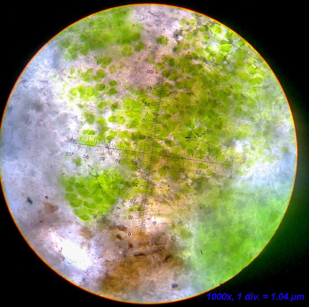
April 10, 2022 at 11:45 am #44758I have been re-examining some British specimens of Verrucaria ochrostoma and this has given me the confidence to confirm your Danish material as such from the information and images you have already provided. Nevertheless, I am going to keep adding to this post, partly to share my observations with the forum, and partly to strengthen our confidence in the identification of the Danish material. First, some readers may want us to go back to first principles and explain why we think we are dealing with a species of Verrucaria. In addition to having perithecioid fruiting bodies and simple ascospores, there are two other useful characters which help us to confirm that such material belongs in the Verrucariaceae. First the photobiont, which is rather small-celled (3-)5-6.5(-9.5) microns across, often with at least one flat side, apparently from division into pairs or in short chains. The algal cells in Verrucaria are usually arranged to give the appearance of (somewhat irregular) vertical columns.

[attachment=6]ochrostoma3122c.jpg[/attachment]
The other useful feature is the absence of interascal filaments (i.e. no paraphyses or similar sterile hyphae between the asci). Instead the hymenial gel has streaks of inclusions (which might be mistaken for paraphyses and perhaps look like paraphyses which have partially disintegrated and now represented by minute, elongated inclusions in disrupted streaks). In the following micrograph the hymenial gel has been stained blue with iodine. Within the gel, to the right of the immature ascus, some of these streaks can be seen).
[attachment=5]ochrostoma2353d.jpg[/attachment] The appearance of V. ochrostoma is rather distinctive, pale grey (rarely as dark as medium grey-brown) algal units, like slightly irregular rounded lumps of putty, appearing as if half a dozen or so lumps have been pushed into place around each perithecium. Young colonist individuals (the most usually encountered) are most distinctive. In old and/or eroded colonies the separate algal units may converge and the most distinctive part of the appearance is partially lost. The apex of the perithecium is at or somewhat below the tops of the algal units and forms a roughly circular dark brown spot usually 100 to 250 microns across. The algal units are typically 150 to 250 microns across (though smaller and larger ones will be found). Each perithecium and its surrounding algal units (perhaps resembling a stylised flower) forms a visual unit typically 500 to 1000 microns across depending on the exuberance of the individual. I have devoted a bit of space to describing the size so that we can be sure that the Danish material is within about the right bounds for this species. Here are a couple of images of typical English material (two different specimens, both collected as scrapings stuck on brown card).
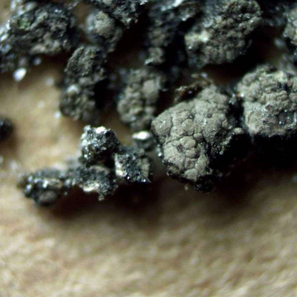
[attachment=4]ochrostoma3122a.jpg[/attachment] 
[attachment=3]ochrostoma2353e.jpg[/attachment] Another feature of V. ochrostoma is the usual presence of a thin epinecral layer to the thallus. Each algal unit is generally filled with algal cells except for a thin layer (e.g. 10 microns) of rounded hyphal cells forming a poorly differentiated cortex (only recognised by an absence of algal cells), these uppermost hyphae usually with diffuse brown pigment. On top of the cortex is a thin (up to e.g. 10 microns thick) epinecral layer which appears amorphous and glassy in section. This epinecral layer gives the algal units a sort of translucent appearance, perhaps slightly waxy, adding subtly to the distinctive appearance. I feel sure that I can see this epinecral layer in the Danish material.
The vast majority of Verrucaria species have an involucrellum so the absence of an involucrellum in V. ochrostoma ought to helpfully narrow the options greatly. Unfortunately it is often difficult to decide whether or not an involucrellum is present due to the layered upper parts of the exciple and the often presence of pigmented tissue in the upper thallus. The best advice is to try to find a young (even immature) perithecium.

[attachment=2]ochrostoma3122d.jpg[/attachment] 
[attachment=1]ochrostoma3122b.jpg[/attachment] The sections above are of the upper part of the exciple of a young perithecium, showing the layering that is present even at this stage, the complete absence of any involucrellum. The first is mounted in water, the second in K, showing that there is a subtle difference in shade of brown between inner and outer layers and they take on an olive tinge in K. The pigmentation, including change in K, has not been much used taxonomically but I suspect it provides a useful subsidiary character in Verrucaria.
The following micrograph shows a section through a more mature part of a colony and here we can see that, although the absence of involucrellum can be discerned with careful examination and experience, the presence of dark tissue in the thallus confuses things.

[attachment=0]ochrostoma2353a.jpg[/attachment] There will be more to come. My main request is that the Danish material is checked for the size of its units, just to confirm that it is about the right scale of thing.
April 19, 2022 at 5:01 pm #44759Thank you for your great effort in confirming this ID! I have rechecked my material and made some follow-up photos with scale-bars attached.
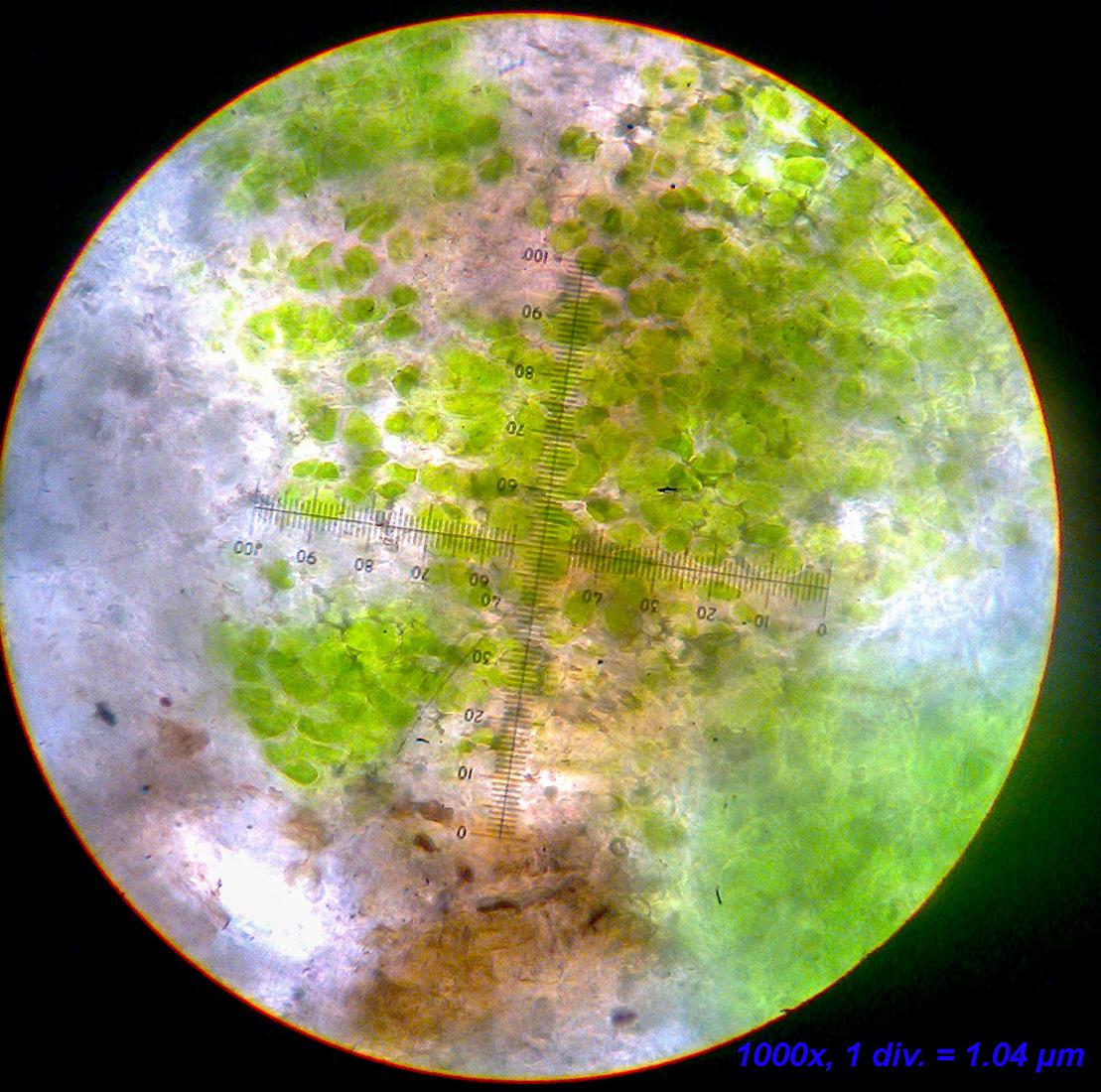
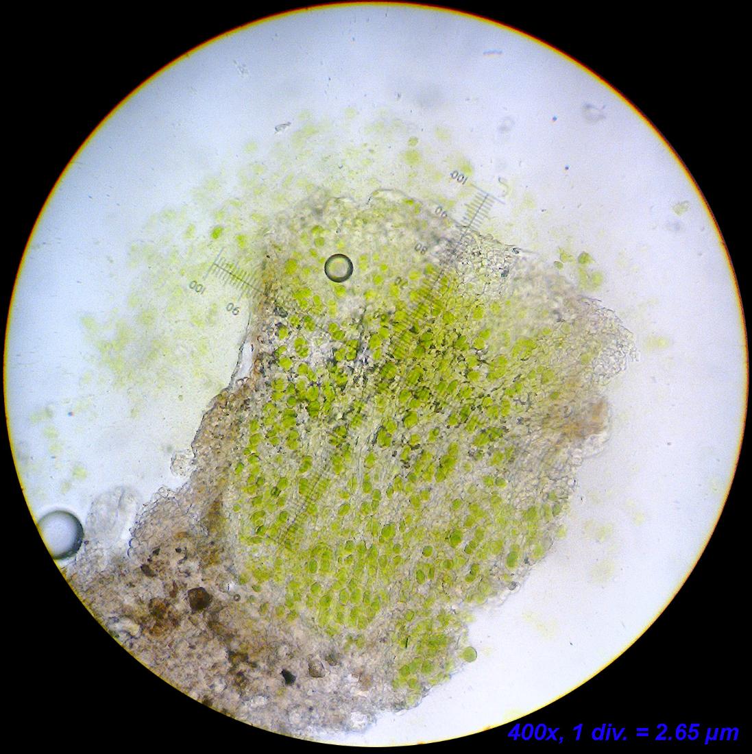
From what I can see, the algae fit fine: they are rather small and generally look as in your attached photo. Furthermore, it looks like they form in vertical “rays” in thin sections.
A few photos of the algae:
[attachment=4]Billede2990.jpg[/attachment] 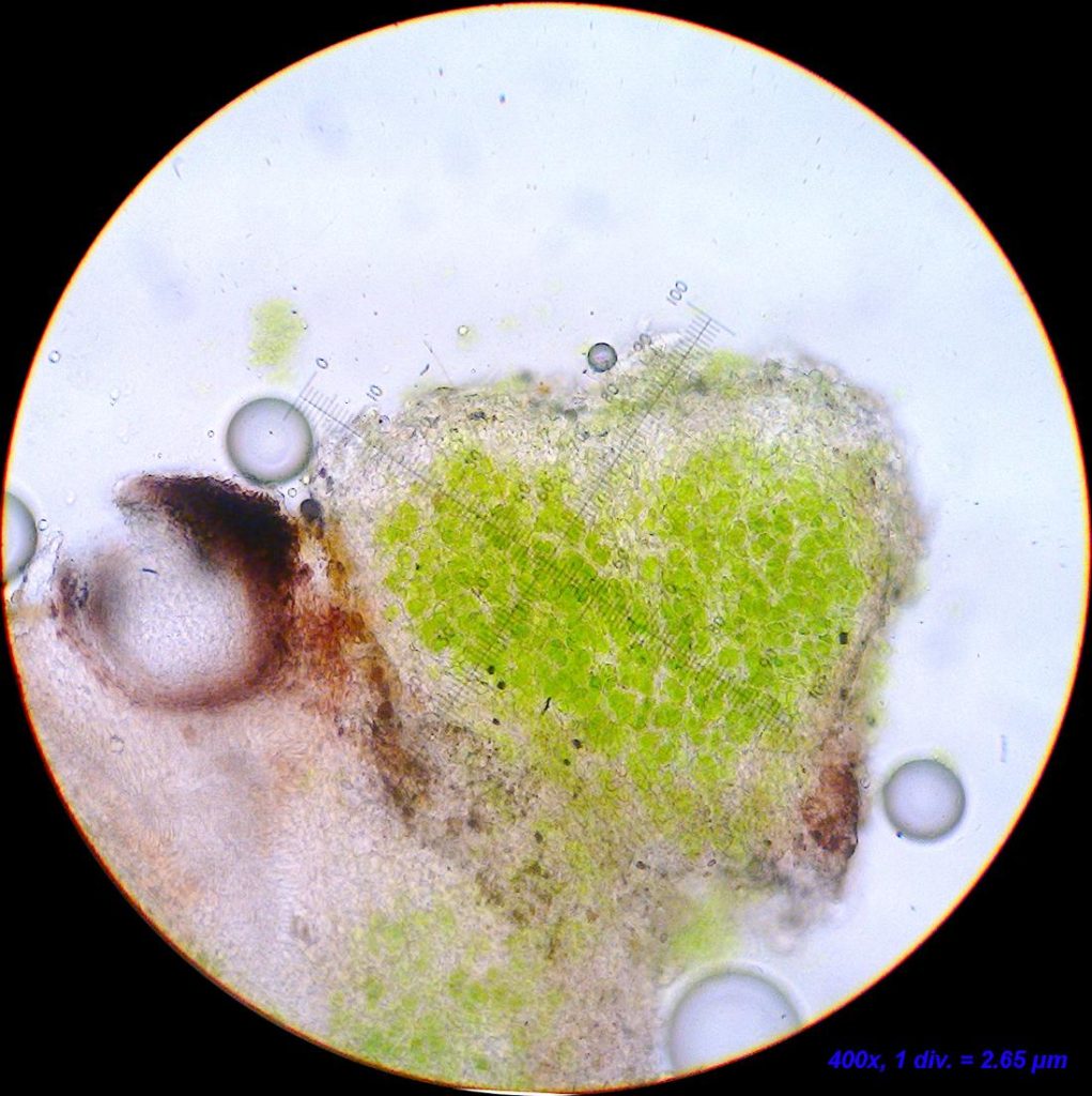
[attachment=2]Billede3000.jpg[/attachment] 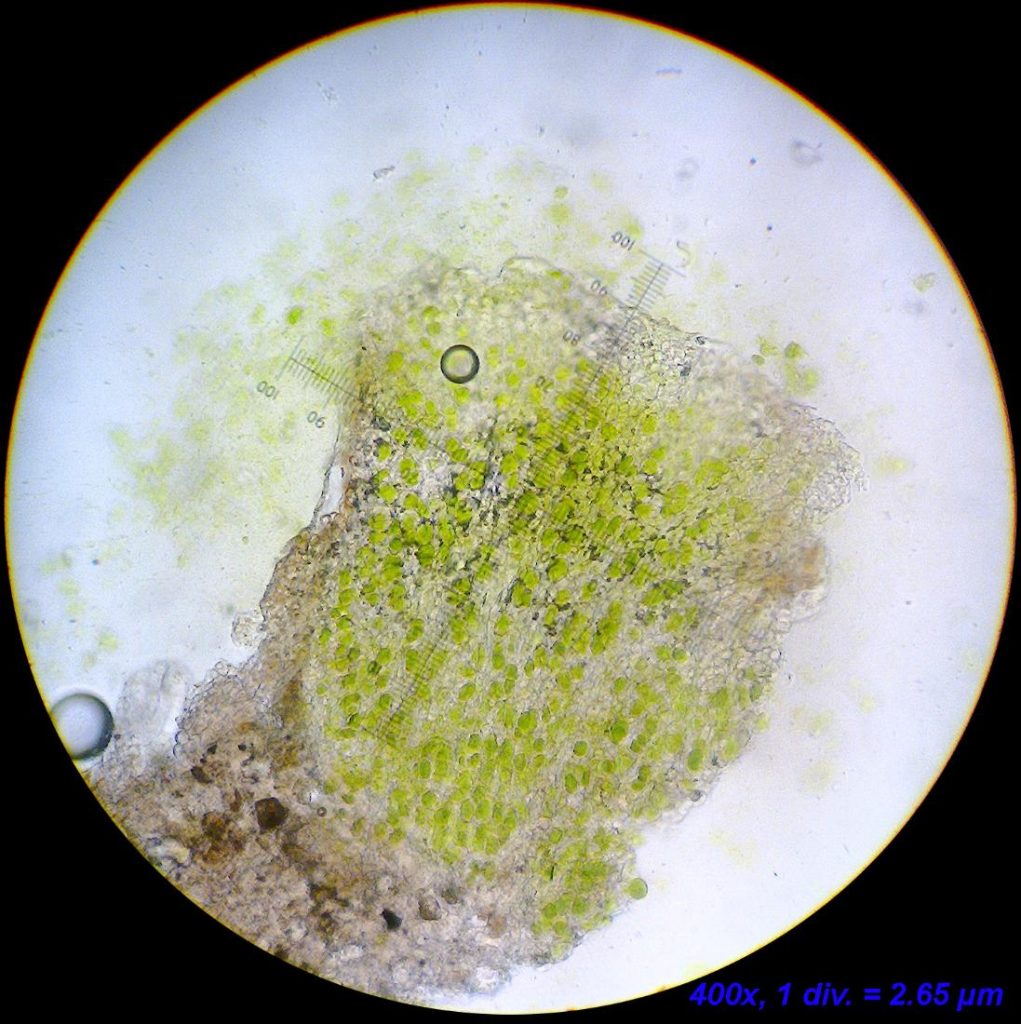
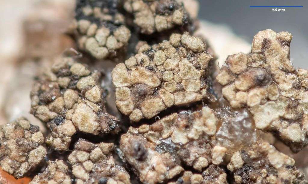
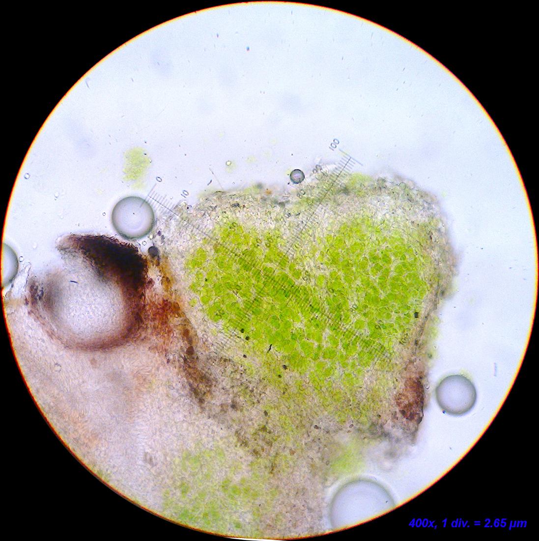
[attachment=1]Billede3001.jpg[/attachment] The algal units seem to fit in size – although a few exceed the range (~350 µm). These are very oblong units so perhaps it is ok?
The size of the units may be judged from this photo. The perithecium in the middle is the one from which the sections in the following have been made.
Note: there’s a small black pycnidium or perithecium (no contents found so far), which I am certain must belong to a lichenicolous fungus.
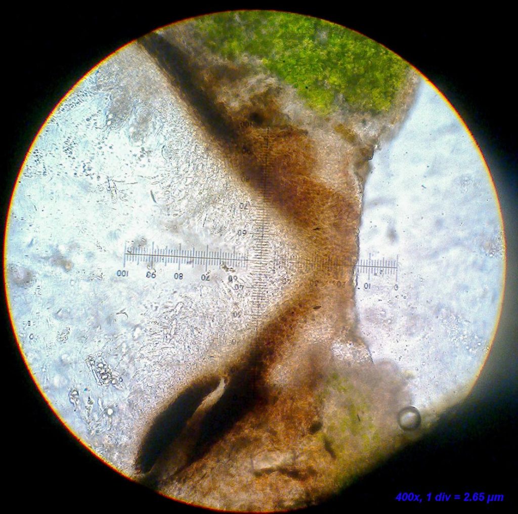
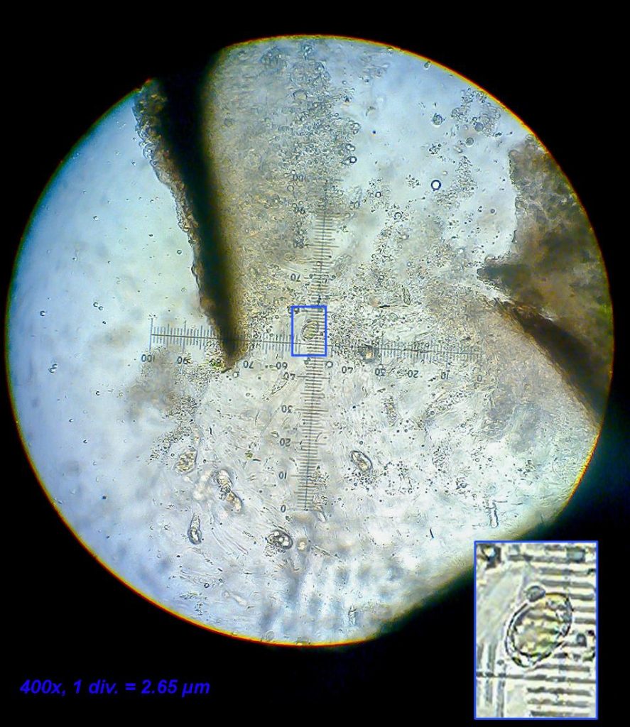
[attachment=0]Verrucaria cf. ochrostoma 1-5.jpg[/attachment] The hamathecium is without interascal filaments (although with “characteristic” Verrucaria-inclusions), but with periphyses and/or periphysoids (not entirely sure how to tell the two apart if I am being honest)
[attachment=7]Billede2987.jpg[/attachment] 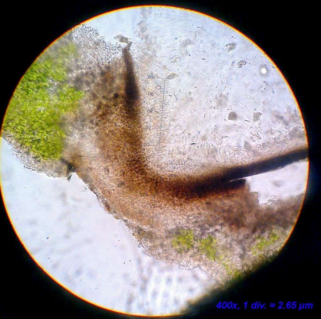
[attachment=5]Billede2989.jpg[/attachment] 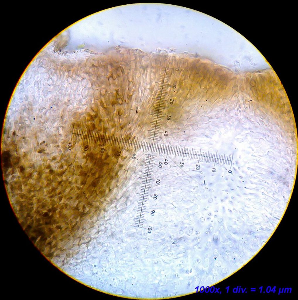
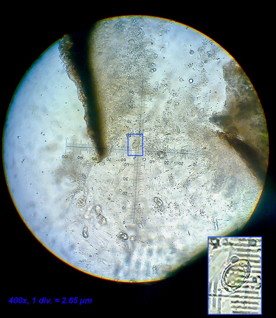
[attachment=3]Billede2992.jpg[/attachment] For whatever reason, I still struggle finding mature and well developed spores. Most asci just contain shriveled spores or simply an amorphous substance. I did find two healthy looking spores in the last section, but they are clearly underdeveloped and rather egg-shaped rather than narrowly ellipsoid. The lack of good spores is my only cause for concern regarding the correct ID, but so much of the other characters fit very well, so I am inclined to overlook this.
[attachment=6]Billede2988.jpg[/attachment] April 24, 2022 at 8:57 am #44760I agree that some properly developed mature spores would have been a nice addition to the information. Although spore shape and (particularly) size is important, I perhaps rely less on spore characters in Verrucaria than some others. I know some people who seem to use the table of spore sizes in LGBI2 as their starting point (which is acceptable) but then fail to appraise all the other characters exhibited by their specimen properly. The spore sizes can be rather variable even within a single individual and don’t always lie neatly within the published ranges. I have a particular issue with V. hochstetteri in this respect as I explain in the following twitter thread: https://twitter.com/obfuscans3/status/1073306196176789509 
[attachment=4]Screenshot 2022-04-24 9.33.18 AM.png[/attachment]
One of my remaining ambitions in lichenology is to study the spore sizes in various specimens of V. ochrostoma, to see how much the size varies depending on individuality and the exuberance of the material.I think I am going to hold my nerve in confirming your Danish material as V. ochrostoma but perhaps we should exchange material at some stage (either you send me a Danish specimen, me send you some British material, or both). There is nothing quite like comparing material side-by-side.
I have come across a couple of V. ochrostoma look-alikes, in both cases I failed to find an adequate name for them. In both cases they were found to have a distinct involucrellum. Along with the overall appearance of your lichen, the absence of any distinct involucrellum is one of the most compelling pieces of evidence pointing to V. ochrostoma in my opinion. This lack of involucrellum is rather a rare trait in the genus. Thanks for providing your observations about the algae and lack of interascal filaments which aren’t of course diagnostic for V. ochrostoma but it may interest people reading this post to know how we are so sure that we are in the right genus.
You mention periphyses and these haven’t been used much in taxonomic studies but I suspect there are useful characteristics to be found if they were studied in the right way. In fact they helped boost my confidence in my original suspicions of British V. ochrostoma.

[attachment=3]Screenshot 2022-04-24 9.46.31 AM.png[/attachment] Here is one of my V. ochrostoma look-alikes:

[attachment=2]Screenshot 2022-04-24 9.45.01 AM.png[/attachment] 
[attachment=1]DtzdTEBWoAA_0dc.jpeg[/attachment]
https://twitter.com/obfuscans3/status/1070974567718490112 Here is another one that looked a bit like an exuberant V. ochrostoma in the field (Chelsea Physic Garden).

[attachment=0]Screenshot 2022-04-24 9.52.26 AM.png[/attachment] To summarise my thoughts so far, I consider your Danish specimen to be Verrucaria ochrostoma but happy continue working on it if you would like further reassurance (particularly perhaps by swapping material).
April 24, 2022 at 10:05 pm #44761Hi again, Mark The french site “Lichens Marins” describe the spores from V. ochrostoma as “ovoid”, which fits the spores from my material, but is seemingly in contradiction to the british material. Wether this is due to taxonomical confusion or intraspecific variation in spore-shape is another matter, that would take some work to sort out. I have found that spore-shape varies quite a bit in some species, but seems conserved in others (V. bryoctona, for instance). I don’t have the largest sample size of any species, so it’s quite possible that they all vary.
I don’t have enough material to split into two, so I propose that I send you all my material for comparison. You are also more than welcome to take a closer look at the proposed LF, should that pique your interest. If at all possible, I would like at least parts of the material returned for storing in the national lichen herbarium.
Once again, I’d like to thank you for your time. You have been a great help.
Here’s a link to the “Lichens Marins” page for V. ochrostoma:
http://www.lichensmaritimes.org/index.php?task=fiche&lichen=690&lang=en -
AuthorPosts







- You must be logged in to reply to this topic.