
Di N

Forum Replies Created
-
AuthorPosts
-
Thanks all for these very interesting follow ups. (The most recent patch that I thought would be B. griseovirens fluoresced bright orange and turned out on testing to be Lecidella scabra. Ugh!)
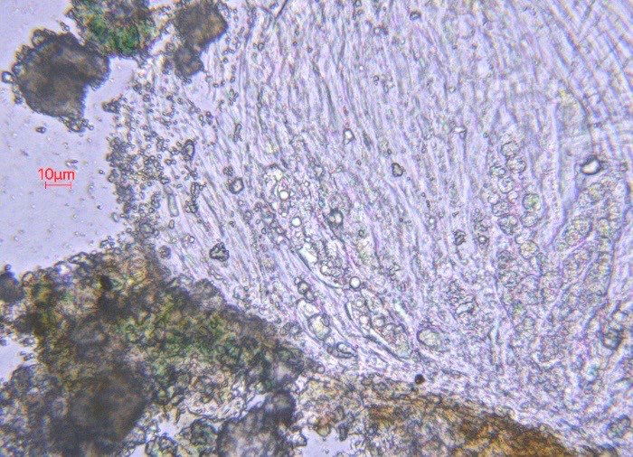
Many thanks for that detailed response. After a lot more investigation, I’m not sure I’m much closer to being sure what it is, but probably Petractis clausa is as close as I’ll get. Further photos just in case they help anyone else. I did get one apothecium section with maybe some evidence of blue-green photobiont.
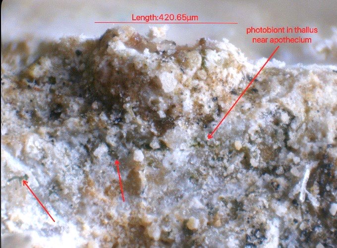
[attachment=7]IMG_6736.jpeg[/attachment] I also looked at the thallus in more detail, and could see photobiont below the apothecia, and within the thallus, which looked more of a blue-green than a green colour, but the thallus was very friable (not really distinct from the substrate) and difficult to sample.
[attachment=6]IMG_6743.jpeg[/attachment]
After several failed attempts, I did get some small patches of thallus, that had what look like cyanobacterial strands below the surface. The surface was orange-flecked. Possibly a thin lithocortex.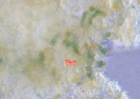
[attachment=5]IMG_6744.jpeg[/attachment] 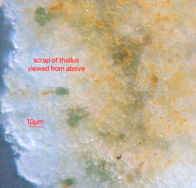
[attachment=3]IMG_6737.jpeg[/attachment]
The scraps mostly seemed to disintegrate when squashed though (I didn’t succeed in getting a cross-section).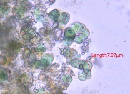
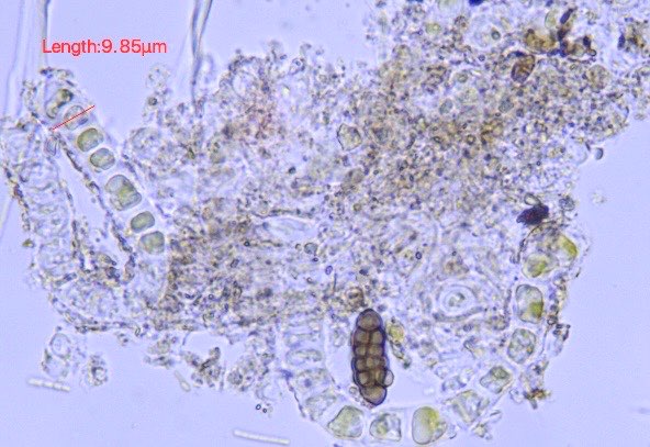
[attachment=4]IMG_6739.jpeg[/attachment] I found a couple of examples where there were strands of cells in chains with maybe a gel covering, but wasn’t sure whether they had blue-green contents (or whether they were contaminants, as there’s clearly a spore from somewhere else in there too).
[attachment=1]IMG_6748.jpeg[/attachment] 
[attachment=0]IMG_6745.jpeg[/attachment] 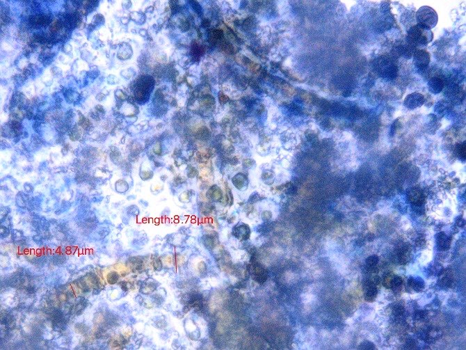
[attachment=2]IMG_6741.jpeg[/attachment] Thank you. That is very helpful, and all very interesting. Di
February 18, 2026 at 10:13 pm in reply to: Lecania (?) in the damp zone on siliceous headstone #46867Thank you so much, Maxine, for the email and photos, that’s great. Yes, the spore size (max 4.2 μm wide) and habitat fit L. suavis much better than the other possibilities. Di
Yes, it was indeed heavily-grazed and then re-grown Lecanora horiza. This patch was further up on the same stone. Thank you. Many thanks. I had wondered about re-growth of something after heavy grazing. We did see Lecanora horiza at the same site. I’ll be going back there, and will look out for more clues. Thanks again. The only pointer against it really is that the largest apothecium we found was only 0.55 mm. Most were O.3 to 0.5 (of about 50 apothecia). Seems best to record it as A. pyracea for now though, with a note in the comment section about the apothecial size. Thank you for the detailed very helpful response. I have since looked at another sample from the same fence with much flatter apothecia, which had no spores wider than 4.2 μm (all outside the asci) and the same reactions with K and C. L. stenotropa again. Thanks
Thank you. That’s really helpful. I collected it rather expecting it to be Buellia griseovirens, and was very surprised by the strong orange UV reaction.
Thank you for the detailed and very helpful reply. Thanks for commenting, Anthony. I did a search in here for merochlorophaea, so I had read your post first, but I don’t think yours showed a red C reaction? Both the C and KC on this were distinctly pink-red visually rather than brownish, though it’s hard to get clear photos. I did have a look round for any more information, and noticed that on the Consortium of LIchen Herbaria page, the C and KC reactions of C. merochlorophaea are both described as ‘pink’ (but those are mostly/all US records, so might be somewhat different of course).
https://lichenportal.org/portal/taxa/index.php?taxon=53435&clid=1009 Just for info. Phaeographis dendritica has been found recently (24.10.24, record only just sent in) at Ryton in Warwickshire, on oak in woodland. Thank you both. The New Forest information is interesting. Very good to hear from you, Mark, thank you. I had seen the photo of the Birmingham outskirts one, but wasn’t sure exactly where it was. I think Sutton Park is VC 38? Our tiny sample from a lobe edge mostly just has punctate pseudocyphellae. I think this shows them beginning to develop, though maybe the lumpier bits are the early stages of isidiomorphs. Unfortunately the light wasn’t too good, and the photos didn’t show the details of the inner lobes very well.

[attachment=0]IMG_7981.jpeg[/attachment] Have you considered Hyperphyscia adglutinata? I find it a lot here (also Warwickshire) on trees, but it’s very easy to overlook as it’s so tiny. Di
-
AuthorPosts
