aspeca

Forum Replies Created
-
AuthorPosts
-
Dear Francesco — Many thanks for your post, and your interest in lichens.
It’s often difficult to identify a lichen from just one or two photos. I’d be willing to wager, though, that your lichen is either
Flavoparmelia caperataor F. soredians. The yellowish colour of the thallus, especially in the centre, would be characteristic of these species, separating them from other similar ‘parmelioid’ lichens. The reproductive structures on your lichen are soredia, not isidia. Isidia have a cortex, so they don’t look as ‘powdery’ as soredia. The soredia of
F. caperataand F. soredianstend to form on the revolute ‘folds’ of the lobes, where they become confluent. That seems to be the case with your lichen. It’s not really possible to be certain of which species you have from these photos. There are some differences, but the two species look quite alike. In the UK generally,
F. sorediansis less common, but it depends on the location. A chemical test using K (potassium hydroxide) would be necessary to separate them definitively: the medulla and soredia of F. caperatawould be K-, while those of F. sorediansK+ yellow turning red. For more detail, see
https://britishlichensociety.org.uk/sites/default/files/Flavoparmelia%20caperata.pdf ” class=”bbcode_url”> https://britishlichensociety.org.uk/sites/default/files/Flavoparmelia%20caperata.pdf Best regards,
Anthony
Dear Sylvia — Thanks for your reply. I was about to send a follow-up to mine, as I saw that I may have misunderstood you in my excitement that another potential
L. pulicarisof the Pd- chemotype might have been found! The epithecial granules in your first photo seem discernible at that magnification, which I assume is 100x or so. That suggests it may not be
L. pulicarisof either chemotype. But it’s perhaps worth checking with Pd, as it does seem to have a bit of the L. pulicaris‘look’ about it, and it’s on lignum. (Perhaps the substrate isn’t entirely significant, assuming the wood is highly eutrophicated. It probably must be to host L. campestris, which I believe doesn’t like acid substrates). It doesn’t seem the most typical example of
L. hybocarpa auct. br.— normally the coarse, descending granules are more abundant, at least in my experience! But it’s also not clearly L. chlarotera s. streither. Some of the granules may have smeared down with the blade — i.e. those scattered deep ‘in’ the hymenium that don’t form a ‘chain of crystals’ back up to the epithecium. A test with Pd, and a section from another apothecium to check the crystals, might help. As for the calcium oxalate crystals — I haven’t seen them like that myself, but I suspect it’s possible for them to take that elongated form, if they’re deposited as the thalline margin grows. So far as I know, no distinction is made in the shape of the thalline margin crystals, but only their size (e.g. small for e.g.
L. campestrisand L. horiza, and large for e.g. L. pulicarisand L. hybocarpa auct. br.). Sorry I can’t be more definitive! Maybe someone else has some insight.
Best regards,
Anthony
Dear Sylvia — Many thanks for sharing this
Lecanora. I’ve found one like that as well, on a birch twig that I collected in Argyll last summer. I posted a Twitter thread about it — please see https://twitter.com/specanatura/status/1740325933310214160 ” class=”bbcode_url”> https://twitter.com/specanatura/status/1740325933310214160 Lecanoraas well. If you haven’t done so already, I’d recommend testing with Pd to determine whether you have the usual form of
L. pulicaris. You may well do, as
L. pulicarisprefers lignum or clean acid bark. In contrast to L. hybocarpa auct. br., L. pulicarishas fine (not individually discernible at <400x magnification) epithecial granules descending between the paraphyses. If your lichen is L. pulicaris, the thalline margin in particular should give a red reaction due to fumarprotocetraric acid. However, research in mainland Europe (Malicek, 2014) has shown that approximately 15% of
L. pulicarisspecimens lack fumarprotocetraric acid and give a Pd- test. This chemotype has not been reported from GB&I, but it may exist here. The only way to be sure would be to subject your specimen (or mine, or Jenny’s) to lab testing (TLC or DNA barcording). In the meanwhile — and assuming you don’t have the usual Pd+ red
L. pulicaris, of course — it would be possible to record your specimen as Lecanora sp., keeping the photos for future reference and study. As for your crystals in K, though — it seems from your photos that you’re referring to the calcium oxalate crystals in the thalline margin? These crystals don’t dissolve in K, whereas the epithecial granules do.
The
Lecanoragenus undoubtedly has more surprises in store for us British lichenologists! Indeed, I’m personally a bit unsure of our British Lecanoraspecies concepts, which I believe Neil Sanderson’s elucidation of L. hybocarpa auct. br.has called into question somewhat. For more on that if you’re interested, please see my recent Fungi Forum post (to which I intend to add some future posts once time allows) — https://www.fungi.org.uk/viewtopic.php?t=4225https://www.fungi.org.uk/viewtopic.php?t=4225” class=”bbcode_url”> Best regards,
Anthony
March 18, 2024 at 11:27 am in reply to: Lecanora hybocarpa auct. br. in France in 1956, and some thoughts #45950Many thanks, Alain. I’d be very interested to hear what you discover from this study. March 17, 2024 at 10:07 pm in reply to: Lecanora hybocarpa auct. br. in France in 1956, and some thoughts #45948Many thanks for your reply, Neil. I hope you don’t mind my inadvertent elevation of your work from ‘elucidation’ to ‘discovery’! But considering that Lecanora hybocarpa auct. br.seems unlikely to be the same taxon as the North American L. hybocarpa, perhaps your work does indeed sit somewhere in between the two levels. Grateful for the pointer to the entry in LGBI2 for
L. hybocarpa. I feel fairly keenly the lack of this volume on my shelf. Three times now I’ve thought I had a used copy in my grasp, but it’s eluded me. If you know of anyone looking to sell their copy, I’d be a willing buyer. It’s amazing to learn that our current concept of
L. chlarotera s. str.may still be obscuring other cryptic taxa. Looking forward to hearing more about your investigations as they develop. LGBI3 suggests that, in addition to the potential relationship to L. sinuosa, the same may be true of L. hybocarpa auct. br.Are you able to shed any further light on that? Thanks again for your interest in my posts above. I hope to add soon those promised further posts about the development of what you call the ‘expansive field concept’ of
L. chlaroterain use here in Britain before 2019. March 16, 2024 at 2:27 pm in reply to: Lecanora hybocarpa auct. br. in France in 1956, and some thoughts #45946Many thanks for your reply, Alain. Yes — despite Brodo’s (1984) work, some question does indeed seem to remain as to whether the size and placement of the epithecial granules are completely reliable for separating species. That said, not even for Brodo is the placement of epithecial granules definitive —
i.e.whether only on the epithecial surface, or descending into the hymenium. Granule placement is just one diagnostic character among others, though it’s the main one separating Lecanora chlaroteraand L. hybocarpa auct. br., at least on current British conceptions. Your findings about the calcium oxalate crystals of
L. chlaroteraare fascinating. Presumably you mean the large crystals in the amphithecium? It certainly seems reasonable that the lichen would deposit more calcium oxalate as it ages, in the same way that our kidney stones — also calcium oxalate — grow larger as we ourselves age. But the small granules ‘inspersing’ the epithecium of Lecanora chlarotera aren’t composed of calcium oxalate. They dissolve in K, whereas calcium oxalate doesn’t. The compound is ‘
chlarotera-unknown’, I believe, and the granules seem to serve an anti-herbivory function (on this point see Malíček, 2014, cited above). My posts above refer to the size and placement of these small crystalline granules in the epithecium, not the large calcium oxalate crystals in the amphithecium. Lucie Kohler (1956) concluded that size of epithecial granules, not placement, was diagnostic. But Magnusson (1932), Poelt (1952) and Brodo (1984) concluded otherwise.
March 10, 2024 at 9:15 pm in reply to: Lecanora hybocarpa auct. br. in France in 1956, and some thoughts #45944In my previous post above, I suggested that William Nylander had changed his mind at least once about the diagnostic characters of Lecanora chlarotera, a species he described somewhat differently three times (1872a, 1872b, 1891). I pointed to Nylander’s differing descriptions as an example of what Lucie Kohler, following August-Marie Hue, called the ‘greatest confusion’ of the ‘old lichenologists’ about the Lecanora subfusca-group, to which L. chlaroterabelongs (Hue, 1903; Kohler 1956, p 167). However, after further research, I’ve revised my opinion: it was Hue who was confused about Nylander’s descriptions. In this post, I’ll argue not only that Hue was confused about Nylander, but also that Nylander might have anticipated one important element of the
subfusca-group classification scheme in use today: the placement of epithecial granules either in a defined layer above the tips of the paraphyses, or also descending between them. Adolf Hugo Magnusson (1932) was the first to develop the modern scheme, which was extended and elaborated by Josef Poelt (1952) and Irwin Brodo (1984). And though Kohler was wrong to follow Hue in ascribing utter confusion to ‘old lichenologists’ such as Nylander, she was right to criticise Hue for rejecting the diagnostic value of epithecial granules in the subfusca-group. I also intend for this post to serve as background for future posts investigating in detail how the British concept of
L. chlaroteracame to be, and how it obscured the existence of another subfusca-group lichen, which was there all along: L. hybocarpa auct. br.But that’s for later. For now, let’s turn back to Hue’s confusion about Nylander and epithecial granules. Hue certainly knew that epithecial granules could be seen in some
subfusca-group species: Quote:Often, it [the epithecium] is bare. Sometimes it is full of small granules of a dark yellow, and then the epithecium is called ‘granular’ —
epithecium granulosum. This is an inaccurate expression, since the granules are not attached to the paraphyses, and they are easily dissolved in potassium hydroxide. Rarely, the granules cover the paraphyses along their whole length. (Hue, 1903, pp 32-33, my translation). Yet as we’ve said, and as Kolher noted, Hue thought that the epithecial granules were not reliable characters for species identification. What’s more, he specifically criticised Nylander for relying upon them to separate some
subfusca-group lichens: Quote:In 1867, in Triana and Planchon,
Prodr. Flor. Nov. Granat.Lich. Supplem., p 543, Mr Nylander attributed to this chlarona-variety [of L. subfuscaAch.] — which he later made a species in its own right — the following diagnostic characters: a whitish, thin, almost smooth or slightly rough thallus; a finely crenulated margin to the apothecia; a granular epithecium and a hymenial gel turning blue in iodine, with the asci taking a violet or reddish-violet tint. Then, in 1872, in his Observ. lichenol. Pyren.-Orient.in Florap. 550 — published in a volume the following year — he [Nylander] separated L. chlaroteraNyl. from [ L. subfusca var.] chlaronaas a new species, distinguishing it from the latter by the more crenulated margin of the apothecia, the non-granular epithecium, the hymenial gel coloured intensely blue by iodine, and the longer spermatia. At first glance, none of these characters could suffice for describing a new species. A few crenulations more or less indicates nothing; the colour of the hymenial gel, i.e.the paraphyses, is the same in both; and lastly, it is not specified how much longer the spermatia are. The only distinctive character that remains, then, is the epithecium, granular or not. But we will see that there are very regularly furrowed apothecia with a granular epithecium, and very little furrowed apothecia with a non-granular epithecium. … What’s more, apothecia with a granular and a non-granular epithecium can be found in the same individual. Consequently, this character, which Mr Nylander regarded as primary, is inconstant, and only one and the same variety exists [ viz. L. subfusca var. chlarona]. (Hue, 1903, p 74, my translation). But Hue was in error. Nylander did not in fact distinguish between a granular epithecium for
L. subfusca var. chlaronaand a non-granular epithecium for L. chlarotera. Nylander distinguished between them in a rather more subtle way: by the placement of granules between the tips of the paraphyses, or only atop them. Hue, who did not think epithecial granules important, failed to grasp Nylander’s meaning. Remarkably, in according such importance to both the presence and the placement of epithecial granules, Nylander was anticipating today’s Magnusson/Poelt/Brodo scheme. For this reason alone, it’s worth examining in detail what Nylander actually wrote, beginning with the description of
L. subfusca var. chlaronathat Hue cites: Quote:Var.
chlarona(Ach., Lich. U., p 397; Nyl., l. c.[ Lich. Scandinav.], p. 160).—Thallus white, thin, sublaevigate or subrugose, determinate; apothecia pale flesh colour or pale dull red (0.5-0.9 mm diam.), with a crenulated thalline margin, constricted; spores 9-15 µm long, 5-8 µm wide; paraphyses slender, inspersed at the tips (at the epithecium) with yellowish granules. (Nylander, 1867, p 543, my translation). Note that Nylander carefully distinguishes between the paraphyses and the epithecium: he uses the latter term to refer only to the surface of the apothecium, i.e. the surface formed by the tips of the paraphyses. Moreover, Nylander says that it’s the paraphyses themselves that are inspersed with granules, albiet at their tips: ‘
paraphyses graciles apice (epithecio) granulis lutescentibus inspersae’. In fact, this translation of the Latin is conservative: another valid translation is ‘paraphyses slender at the tips (at the epithecium), inspersed with yellowish granules’. But Hue failed either to notice this distinction, or to consider it significant, saying only that Nylander described the chlarona-variety as having a ‘granular epithecium [ épithécium granulé]’. Further evidence that Nylander intended a more subtle distinction between granular epithecium and granular paraphyses comes from his descriptions of
L. chlarotera, which as Hue noted he distinguished from the chlarona-variety. According to Hue, Nylander wrote that the epithecium of L. chlaroterawas non-granular, citing his December 1872 description in the journal Flora. But Hue was incorrect here, and perhaps he was even being a bit disingenuous. He knew that Nylander had described L. chlaroterathree times, having cited all three descriptions in his 1903 paper, and what’s more he knew that: [list]- Nylander had published his protologue for
L. chlaroterain January 1872 in the journal Bullétin de la Société de Normandie, in which he said that the paraphyses, not the epithecium, lacked granules (‘ paraphyses non inspersae’) (1872a, p 274); [/list] [list]- Nylander had offered no information about granules whatsoever in his December 1872 description of
L. chlaroterain the journal Flora(1872b, p 550), the one Hue cited to support his criticisms of Nylander; [/list] [list]- Nylander had asserted that
L. chlaroterahad a granular epithecium (‘ epithecium inspersum’) in his third and last re-description (1891, p 44), while explicitly describing other species in the same publication as having granular paraphyses (e.g. L. chlarodes, of which Nylander wrote ‘paraphyses tapering upwards, finely inspersed [ paraphyses gracilescentes sursum subtiliter inspersae]’). [/list] The only way to make consistent sense of these three descriptions is to understand Nylander to mean that the two lichens differ not in having epithecial granules — since they both do — but in the placement of the granules. Unlike in the
chlarona-variety, the paraphyses of L. chlaroteraare not inspersed with granules (‘ paraphyses non inspersae’), yet its epithecium is still formed of a layer of granules atop the paraphyses (‘ epithecium inspersum’). But doesn’t this conclusion amount to absolving Nylander of an error while simultaneously attributing one to Hue? Even so, they cannot both be correct, especially considering that Hue claimed to have examined two specimens that Nylander determined as
L. chlarotera— including one from the Canary Islands to which Nylander explicitly referred in his 1891 description of L. chlaroterawith epithecium inspersum— and found them both to lack any granules (Hue, 1903, p 77-78)! Ultimately, the balance of evidence appears to me to favour Nylander over Hue — not only the evidence above, but also the following evidence:
[list]- Nylander’s own herbarium specimen for
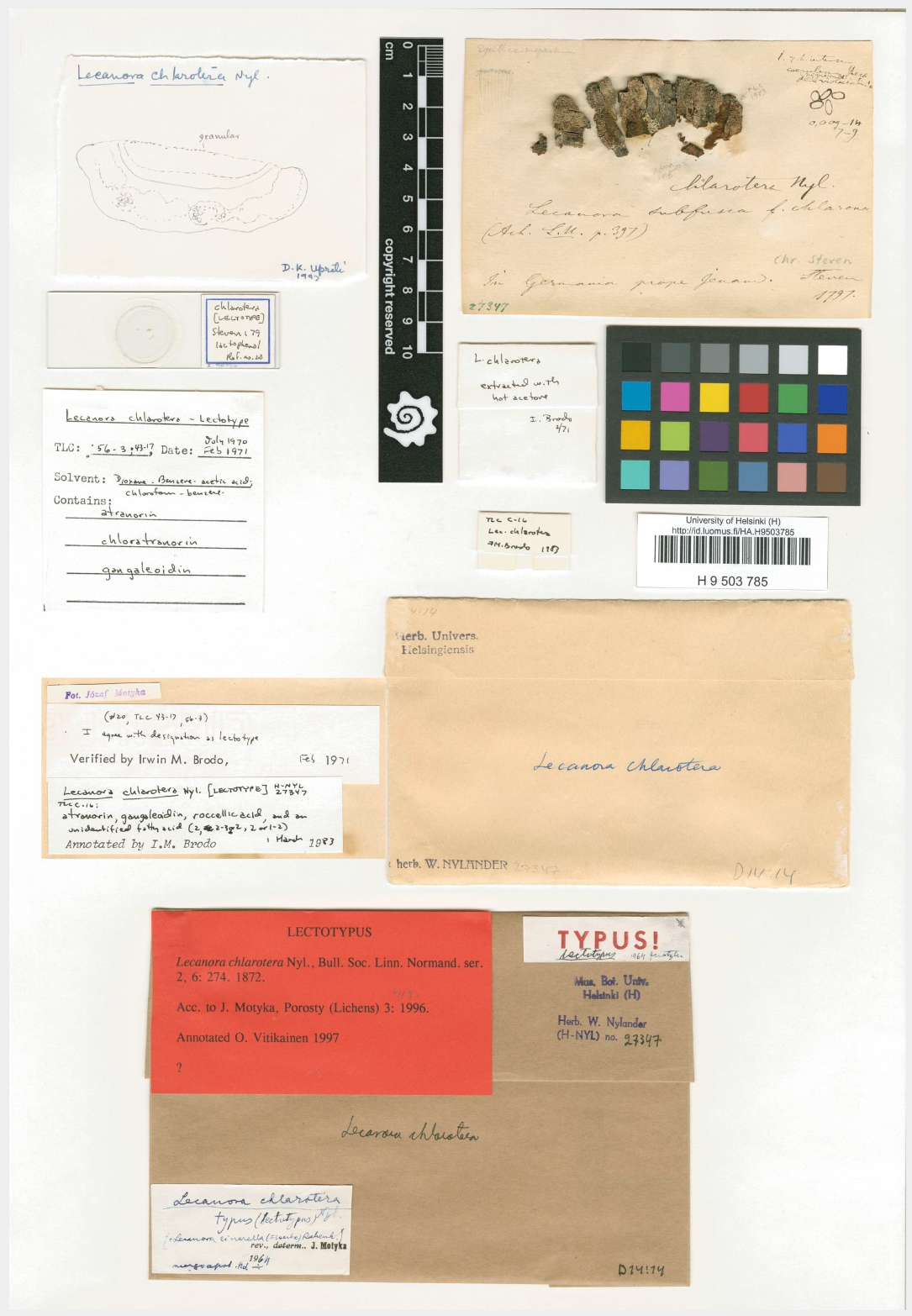
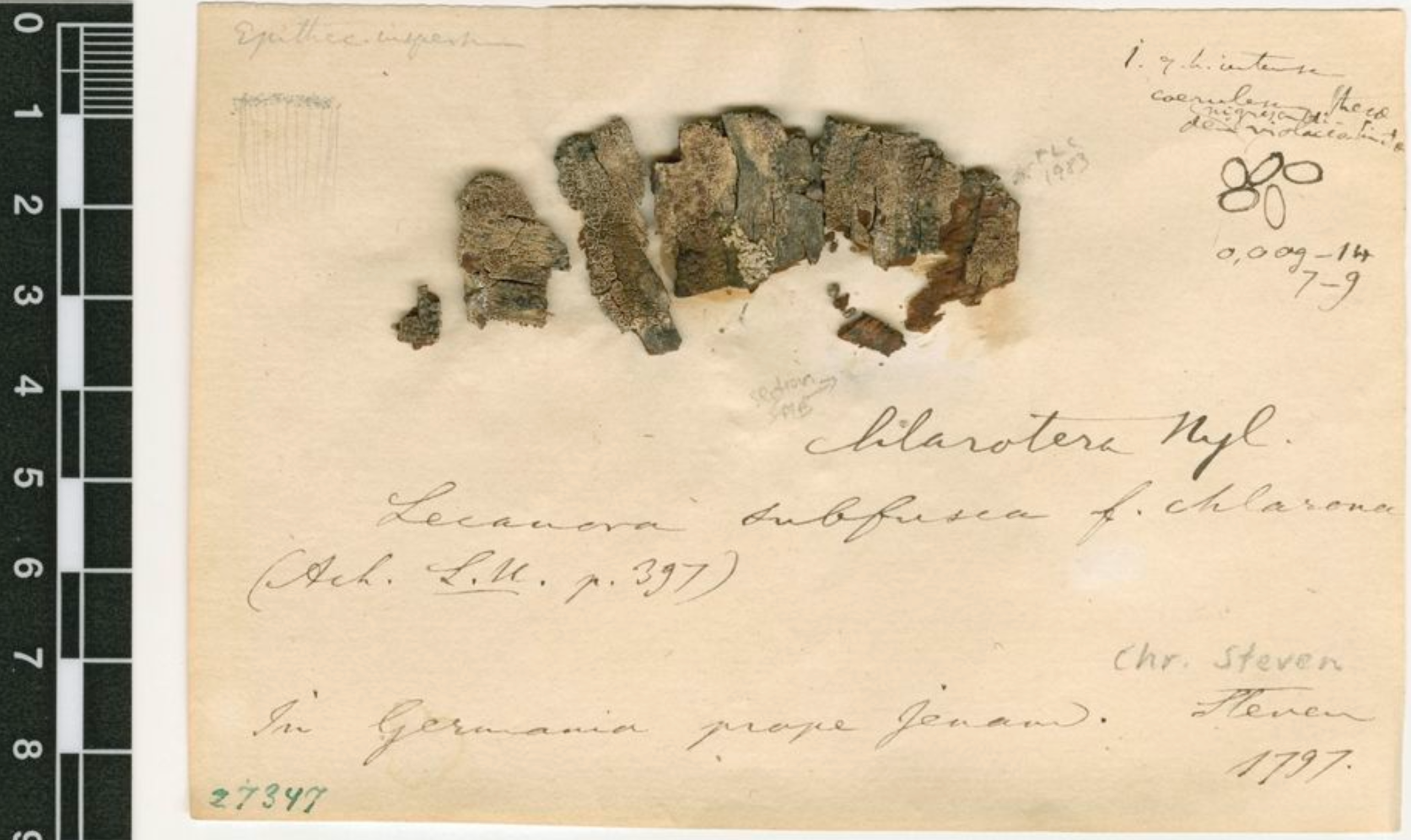
L. chlarotera— collected in Germany near Jena in 1797 by Christian von Steven — has granules only atop the paraphyses (‘ epithecium inspersum’) and not between them (‘ paraphyses non inspersae’). See Figures 1 and 2 below for photographs of the specimen, now held at the University of Helsinki as H-NYL 27437 and considered to be the L. chlaroteralectotype. Note especially the pencil sketch at the top left of Figure 2, labelled ‘ Epithecium inspersum’ and showing clearly that the granules form a layer atop the paraphyses only. [/list] - Nylander’s distinction between the epithecium, in the sense of the very surface of the apothecium, and what’s sometimes called the epihymenium, in the sense of the topmost part of the hymenium that also includes the epithecium, is often elided in lichenology. The term ‘epithecium’ is often used to mean the epihymenium, and it’s not hard to imagine that Hue, who didn’t consider epitheical granules or their placement diagnostically important, might use the term in that way for simplicity’s sake.
[list][/list] - The
chlarona-variety of L. subfuscahas since been circumscribed as L. pulicaris, the classic subfusca-group species with granules between the paraphyses (Brodo, 1984; Brodo & Vitikainen, 1984).
[list][/list] I’d like to add yet a further piece of evidence, which would conclusively decide the case: an examination of the Canary Islands
L. chlaroteraspecimen, which Nylander described as having an epithecium inspersum, and Hue described as having an épithécium non granulé. The specimen was collected in 1845 or 1846 by Eugène Bourgeaux on behalf of Philip Barker Webb (Nylander, 1891; Raymond, 1972). I believe it can be found today in the Webb collection in the Herbarium Centrale Italicum in Florence, but I don’t have any access to it. Be that as it may, Hue’s own
subfusca-group classification scheme, which prioritised external morphological characters such as the shape of the thalline margin, was superseded by the Magnusson/Poelt/Brodo scheme that I argue Nylander anticipated in a small way. For more, see my previous post above. And as I discussed in that post, Neil Sanderson’s 2019 discovery of
L. hybocarpa auct. br.suggests that the Magnusson/Poelt/Brodo scheme might require further revision, at least for the British Isles. In future posts, I’ll turn back to that point, trying to shed some light on it by investigating how the British concept of
L. chlaroteracame to be. REFERENCESBrodo IM (1984).The North American species of the Lecanora subfuscagroup. Beihefte zur Nova Hedwigia79: 63–185. Brodo IM & Vitikainen O (1984).The typification of Lecanora subfusca(L.) Ach., its varieties and some of its related taxa published before 1850. Mycotaxon21: 281-298. https://www.researchgate.net/publication/285029816_The_typification_of_Lecanora_subfusca_L_Ach_its_varieties_and_some_of_its_related_taxa_published_before_1850. ” class=”bbcode_url”> https://www.researchgate.net/publication/285029816_The_typification_of_Lecanora_subfusca_L_Ach_its_varieties_and_some_of_its_related_taxa_published_before_1850. Hue A-M (1903).Causerie sur le Lecanora subfuscaAch. Bulletin de la Société Botanique de France50: 22-86. https://www.biodiversitylibrary.org/item/272970https://www.biodiversitylibrary.org/item/272970” class=”bbcode_url”> Kohler L (1956).Remarques sur les Lecanoracorticoles du group subfusca: Leur répartition dans les Alpes du Dauphiné. Revue bryologique et lichenologique25: 167-182. https://www.biodiversitylibrary.org/bibliography/169357https://www.biodiversitylibrary.org/bibliography/169357” class=”bbcode_url”> Magnusson AH (1932).Beiträge zur Systematik der Flechtengruppe Lecanora subfusca. Meddelanden Från Göteborgs Botaniska Trädgård7: 65–87. Nylander W (1867).Lichenes. In J Triana & JE Planchon, Prodromus Florae Novo-Granatensis: Cryptogamie. https://bibdigital.rjb.csic.es/records/item/9742-prodromus-florae-novo-granatensis-cryptogamie ” class=”bbcode_url”> https://bibdigital.rjb.csic.es/records/item/9742-prodromus-florae-novo-granatensis-cryptogamie Nylander W (1872a).Observata lichenologica in Pyrenæis Orientalibus. Bulletin de Société Linnéene de Normandie2(6): 256-328 https://www.biodiversitylibrary.org/item/37520https://www.biodiversitylibrary.org/item/37520” class=”bbcode_url”> Nylander W (1872b).Observata lichenologica in Pyrenæis Orientalibus. Flora55: 424-431 & 546-554. https://www.biodiversitylibrary.org/item/971https://www.biodiversitylibrary.org/item/971” class=”bbcode_url”> Nylander W (1891).Lichenes Pyrenæorum Orientalium: Observatis Novis. https://catalog.hathitrust.org/Record/100653227https://catalog.hathitrust.org/Record/100653227” class=”bbcode_url”> Poelt J (1952).Die Lecanora subfusca-gruppe in Süddeutschland. Berichte der Bayerischen Botanischen Gesellschaft29: 58–69. https://www.bbgev.de/_files/ugd/f05de5_d67aa0972cb04a418c12edec88586a2f.pdf ” class=”bbcode_url”> https://www.bbgev.de/_files/ugd/f05de5_d67aa0972cb04a418c12edec88586a2f.pdf Raymond M (1972).Bourgeaux, Eugène. In Dictionary of Canadian Biography, vol 10. http://www.biographi.ca/en/bio/bourgeaux_eugene_10E.html ” class=”bbcode_url”> http://www.biographi.ca/en/bio/bourgeaux_eugene_10E.html 
[attachment=1]H-NYL 27347 Lecanora chlarotera lectotype 1.png[/attachment] 
[attachment=0]H-NYL 27347 Lecanora chlarotera lectotype 2.png[/attachment] That’s an odd-looking one, Jenny. But I’d agree with John that it’s probably not L. sinuosa. It seems to lack the ‘incurved apothecium margin and thallus warts with a sinuous outline’ that give L. sinuosaits name, and which jump out clearly from John’s photo (van Herk & Aptroot, 1999, p 548). So is it
L. hybocarpa auct. br.then? If you can resolve a sharper image of the epithecial granules, it would be interesting to check their size against obvious specimens. According to the dichotomous key for Lecanorain the LGBI3 volume on the Lecanoraceae, both L. sinuosaand L. hybocarpa auct. br.have fine granules. But the L. hybocarpa auct. br.I see have coarse granules — notwithstanding some strange observations such as this one (and I recall you said you’ve also made a similar observation): https://twitter.com/specanatura/status/1740328559712043044 ” class=”bbcode_url”> https://twitter.com/specanatura/status/1740328559712043044 LGBI3 also suggests that
L. hybocarpa auct. br.might be a morph of L. sinuosa. Presumably there’s a continuum of characteristics between them. Here’s the link to van Herk and Aptroot’s 1999 article containing their protologue for
L. sinuosa: https://www.researchgate.net/profile/Kok-Van-Herk/publication/230604285_Lecanora_compallens_and_L_sinuosa_two_new_overlooked_corticolous_lichen_species_from_Western_Europe/links/0f3175314c957b506a000000/Lecanora-compallens-and-L-Sinuosa-Two-New-Overlooked-Corticolous-Lichen-Species-from-Western-Europe.pdf ” class=”bbcode_url”> https://www.researchgate.net/profile/Kok-Van-Herk/publication/230604285_Lecanora_compallens_and_L_sinuosa_two_new_overlooked_corticolous_lichen_species_from_Western_Europe/links/0f3175314c957b506a000000/Lecanora-compallens-and-L-Sinuosa-Two-New-Overlooked-Corticolous-Lichen-Species-from-Western-Europe.pdf Happy to hear from others who might disagree, but for what it’s worth, I wouldn’t hesitate to record your lichen as L. hybocarpa auct. br.The only other British lichens with the same pattern of epithecial granules atop and descending between the paraphyses are
L. pulicarisand L. sinuosa. The granules of both of those species are defined as fine (without discernible shape at 400x magnification), whereas the granules in your lichen seem to be coarser, in keeping with the specimens of L. hybocarpa auct. br.that I’ve observed. You probably know that
L. pulicarisand L. sinuosaalso have somewhat different external morphologies as well. Your lichen doesn’t seem a candidate for either species from its appearance, especially not L. sinuosawith its very warty thallus dominated by semi-enclosed apothecia. You could always see if your lichen is Pd- just to rule out
L. pulicaris, though given the coarse granules and fawn-brown discs I personally wouldn’t think it necessary. And it wouldn’t be 100% foolproof anyway! No-one has demonstrated conclusively that the Pd- chemotype of L. pulicariscan be found in Britain as it is on the Continent, but there have been some strange observations: https://twitter.com/specanatura/status/1740325933310214160 ” class=”bbcode_url”> https://twitter.com/specanatura/status/1740325933310214160 Thanks, Alain — that’s an important point. But I think in this particular case we can be fairly confident that the K+ brown reaction that I’ve observed in my C. furcataspecimen is due to fumarprotocetraric acid, for the following reasons: [list]- My specimen certainly contains fumarprotocetraric acid, as the Pd+ red reactions show.
[/list] - We know from current and historical North American sources that K+ brown is indicative of high levels of fumarprotocetraric acid.
[list][/list] - My specimen appears to contain no atranorin, or at least any amount detectable with the standard K-test.
[list][/list] - K+ brown would be an unusual reaction for atranorin, even in the presence of fumarprotocetraric acid.
[list][/list] - It’s highly likely that my specimen, collected in Scotland, would be one of only two chemotypes: with fumarprotocetraric acid only, or with fumarprotocetraric acid and atranorin —
i.e.Chemos I and IV following the very helpful typology on your Lichens Marins website (for other readers, the relevant webpage is: http://www.lichensmaritimes.org/index.php?task=fiche&lichen=190&lang=en ” class=”bbcode_url”> http://www.lichensmaritimes.org/index.php?task=fiche&lichen=190&lang=en
[list][/list] - Again, following your chemotypology, the other possible major lichen substances would be bourgeanic acid, psoromic acid and conpsoromic acid.
[list][/list] - But as you note on your website, chemotypes containing psoromic/conpsoromic acid (Chemos III, V and VI) are Continental, and chemotypes containing bourgeanic acid (Chemos II and VII) are rare (and I might add they’re not mentioned in the British flora).
[list][/list] - As you also note, bourgeanic acid doesn’t give a colour reaction with K, and psoromic/conpsoromic acid gives a Pd+ yellow and not Pd+ red reaction (in the absence of fumarprotocetraric acid).
[list][/list] I certainly agree we must certainly interpret any new colour spot-test reactions carefully — though given North American practice, K+ brown for fumarprotocetraric acid isn’t really a new spot-test. And in this case it seems hard not to conclude that my Scottish
C. furcataspecimen is Chemo I, with fumarprotocetraric acid the only lichen substance detectable by colour spot-tests (Pd+ red most effectively, but also K+ brown). Following up further on the K+ brown reaction for fumarprotocetraric acid that Neil mentioned in this thread. In search of a more convincing result, I applied K to another spot on my specimen of Cladonia furcataabove. Rewarded with a clear K+ brown reaction, I applied Pd directly nearby. The strong Pd+ red demonstrates the presence of fumarprotocetraric acid, as expected. And the lack of any K/UV+ yellow flurorescence on the K-spot suggests fairly unmistakeably that the K+ brown is due to fumarprotocetraric acid, and not by strange chance to any trace of atranorin, which C. furcatacan sometimes contain. 
[attachment=3]20240204-P2040014.jpg[/attachment] 
[attachment=2]20240204-P2040016.jpg[/attachment] 
[attachment=1]20240204-P2040017.jpg[/attachment] An interesting note: where Pd overlapped K, the brown reaction became yellow. I wonder what Pd+ yellow compound results from the decomposition of fumarprotocetraric acid in K?
Another interesting note: a Google search on the K+ brown reaction for fumarprotocetraric acid turned up this 1935 article by American lichenologist RH Torrey entitled ‘Paraphenylenediamine: A new colour test for lichens’:
https://www.jstor.org/stable/40597010https://www.jstor.org/stable/40597010” class=”bbcode_url”> It appears from Torrey’s article that, until Pd came along, some lichenologists relied on the K+ brown reaction for fumarprotocetraric acid to help separate
Cladoniaspecies. Neil noted above that the reaction is still mentioned in North American Cladonialiterature. And the table of colour reactions in IM Brodo’s 2001 North American flora lists fumarprotocetraric acid as ‘K+ brownish’. 
[attachment=0]Screenshot 2024-02-08 at 22.43.30.png[/attachment] Torrey suggests that the K+ brown reaction is also detailed in what he calls Annie Lorrain Smith’s ‘Manual of British Lichens’. That’s not a title of Smith’s of which I’m aware. So far as I know, Smith published a field guide entitled
A Handbook of the British Lichens(1921), a flora in two volumes entitled A Monograph of the British Lichens(1911 and 1918), and a treatise entitled simply Lichens(1921). In none of those works by Smith could I find any reference to K+ brown for fumarprotocetraric acid. She mentions only K+ yellow for atranorin in her descriptions of
Cladoniaspecies (unless I’m missing something). I also couldn’t find it in the 1913 beginner’s flora by Gustav Lindau that Torrey calls ‘Die Flechten für Anfänger’, though the electronic copy I found online was hard to navigate. (Apparently, the proper title is Kryptogammenflora für Anfänger, Band 3: Die Flechten.) That isn’t to say the K+ brown reaction isn’t found in any British work: it’s mentioned in in Orange, Smith & James’s 2010 monograph
Microchemical Methods(p 12), for example. I also found it mentioned alongside Pd+ red as a diagnostic for Usnea antarcticain this 1964 flora of Antarctic lichens published by the British Antarctic Survey: https://core.ac.uk/download/pdf/33451352.pdfhttps://core.ac.uk/download/pdf/33451352.pdf” class=”bbcode_url”> U. antarcticain some online descriptions from Australia and New Zealand that I found in my Google search.) But anyway: assuming that there isn’t a ‘Manual of British Lichens’ by Annie Lorrain Smith of which I’m unaware, the K+ brown reaction for fumarprotocetraric acid on which Torrey and other (presumably mainly North American) lichenologists relied doesn’t seem to have been much used by British lichenologists before being obviated by the more effective Pd+ red.
Neil Sanderson wrote:
GerraultHow yellow is the reaction? I looks quite brownish to me. Potentially the K+ brownish reaction to fumarprotocetraric acid; this is often weak and is clearest when there is a lot of fumarprotocetraric acid in the lichen. As it is inconsistent it tends not to be mentioned in descriptions, although American
Cladonialiterature does mention it more. We should have added a note on this reaction in the Cladoniaceae volume of the LBGI, but I forgot. Neil
Apropos of Neil’s comment above about a K+ brownish reaction to fumarprotocetraric acid, I believe I may have observed it on a
Cladonia furcataspecimen that I collected this summer in Scotland (VC101 Kintyre : NR77 Ormsary : August 2023 : In pocket of humus on siliceous rock). The first photo below shows a Pd-test with a strong red reaction as expected. The second shows a K-test presumably with Neil’s weak brown reaction. Many thanks to Mark for pointing out to me the connection to the discussion in this thread. 
[attachment=1]20240121-P1211315.jpg[/attachment] Many thanks, Mark, for bringing this LF to the attention of Brian C. It’s exciting to hear that he recognises having seen it before, and that he was easily able to find a likely specimen. It’s also good to know that our finds can be more publicly disseminated via the BLS database as Acremonium sp, at least until a proper name is found. I’ve emailed Brian, and copied you, bringing some further micrographs to his attention. These micrographs relate to the Norfolk specimens that I found, one of which fairly clearly seemed to be our hyphomycete LF, and the other of which may or may not be. Perhaps the additional evidence will help with identifying the LF — and at the very least, Brian may discount it as relating to a different LF, which would be equally valuable.
I sent him the micrographs by way of a link to the presentation about this LF that I recently gave to the BLS Lichen Chat and Improvement Group. Having published that link on Twitter some time ago, it occurred to me that I should just as well have done so here! —
https://drive.google.com/file/d/1bTuHFgsKoJo_XnTPlgr-qm7Kz1vs23tf/view Many thanks, Fay — it’s fantastic to know that you agree on T. delicata. I’m thrilled to be able to add to the small number British records of this species! It’s also good to know about the record on
L. hybocarpafrom Hampshire. Heuchert et alsay that the 1977 holotype from Buckinghamshire is on ‘ L. cf chlarotera‘, which I assume is most probably L. hybocarpaas well. As for
T. pertusariicola, I went by the lack of a BLS distribution map for the species. But I somehow overlooked the mention on the FGBI website, as well as your own BLS species description for it, both of which I now see! Not sure how I managed that oversight, especially as I saw your BLS species description for T. delicata. Grateful for the correction. Thanks for your question, Bob — lovely photo. It would be helpful to have more of an overview, but your lichen gives the strong impression of Physcia tenella. It’s a very common leafy or ‘foliose’ lichen on bark and other substrates, especially in lowland UK. Indeed, it’s probably fair to say that it’s been on the increase, as it thrives on the excess nitrogen that our agricultural activities have injected into the environment. You can read more about P. tenella on the British Lichen Society website at
https://britishlichensociety.org.uk/sites/default/files/Physcia%20tenella.pdf ” class=”bbcode_url”> https://britishlichensociety.org.uk/sites/default/files/Physcia%20tenella.pdf https://britishlichensociety.org.uk/resources/species-accounts/physcia-tenella ” class=”bbcode_url”> https://britishlichensociety.org.uk/resources/species-accounts/physcia-tenella P. tenella tends normally to reproduce asexually or ‘vegetatively’, through powdery grains called ‘soredia’, which contain cells from both the fungus and the alga that make up the symbiotic organism. These soredia develop in masses called ‘soralia’, which on P. tenella tend to form on the tips of the lobes shown in your photo. They disperse in wind and rain to colonise other surfaces and become full lichen bodies or ‘thalli’.
One must be careful to distinguish between P. tenella and the look-alike P. adscendens. They’re best told apart by the shape of their lobe tips and their soralia. The lobe tips of P. adscendens tend to be closed and ‘hooded’ rather than open and ‘fingery’, and the soralia develop in the ‘hoods’ rather than on the tips of the ‘fingers’.
Your dark ‘claws’ are called ‘cilia’, which are hair-like structures or outgrowths of the thallus. P. adscendens has them as well, as do some other lichens. Their purpose isn’t entirely clear — they don’t help with attachment to the substrate, as similar outgrowths on the underside called ‘rhizines’ do. Perhaps they help with the condensation of moisture, as lichens take their water from the air or from rain. Perhaps they have some other purpose as well.
By the way — the whitish spots on the lobes of your lichen are called ‘pseudocyphellae’. They represent a thinning of the lichen’s surface layer or ‘cortex’, which helps to facilitate gas exchange. The alga inside the fungus body draws in carbon dioxide to photosynthesise sugars for itself and for the fungus, and oxygen must be released.
If you could post a photo showing a bit more of the lichen, it might be possible to confirm that you have P. tenella. That said, immature thalli of P. tenella and P. adscendens, which haven’t yet developed soralia, are generally indistinguishable.
- Nylander had published his protologue for
-
AuthorPosts