aspeca

Forum Replies Created
-
AuthorPosts
-
Many thanks, Neil — very helpful as always. Many thanks, Neil — it’s very helpful to see those photographs. Very helpful in more ways than one! Quite apart from
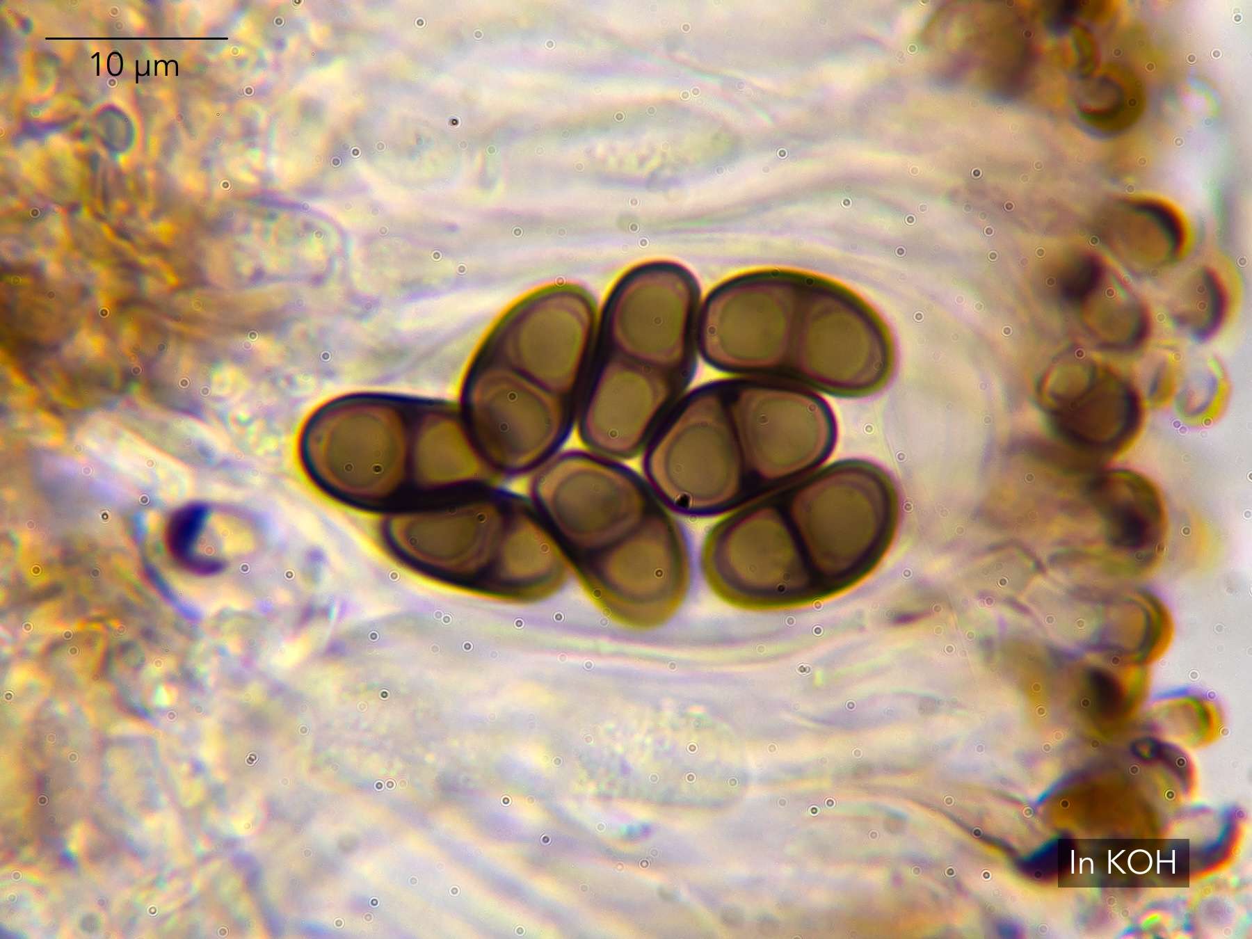
Myriolecis salina, I immediately twigged from your third photo that I might have concluded Amandinea punctatatoo quickly, and that I should have checked more carefully to see whether I had A. pelidna (lecideina)on my pebbles instead. Having looked again at the spores, it seems I do. They appear to have the characteristic ‘girdle’ from median thickening at the septum. Let me know if you disagree.
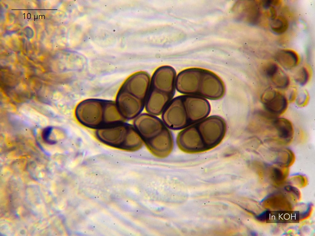
[attachment=0]Amandinea pelidna 20260704-A-7.jpg[/attachment] And as it happens, yesterday I was looking at a lichen on a siliceous gravestone that I couldn’t place, but which looked inexplicably like
Rinodina aspersaand tested C+ red. Thanks to your photo, I now know it’s just a very scattered Trapelia obtegens. Best regards,
Anthony
Many thanks, Paul. I had discounted Myriolecis dispersadue to the hard flint substrate, though I suppose it’s possible that the pebble is somewhat buffered by dust. For comparison, I’ve looked at some
M. dispersagrowing on a fragment of concrete slab that I took from the same location. For what it’s worth, its hymenium is taller but not hugely so — more like 70 µm rather than 60 µm. LGBI3 indicates that
M. salinahas noticeably wider paraphyses than M. dispersa, and that this is a distinguishing feature. Best efforts to measure the paraphyses of my M. salinaput them generally over 2 µm wide, and up to 3 µm — which is in the range for M. salinabut not M. dispersa. The paraphyses of my M. dispersameasure up a bit narrower, but there’s not much in it. And as you say — I wonder how often these things are measured. ITALIC doesn’t give any figure for
M. dispersaparaphyses width, but the ITALIC figure for M. salinaparaphyses is the same as the LGBI3 figure for M. dispersaparaphyses! My
M. dispersawas also clearly UV-, whereas my putative M. salinaappears to be UV+ dull orange. Apart from indicating a difference between the two, though, I’m unsure what to make of it, inasmuch as M. salinais supposed to be UV- as well. There are other southern records for
M. salinafrom Hampshire and Somerset — presumably also from shingle. But as this would be a first record for Suffolk, I think I may hold out for now in the hope of finding a clearer specimen. Thanks again and best regards,
Anthony
Many thanks, Neil — that’s extremely helpful. I’ll let you know of any further developments from either the management meeting or a further site visit. This link should give you five full-sized high-resolution photos. Please feel free to use any of them that you like — all Creative Commons BY-SA 4.0.
https://aspenecology.com/wp-content/uploads/2026/03/Anaptychia-ciliaris.zip ” class=”bbcode_url”> https://aspenecology.com/wp-content/uploads/2026/03/Anaptychia-ciliaris.zip Best regards,
Anthony
Dear Neil — Thanks for this. Sad to hear that
A. ciliarisis still in free fall. Will you continue to classify it as EN, or has it sadly become a candidate for the CR category? Janet has gone back over the historical Suffolk records, and she confirms that this is indeed the first sighting in the county since 1985. (The 1993 date in the comment of one of the Sotterley records referred to a publication, not an observation. And the 2005 date referred to the year an undated record was received by the recording scheme, which turned out to refer back to the 1985 observation.)
A quick question about ammonia pollution. The BLS website species description previously indicated that
A. ciliariswasn’t ammonia-sensitive per se, and indeed that it liked a bit of nutrient enrichment when on e.g.lime or oak. But it also warned that particulate matter from inorganic fertiliser dust would become trapped in the tomentum and encourage the growth of algae over the cortex, smothering the lichen. The BLS website species description now matches LGBI3, removing the reference to particulates and citing high ammonia levels in general. Has
A. ciliarisbeen found to be more ammonia-sensitive than previously thought, or are background ammonia levels simply so high now that they exceed the lichen’s tolerances? Or perhaps a bit of both? I’m meeting with the landowners tomorrow to discuss management. The local Natural England team is now also aware. I’d like to go back to the site for a more thorough search (with binoculars!) — this lichen was on a low branch, but the tree itself was elevated on a mound, which gave it some height. I wonder if there may be other thalli higher up in the same tree, or in other trees.
Please do feel free to use the photos — but ignore the ones here. I’ll send you a link to download full resolution versions, which will enlarge better for the BLS website.
Thanks again and best regards,
Anthony
Many thanks for this, Neil — very helpful. That sounds like just the obvious thing I was missing. I’ll have a further look at the material with that in mind. Dear Neil — Many thanks for this. I’ll be keen to hear what Brian eventually decides about the pragmatic name we should use here in Britain. It feels as though we have the name
hybocarpaon a sort of loan from North America! So
L. pseudoargentatais definitely here in the UK then. It’s good to hear your confirmation that it wouldn’t be confused with L. sinuosa/hybocarpas. lat. But it seems that it might not be reliably separable from
L. chlaroteras. str.? You mention it’s slightly morphologically distinct, so there’s some hope there. But if not, will we possibly end up with two pragmatic taxon names — not only
L. sinuosa/hybocarpas. lat., but also some pragmatic name that covers both L. chlaroteras. str. and L. pseudoargentata? Best,
Anthony
Many thanks for this, Neil — fascinating and exciting stuff! I’ve only read Arup
et al.(2025) quickly, so I might have missed something. But it raised a sort-of ‘field lichenological’ question in my mind, which I’d be keen to hear your views on. What you think about their suggestion to use ‘
Lecanora hybocarpas. lat.’ as the working taxon name? It certainly seems the right taxon name for comprehensive research, but is it the best pragmatic recording taxon for Britain? Arup and his co-authors didn’t find any
L. hybocarpa(Tuck.) Brodo (1984) in Europe despite a substantial search. And as you’ve pointed out, it would have been unexpected if they had done. So while there may be no reliable morphological separation between L. hybocarpa(Tuck.) Brodo (1984) and L. sinuosa/subsinuosa/cryptosinuosa, isn’t there a fairly reliable biogeographical separation? And if so, wouldn’t that suggest that we should use ‘
L. sinuosas. lat.’ as the pragmatic recording taxon, at least for British recording purposes ( i.e.to encompass what we’ve thus far been calling L. hybocarpaauct. br. and L. sinuosa)? I suppose the confirmed presence of
L. pseudoargentatain Europe complicates matters a bit. But insofar as that species appears to have a chlarotera-type epihymenium, not a pulicaris-type, it should be pragmatically distinguishable from what I’m calling ‘ L. sinuosas. lat.’ There might be difficult cases to judge, as you and Pier-Luigi have pointed out already exist between
L. hybocarpaauct. br. and L. chlaroteras. str. But LGBI3 accepts that it’s pragmatically possible to differentiate between the chlarotera– and pulicaris-type epihymeniums in most(?) cases, so perhaps the presence of L. pseudoargentatain Europe doesn’t pose an insuperable difficulty. So far as practical field recording goes, it seems best to use the narrowest taxon name possible. So while we might have to use ‘
L. sinuosas. lat.’ just as we use ‘ Cladonia chlorophaeas. lat.’ (or perhaps ‘ C. grayis. lat.’), that name would seem a bit closer to the actual European (or British) species than ‘ L. hybocarpas. lat.’ Again, I could be missing something here. I’d be grateful for your views, or clarification.
Many thanks,
Anthony
Many thanks, Neil. Glad to have your confirmation. Are there any plans afoot to sequence and compare European and American
L. hybocarpa? Thanks again, Neil — that’s really helpful. I’m pretty confident I saw
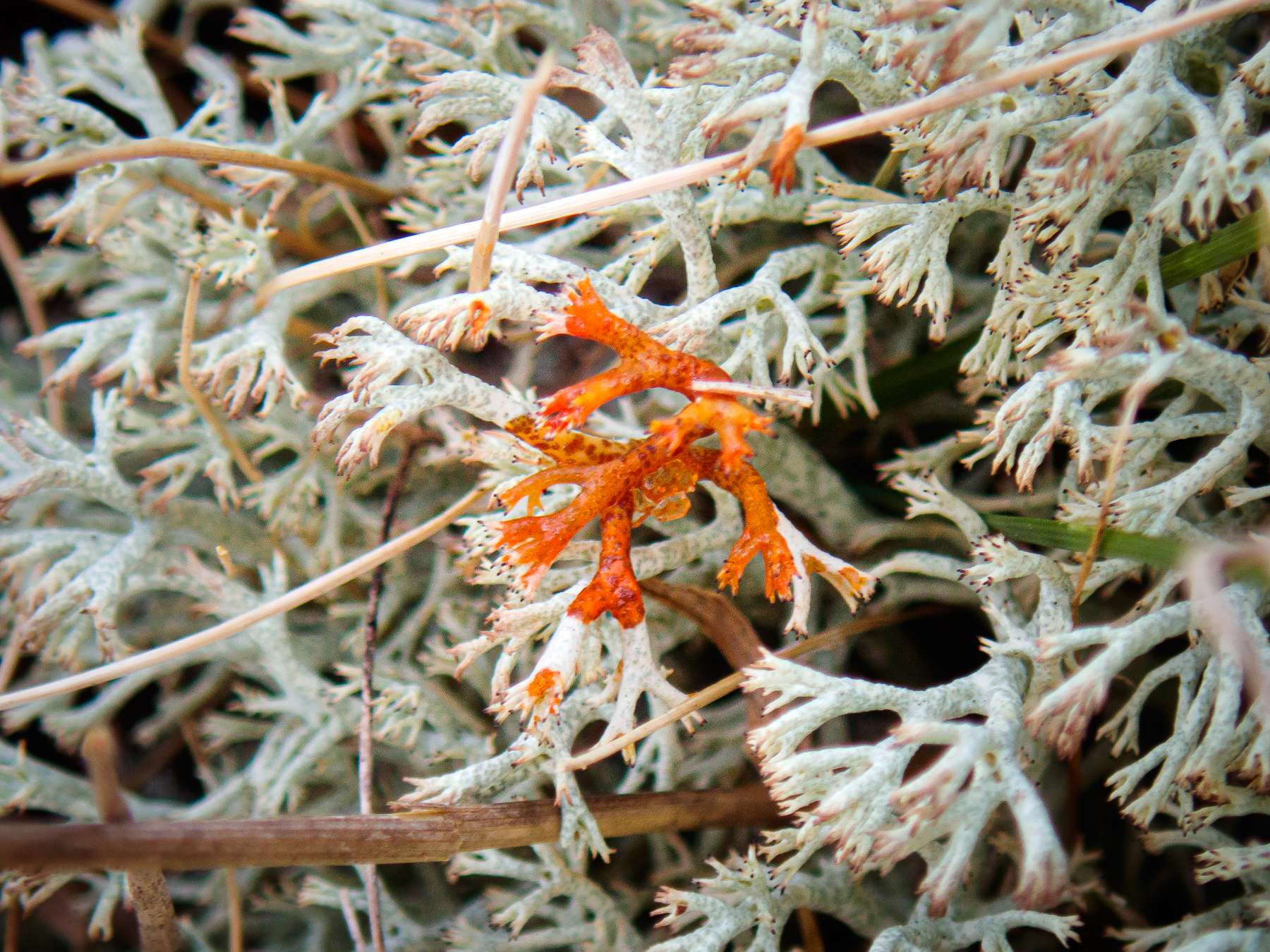
Cladonia arbusculaon another lowland heath in Suffolk (Wortham Ling, at VC25/TM0975), which tested a clear Pd+ red, and which has been recorded there before. See photo below (please do let me know if you disagree). However, I realise now that the sample I collected for my herbarium is mostly if not all
C. portentosa, with which the C. arbusculamust have been growing in something of an entangled fashion. This would explain the UV-photos above, which must be showing the UV+ blue fluorescence of C. portentosain all three cases. If your
C. arbusculawas quite dull under UV-light, then it seems that distinguishing between UV+ blue fluorescence on the one hand, and mere UV-reflectance on the other, is less problematic than I’d assumed. How frustrating! But also highly instructive. One expects one’s mistakes would result from thinking something is easier to recognise than it really is, rather than from thinking it’s harder!
Best regards,
Anthony
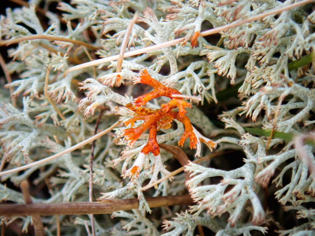
[attachment=0]Cladonia arbuscula subsp squarrosa 20250412-A.jpg[/attachment] Many thanks, Neil. I can see the differences in the surface that you mean. Together with Alain’s comments, I think this points fairly clearly to C. portentosa. A fairly ‘exuberant’ form with which I wasn’t previously familiar. I’m a bit concerned now, though, about the fluorescence question, so any comments you might have on the photos below would be much appreciated. Does perlatolic acid degrade over time in the herbarium?
Best regards,
Anthony
Many thanks, Neil — that’s good to hear. Westleton Common seems to be proving more and more interesting! Best regards,
Anthony
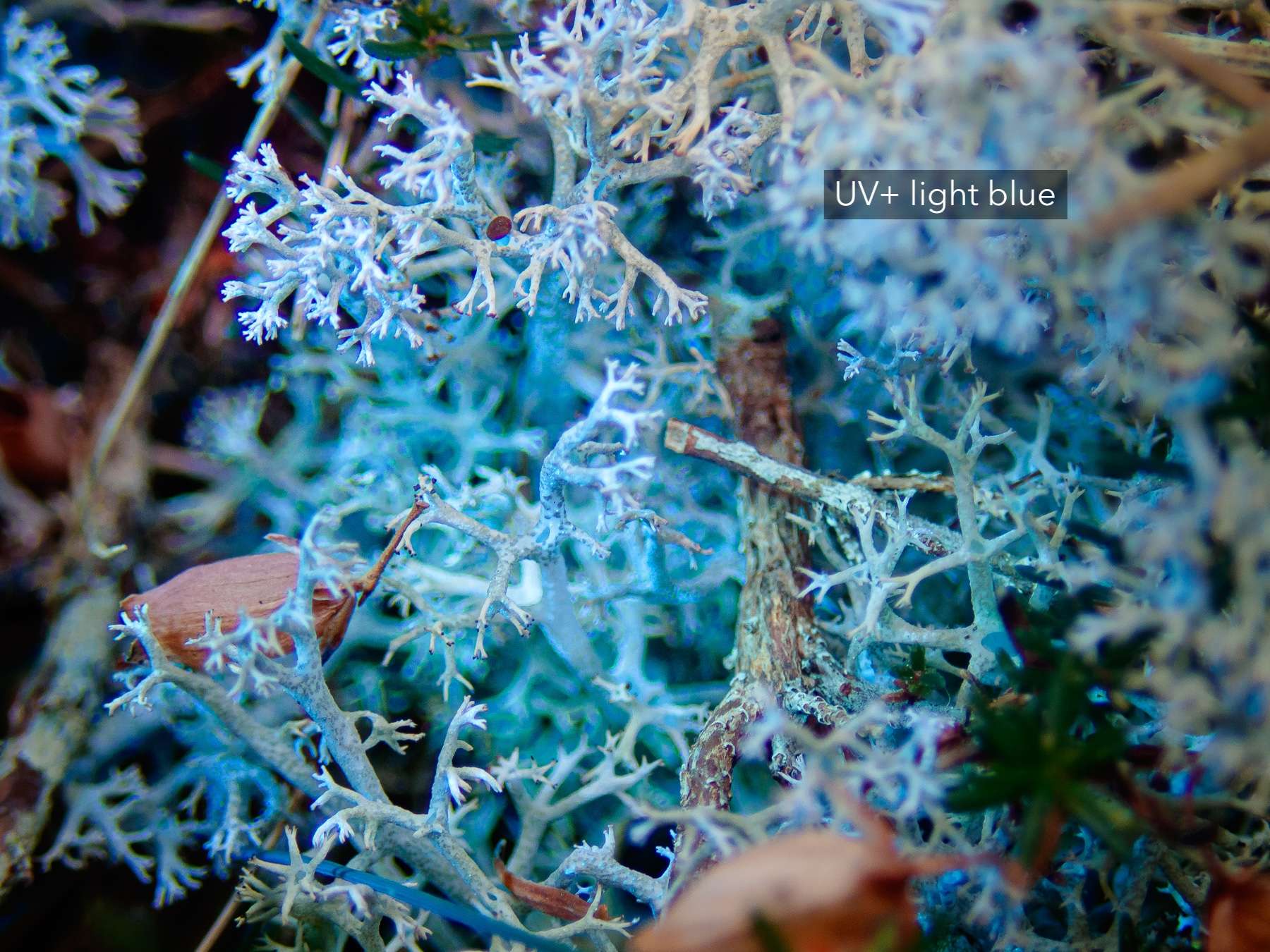
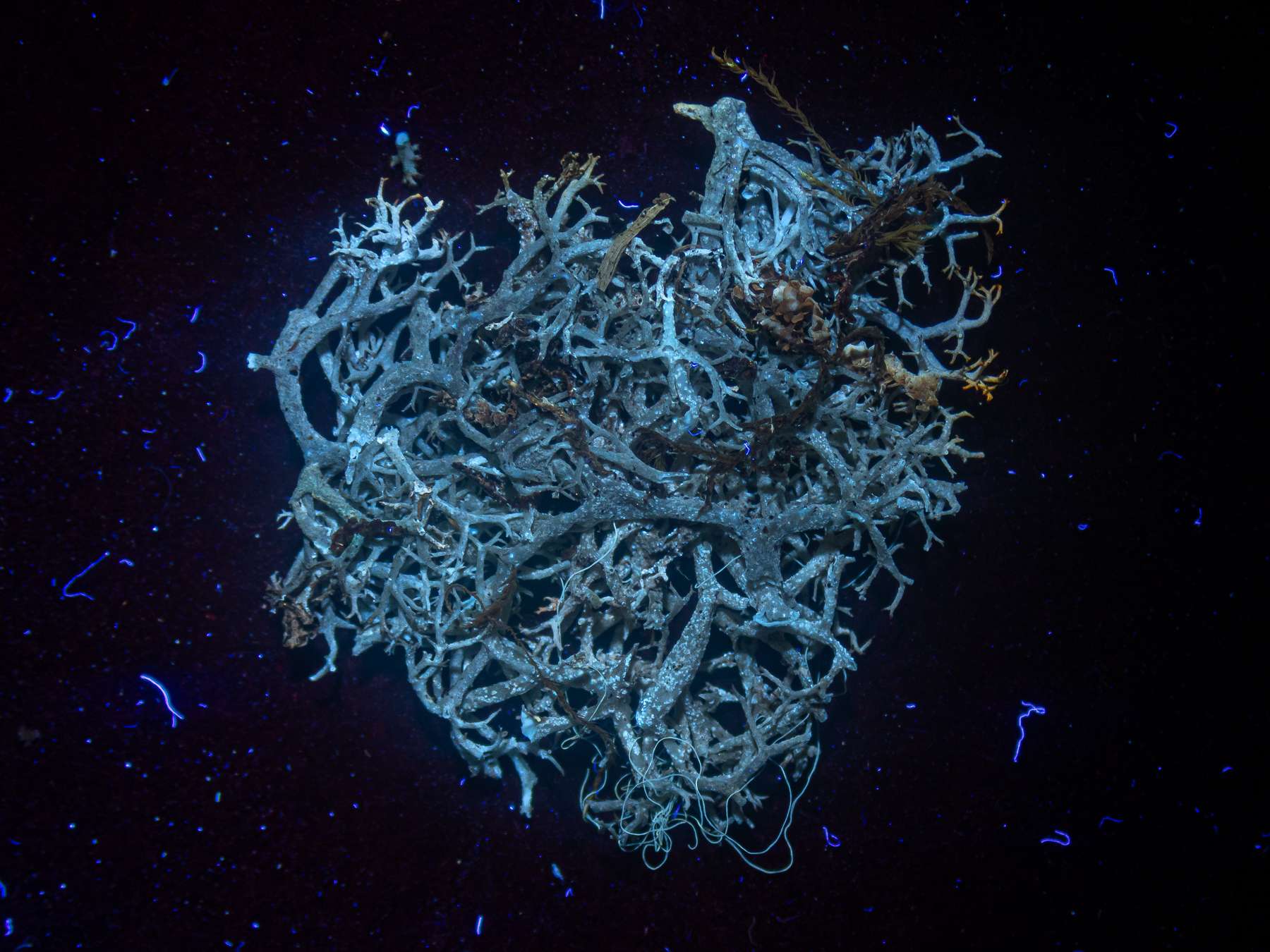
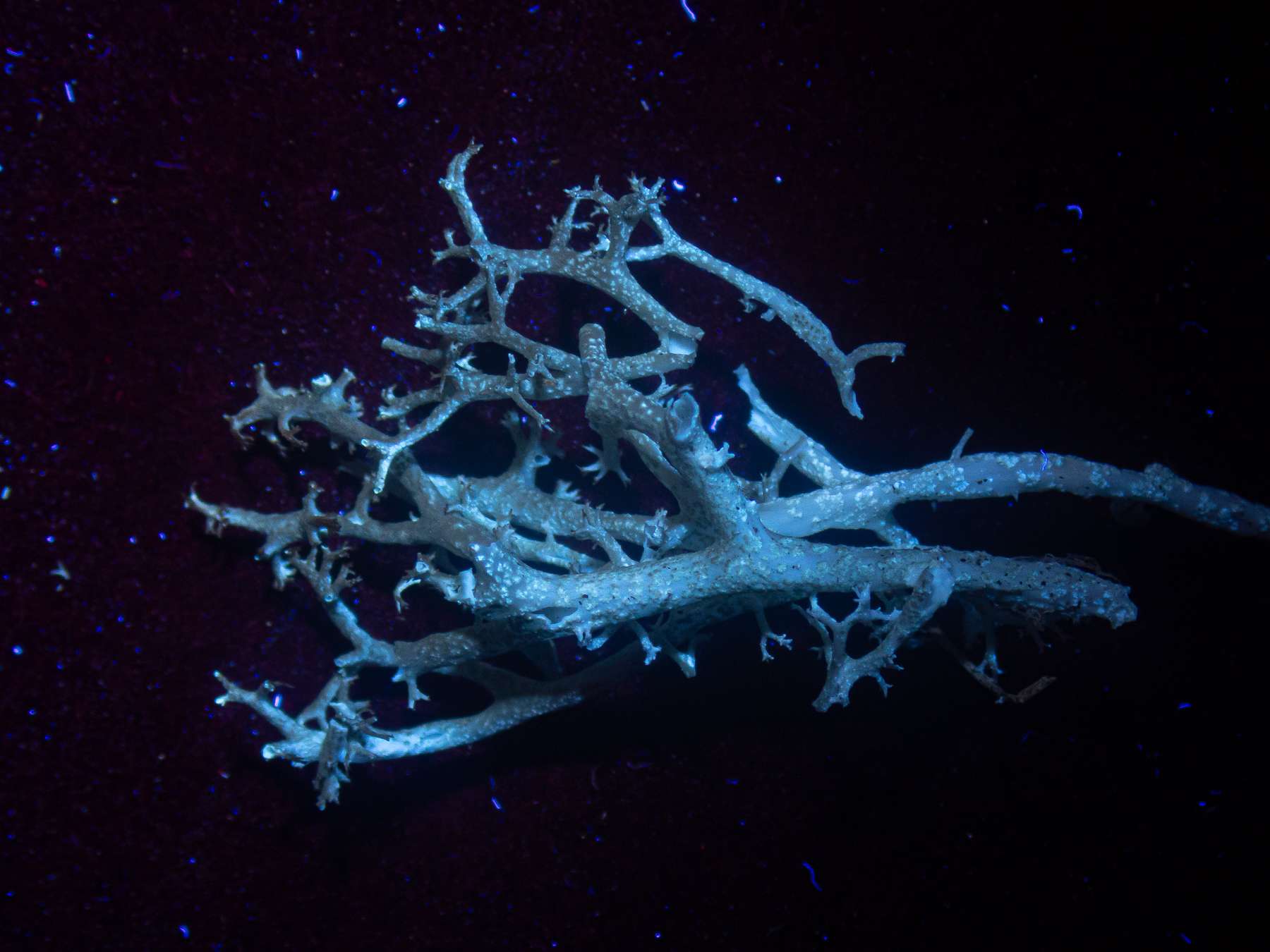
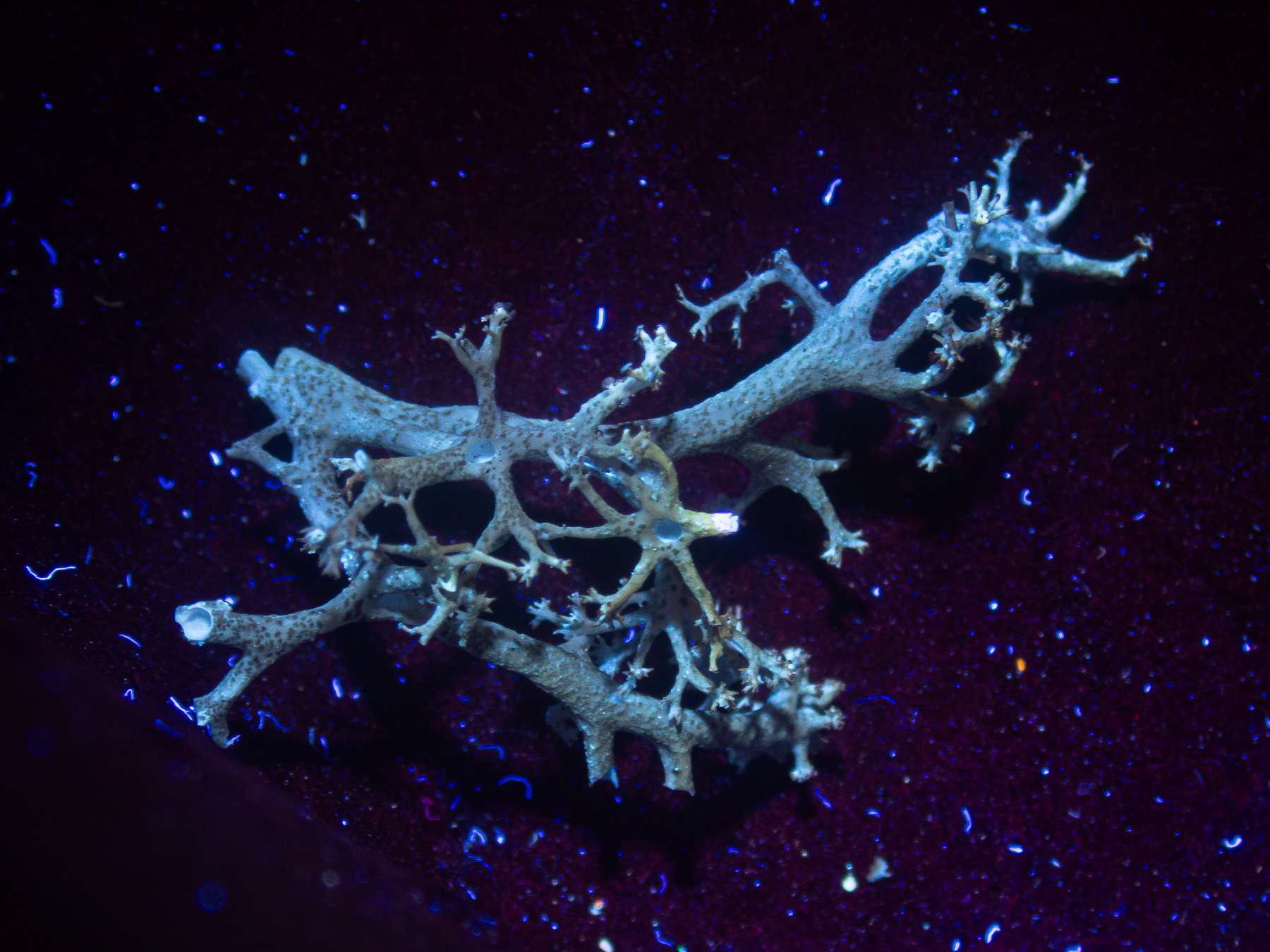
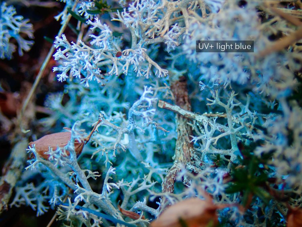
Many thanks, Alain. You may be right. With pale Cladonia, it can be difficult to distinguish true blue UV-fluorescence from mere blue UV-reflectance! Below are some comparison photos that may help:
C. portentosa, C. arbusculasubsp. squarrosa(reacts Pd+ red), and the possible C. mitis. All the collected podetia are dry, and either months or years old. But just in case perlatolic acid degrades over time, I’ve also included a photo of fresh
C. portentosafluorescing clearly in the field. (If I can get sufficient shade on the specimen, I haven’t yet found it too problematic to capture the UV+ blue fluorescence of C. portentosafrom fresh lichens.) In all cases, I’m using a front-filtered 365nm UV-torch (Alonefire). I also equalised the white balance, tint and saturation of the three photographs of the collected podetia to allow for better comparison.
Considering that
C. arbusculais UV-, it stands to reason that it must be merely reflecting the UV-light (a bit like white paper), not fluorescing on its own. The C. portentosa, however, must be fluorescing UV+ blue from perlatolic acid. Perhaps that accounts for its somewhat more even blueness, especially in the photo of the fresh lichen in the field? To my eyes — and this is just my own colour perception of course! — the possible
C. mitislooks more like the C. arbuscula(i.e. merely reflecting) than it does the C. portentosa(i.e. fluorescing). But I accept that others may see the colours differently, and that the morphological similarities and differences may be better evidence one way or the other. Thanks again,
Anthony
[attachment=3]Cladonia portentosa 20240921-B01 Westleton.jpg[/attachment] 
[attachment=2]Cladonia portentosa 20230802-A.jpg[/attachment] 
[attachment=1]Cladonia arbuscula subsp squarrosa 20250412-A.jpg[/attachment] 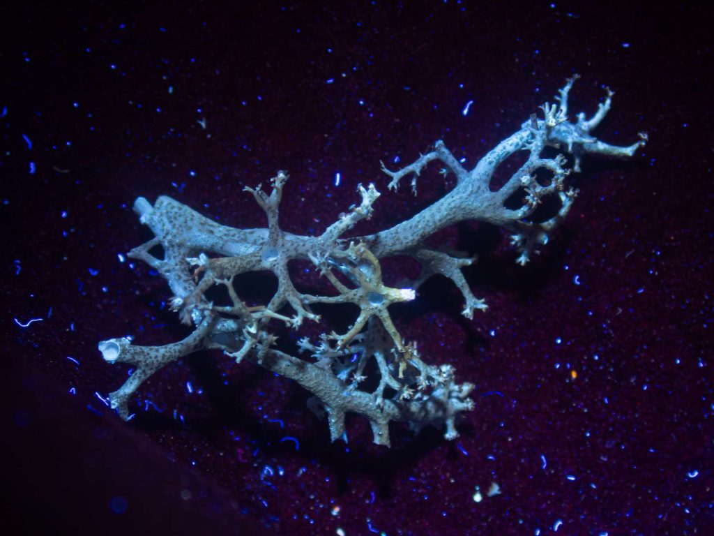
[attachment=0]Cladonia mitis sp inc 20250628-B-5.jpg[/attachment] Many thanks, Alain. Yes — I wondered if it might be V. halizoa, not least because of the colour. But there don’t seem to be any of the fairly abundant, and relatively large and prominent perithecia that seem to characterise that species — and which are shown so well in lichen image banks (including your Lichens Marins). But not having seen V. halizoamyself, I can’t be sure. In any case, it seems it must remain something of a mystery, at least to me! Thanks again for your input.
Many thanks, Alain. My apologies — I think I unfortunately created confusion with my speculations. I didn’t mean to suggest that orange body in the third photo is part of
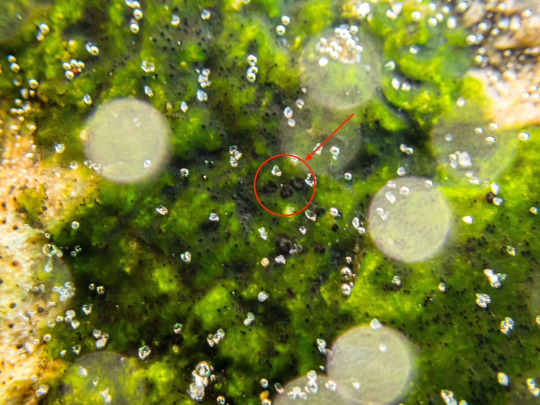
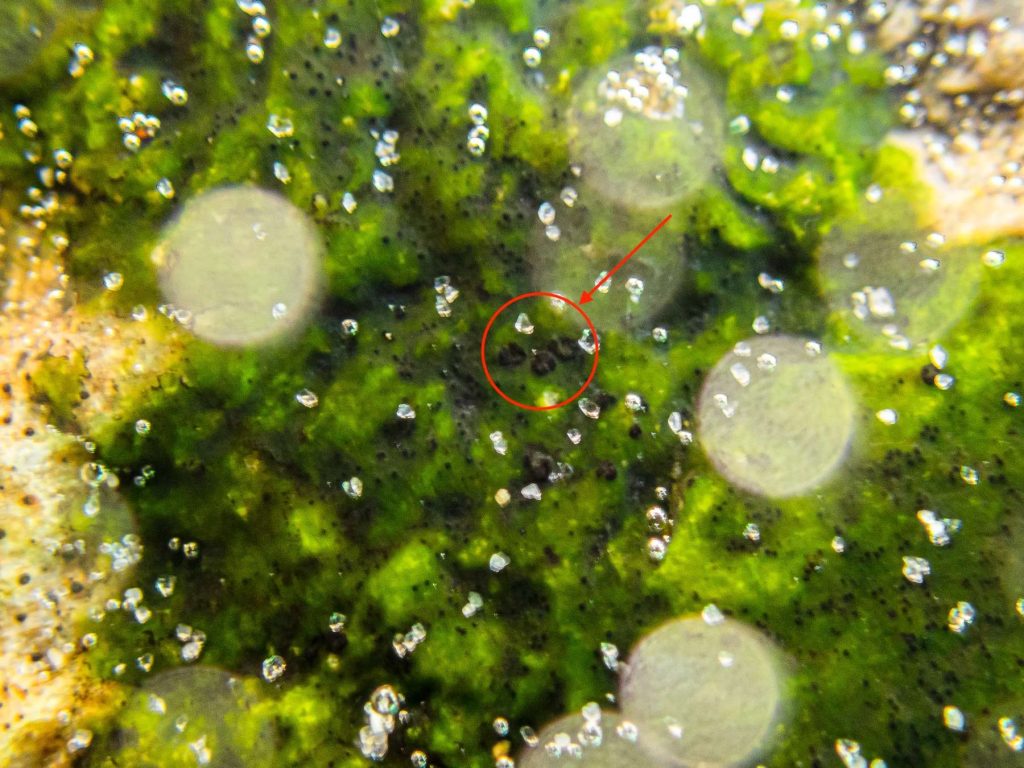
S. marinum. As mentioned, I believe it’s an encrusting sponge. I was only struck by the ‘black dots’ on it that looked at least superficially similar to S. marinum, and that put me in mind of Andre Aptroot’s contention that S. marinumisn’t (or isn’t only) an LF, and that it occurs on various substrates. But it’s perhaps best to leave all that aside. I’ve attached an annotated photo here, encircling what appear to be some of the
S. mariumperithecia on W. mucosa. As you can see from the sand grains in the water, they’re very small. They look very much like some of the structures in your photo above, as well as in the following photos of S. marinum: [list]https://www.afl-lichenologie.fr/Photos_AFL/Photos_AFL_S/Text_S3/Stigmidium_marinum.htm ” class=”bbcode_url”> https://www.afl-lichenologie.fr/Photos_AFL/Photos_AFL_S/Text_S3/Stigmidium_marinum.htm [/list] [list]https://www.researchgate.net/publication/321054505_Ocean_view_A_first_assessment_of_the_littoral_crustose_lichen_biota_of_south_Brazil ” class=”bbcode_url”> https://www.researchgate.net/publication/321054505_Ocean_view_A_first_assessment_of_the_littoral_crustose_lichen_biota_of_south_Brazil [/list] As for the appearance of the
W. mucosa— I should have explained better what I meant by ‘underwater photos’. I took them quite literally underwater, with a submersible camera. The water was in motion as the tide was coming in, creating some softness in the image. The thalli were also in strong light, enhanced by the photo settings, and the thalli were fully hydrated. These factors explain (I think) the unusual appearance. I admit again that there’s unlikely to be any conclusive interpretation of my photos, especially as I have no sample. But I thought it worth posting for interest, and in case anyone’s seen something similar. I know you don’t seem to recognise
S. marinumhere, and it would be really helpful for me to know more precisely what I might be missing given the similarities mentioned above. Thanks again,
Anthony
[attachment=0]Stigmidium marinum sp inc 20250820-A-1.jpg[/attachment] -
AuthorPosts