
Myriolecis salina?
- This topic is empty.
-
AuthorPosts
-
July 15, 2026 at 11:59 am #33184
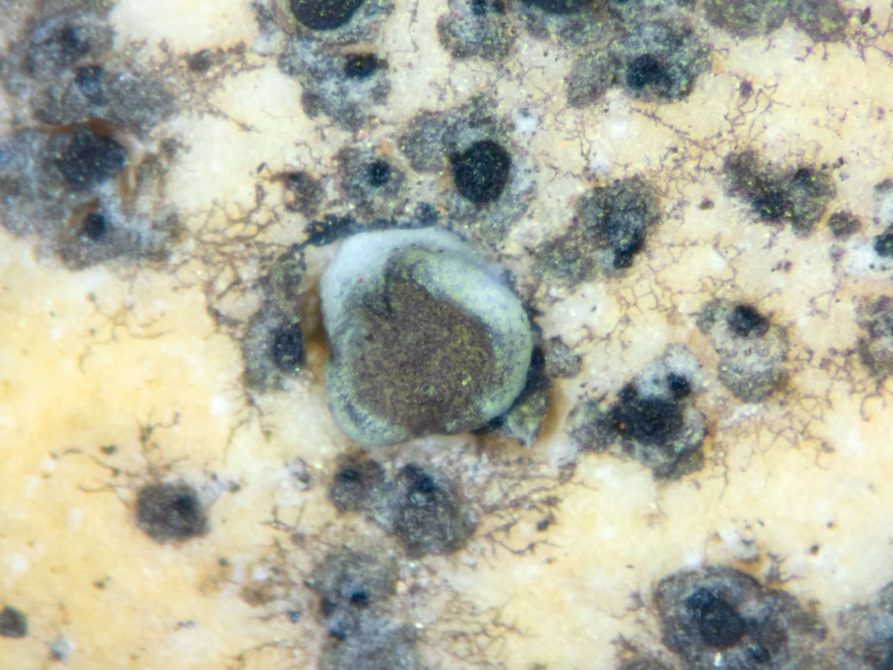
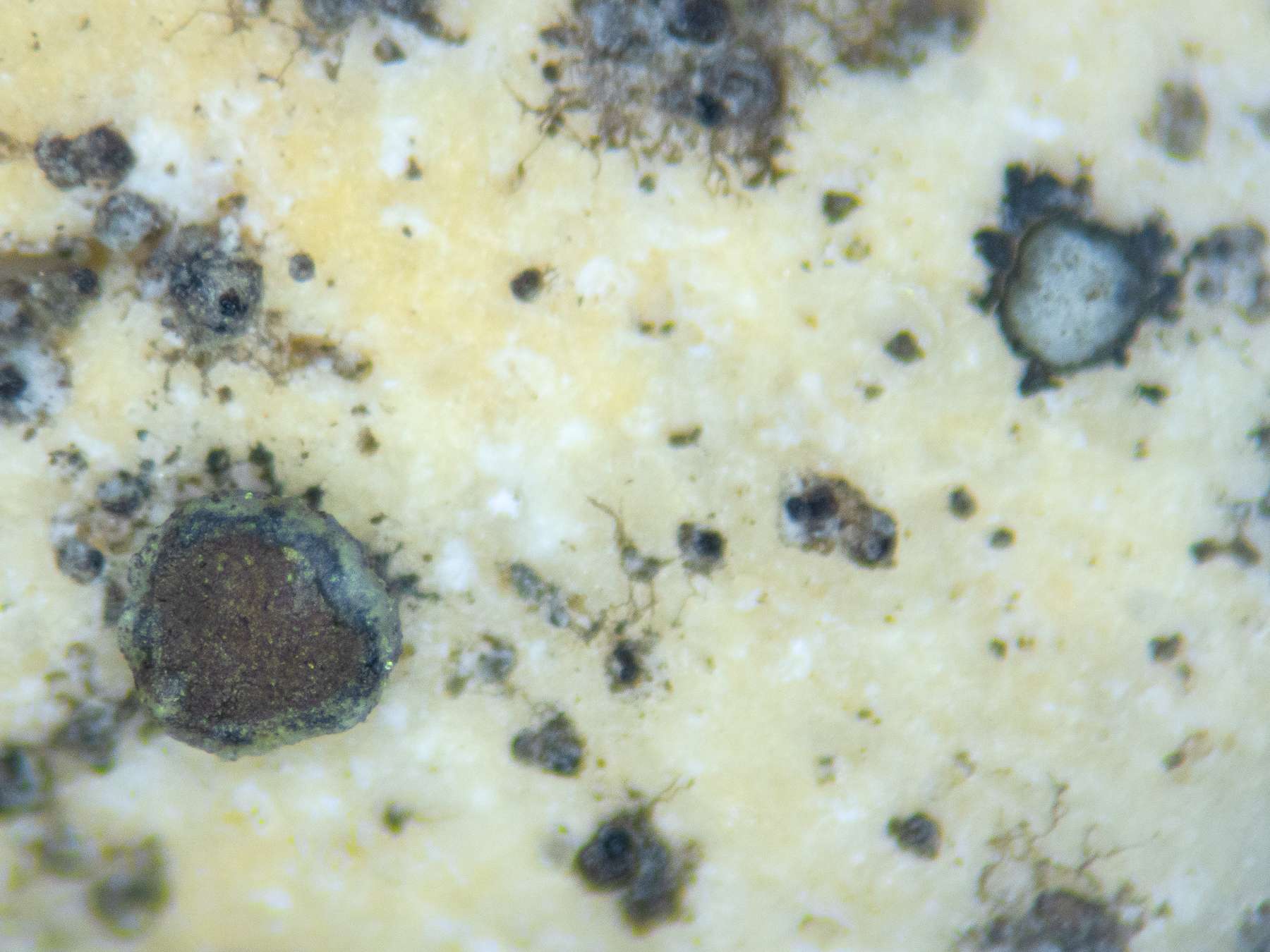
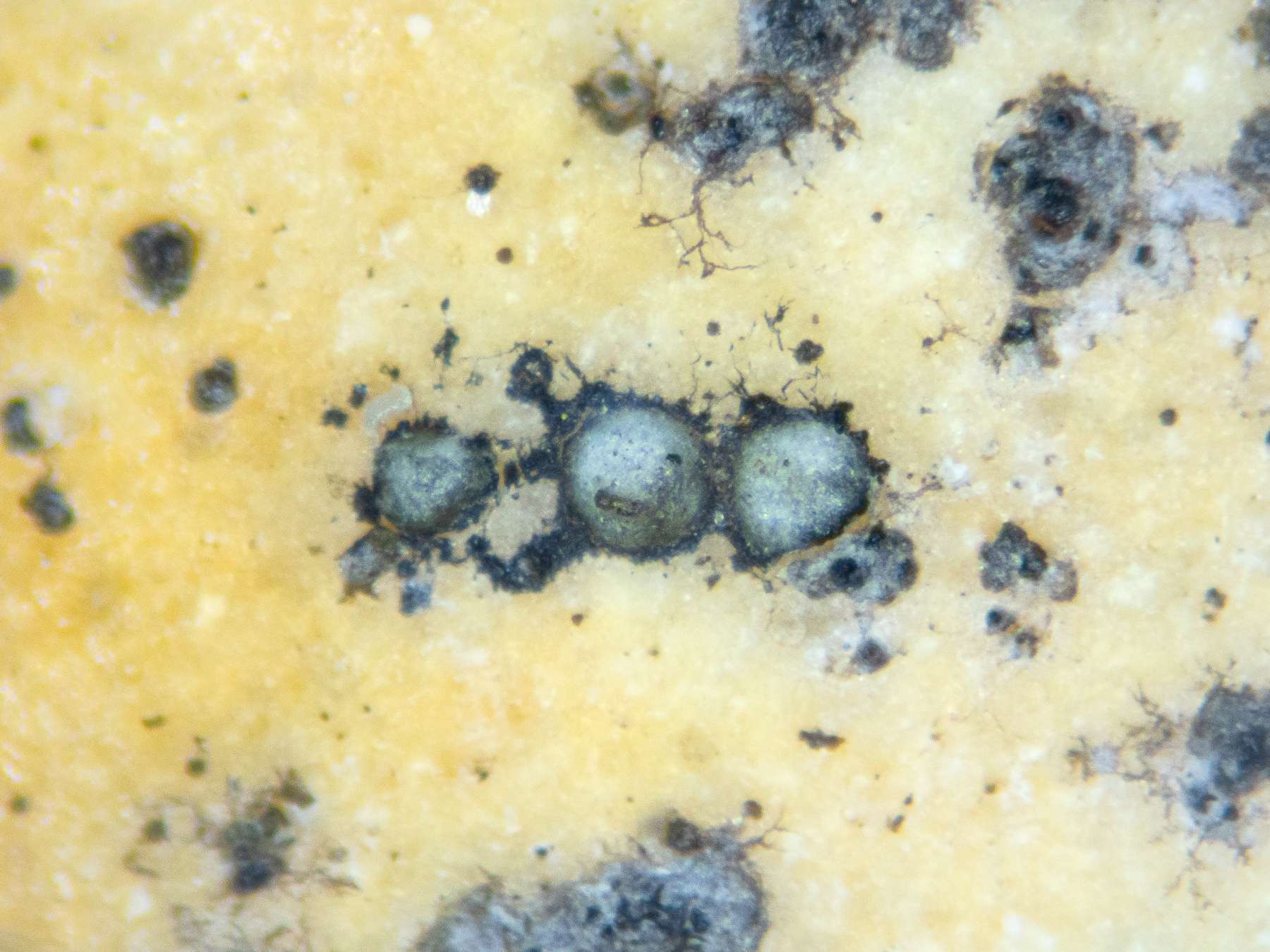
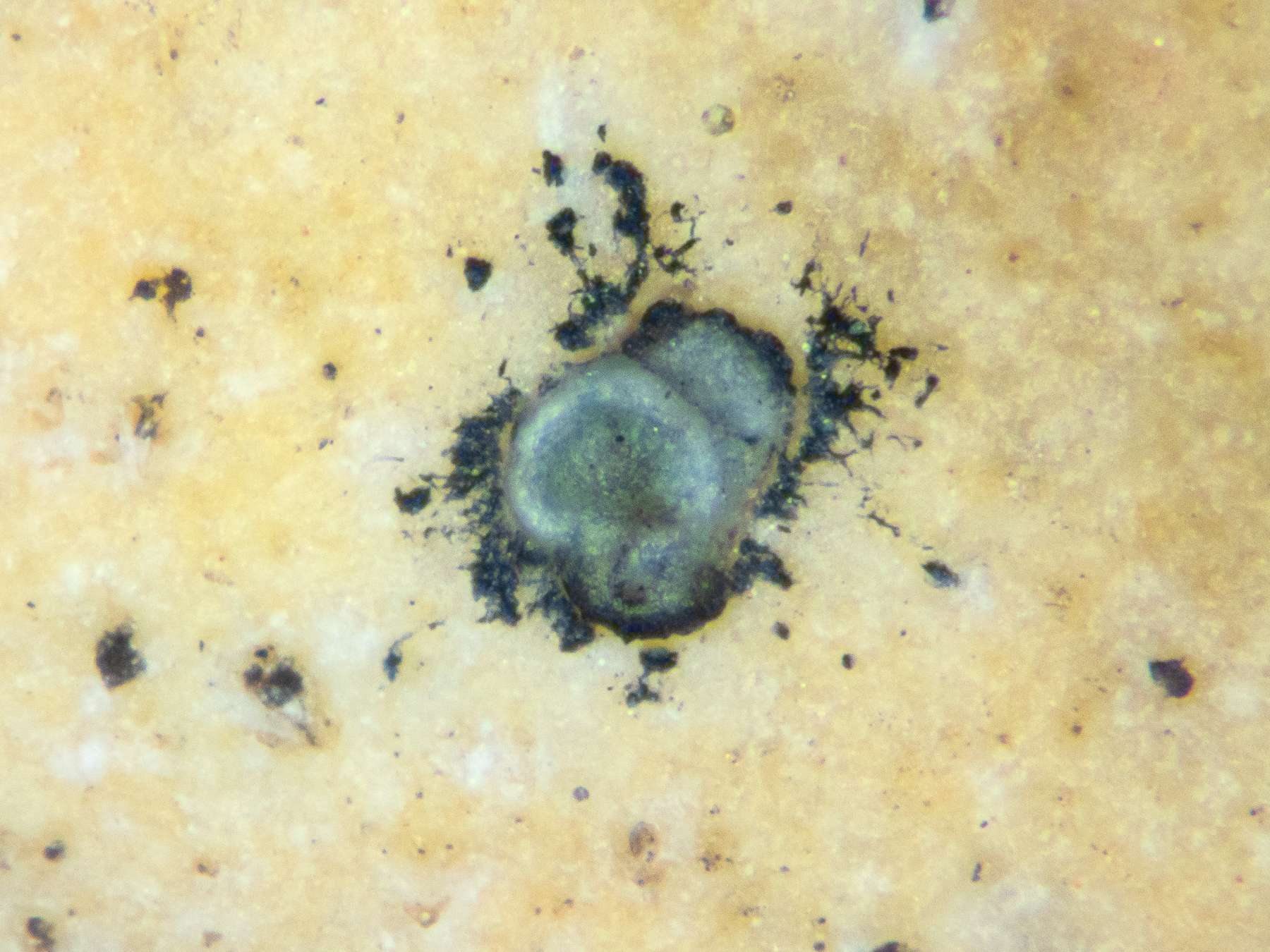
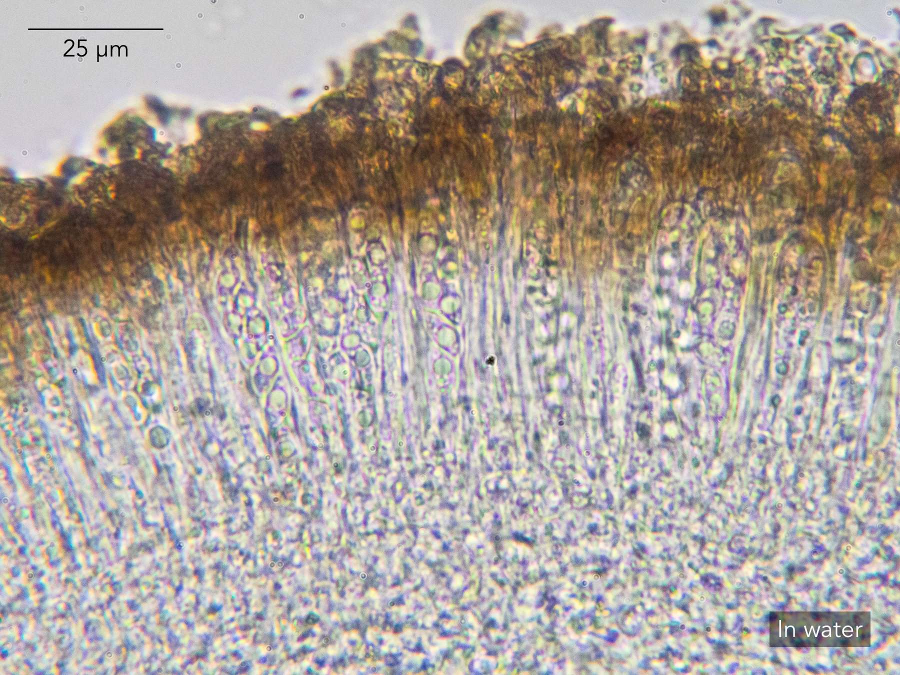
I’d appreciate a steer on this one. Found on a flint pebble in vegetated shingle at Shingle Street, an extensive shingle beach in VC25 East Suffolk. Growing among abundant Amandinea punctata(confirmed microscopically). Apothecia small (all >1 mm and some >0.5 mm), lecanorine and dispersed. Discs ranging from reddish brown to pale brown-grey, but both apothecium types looking identical in section. Thallus apparent only around some apothecia, and then only as small convex swellings. Prothallus apparently black, but not much of it. Hymenium short at ~60 µm. Spores colourless, aseptate, right for
Myriolecis, at ~10-12 µm by ~5 µm. Mainly fine POL+ crystals in epithecium and amphithecium, and some inspersed in hymenium, but no ‘massive’ calcium oxalate crystals in amphithecium. No reactions so far as I can tell, apart from a faint UV+ dull orange (not at all like the distinct bright orange of
M. fugiensor M. andrewii). LGBI3 is leading me to
M. salina, which it describes explicitly as a coastal lichen of siliceous pebbles. And though I haven’t detected any C+ pink reaction (at least macroscopically, as I haven’t yet tried in section), M. salinais described in LGBI3 as C± pink. The lichen also seems to agree with the description of
Polyozosia salinaon ITALIC, especially in the POL+ crystals, which don’t appear in the LGBI3 description. (That said, ITALIC doesn’t seem to hedge on the C+ pink reaction in the same way as LGBI3 does.) The UV+ reaction stands out as potential counter-evidence to both LGBI3 and ITALIC, however. At the same time, I understand from a bit of digging around that
M. salinais poorly understood as well as rarely recorded. Perhaps there’s a clearer identification that I’m just missing? Photos below.
BTW — the map and text in LGBI3 suggests that M. salina has been recorded in East Suffolk before, but there are no such records in the BLS database. Does anyone know if the records have been re-assigned to another species, and if so which one?
Many thanks,
Anthony

[attachment=9]Amandinea punctata & Myriolecis salina sp inc 20260704-CA-1.jpg[/attachment] 
[attachment=8]Amandinea punctata & Myriolecis salina sp inc 20260704-CA-2.jpg[/attachment] 
[attachment=7]Amandinea punctata & Myriolecis salina sp inc 20260704-CA-3.jpg[/attachment] 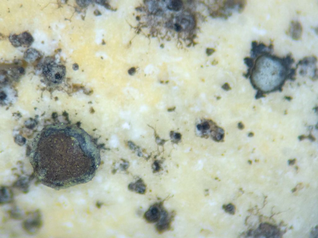
[attachment=6]Myriolecis salina sp inc 20260704-A-1.jpg[/attachment] 
[attachment=5]Myriolecis salina sp inc 20260704-A-2.jpg[/attachment] 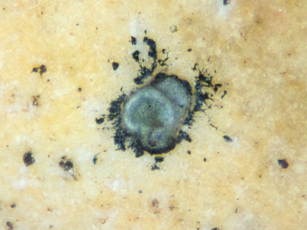
[attachment=4]Myriolecis salina sp inc 20260704-A-3.jpg[/attachment] 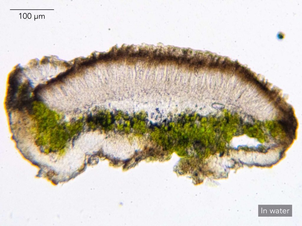
[attachment=3]Myriolecis salina sp inc 20260704-A-5.jpg[/attachment] 
[attachment=2]Myriolecis salina sp inc 20260704-A-6.jpg[/attachment] 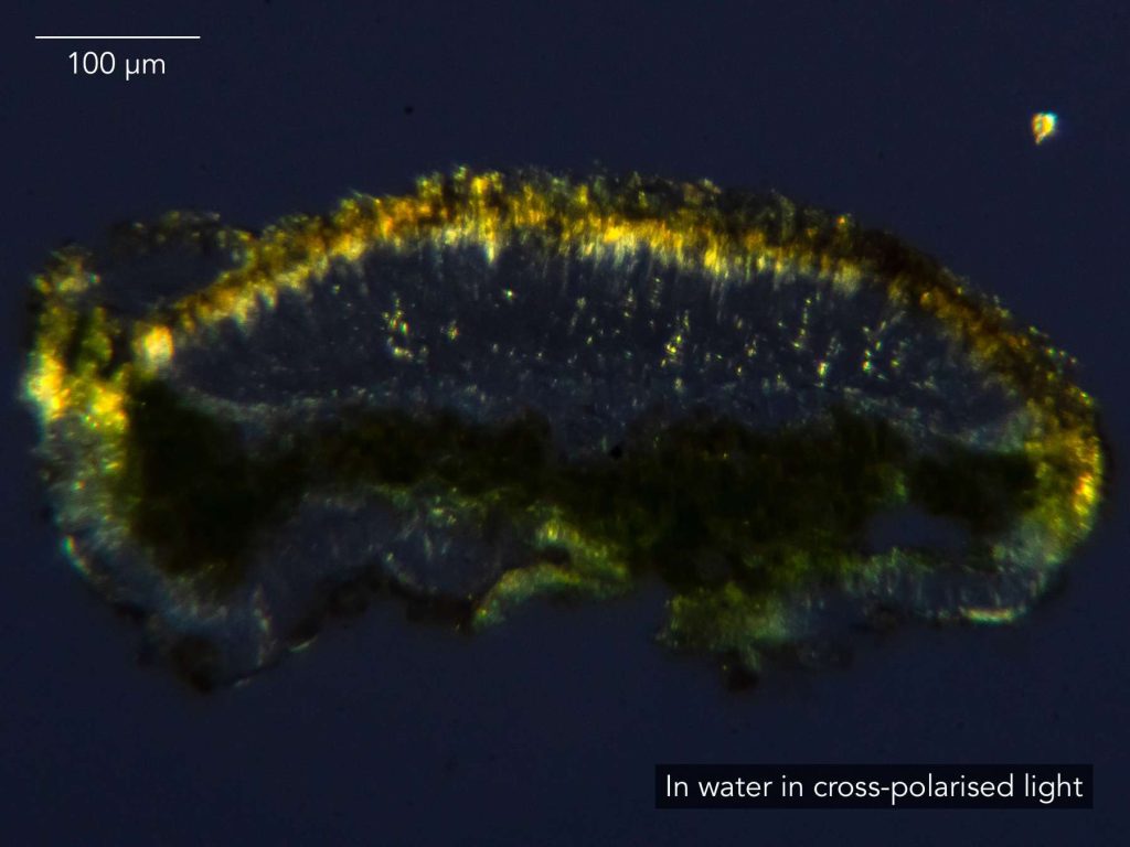
[attachment=1]Myriolecis salina sp inc 20260704-A-7.jpg[/attachment] 
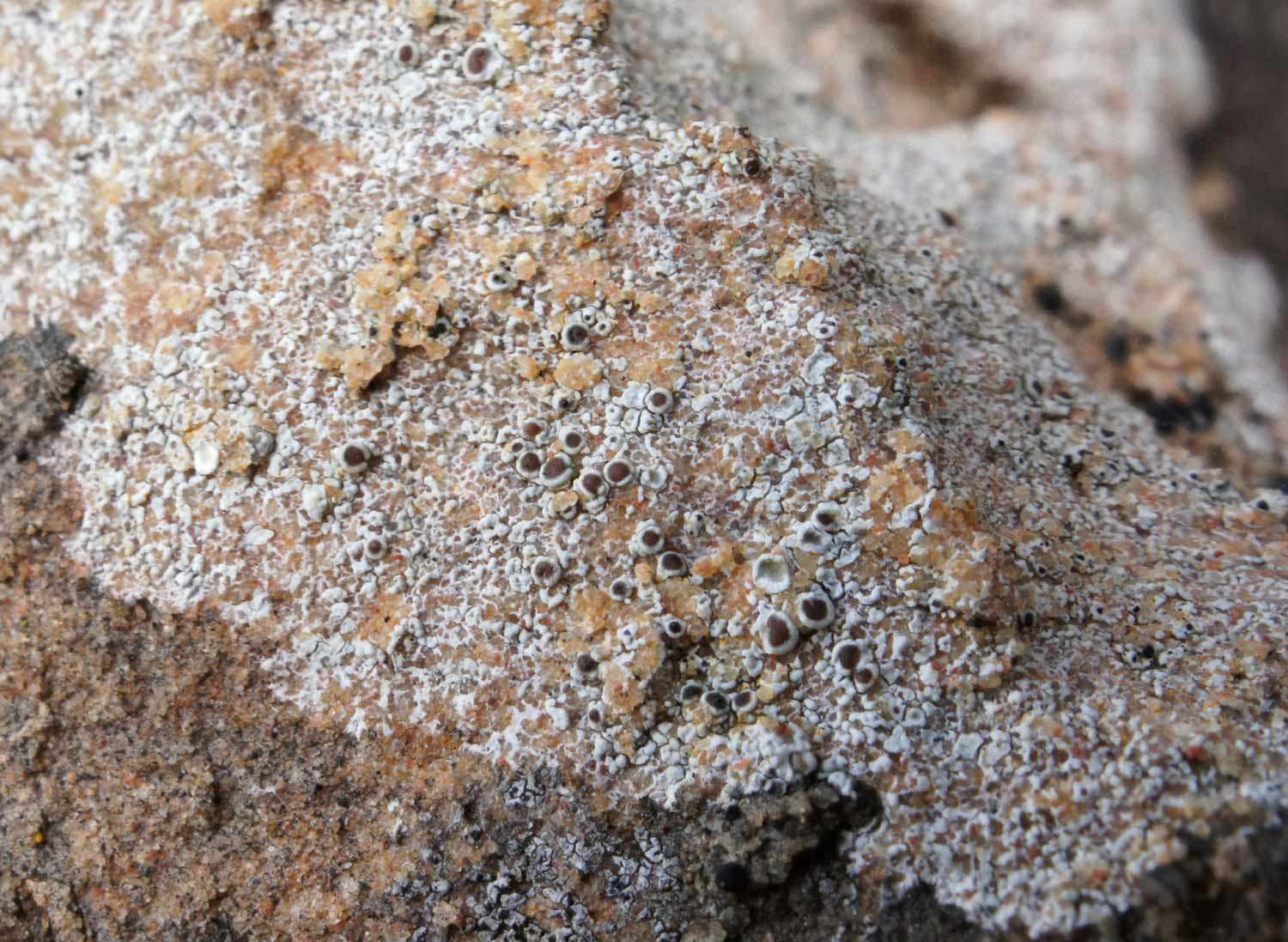
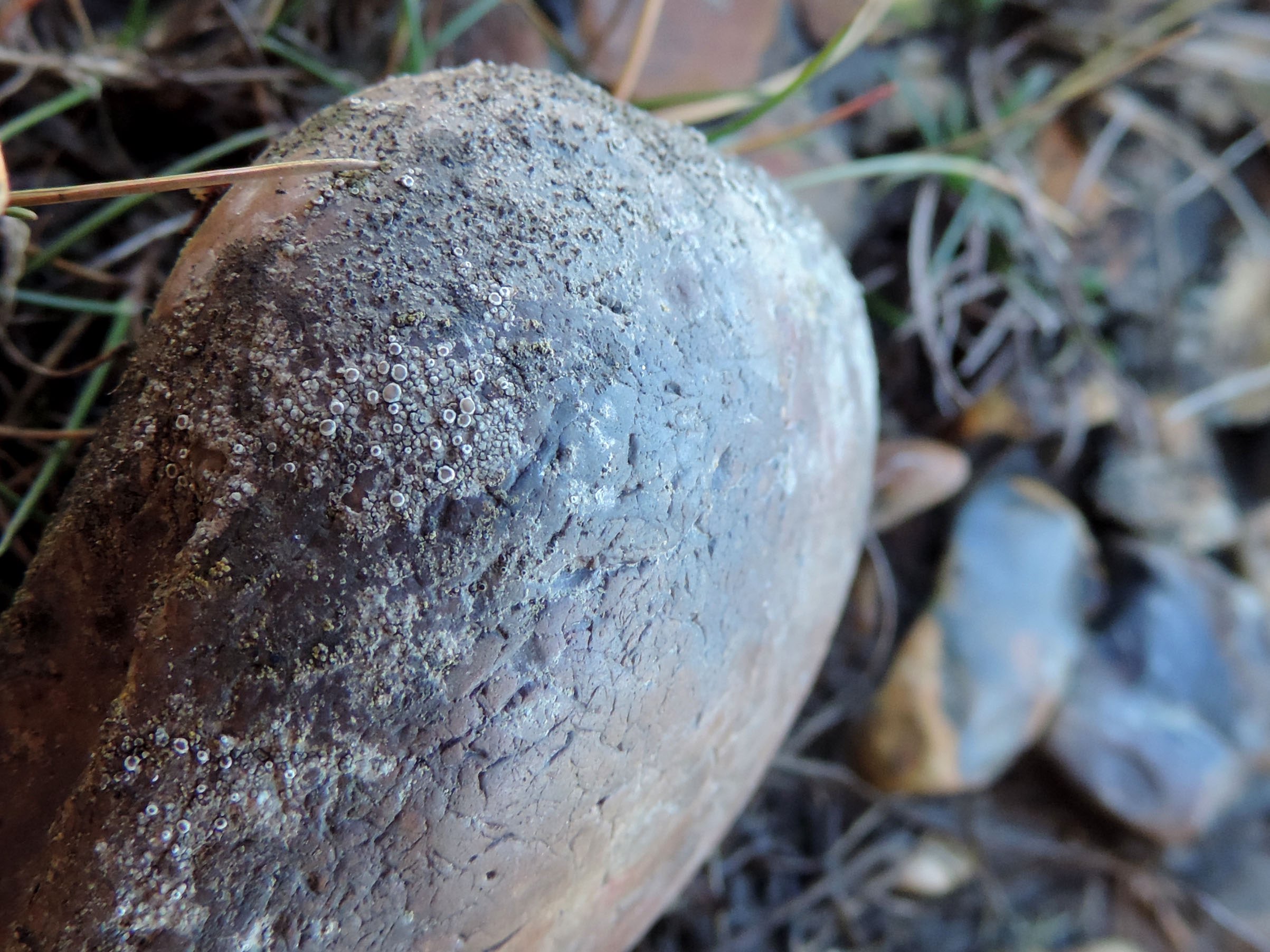
[attachment=0]Myriolecis salina sp inc 20260704-A-8.jpg[/attachment] July 15, 2026 at 3:59 pm #46972Firstly, I’m no expert in this group, so let the buyer beware… This is mostly a northern species and while it always appears to be maritime it’s not restricted to shingle. The BLS record for Pabbay came from coastal gneiss and I’ve seen it on maritime old red sandstone from Skye (see image). Collections on shingle always look odd, presumably it’s the extremely hard substrate making it difficult for fungal hyphae to penetrate. There doesn’t seem to be any reason not to record this as
M. salina, but it could possibly also be an odd form of M. dispersa, I suppose. That seems to have a taller hymenium than yours, but I do wonder how many measurements have been made of such features. 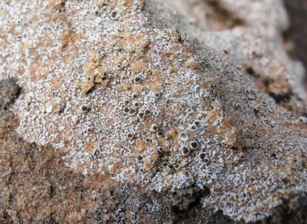
[attachment=0]A08936a.jpg[/attachment] The inland Suffolk records appear to be errors (possibly for
Lecanora saligna?) but Janet would have to confirm this. They are missing from current BLS maps but still accessible in GBIF via an old version of the BLS database – and also from the Fungus Conservation Trusts’s CATE2 database. Not sure how they got hold of them. Caveat emptor here too… Best wishes
Paul
July 16, 2026 at 5:01 pm #46973Many thanks, Paul. I had discounted Myriolecis dispersadue to the hard flint substrate, though I suppose it’s possible that the pebble is somewhat buffered by dust. For comparison, I’ve looked at some
M. dispersagrowing on a fragment of concrete slab that I took from the same location. For what it’s worth, its hymenium is taller but not hugely so — more like 70 µm rather than 60 µm. LGBI3 indicates that
M. salinahas noticeably wider paraphyses than M. dispersa, and that this is a distinguishing feature. Best efforts to measure the paraphyses of my M. salinaput them generally over 2 µm wide, and up to 3 µm — which is in the range for M. salinabut not M. dispersa. The paraphyses of my M. dispersameasure up a bit narrower, but there’s not much in it. And as you say — I wonder how often these things are measured. ITALIC doesn’t give any figure for
M. dispersaparaphyses width, but the ITALIC figure for M. salinaparaphyses is the same as the LGBI3 figure for M. dispersaparaphyses! My
M. dispersawas also clearly UV-, whereas my putative M. salinaappears to be UV+ dull orange. Apart from indicating a difference between the two, though, I’m unsure what to make of it, inasmuch as M. salinais supposed to be UV- as well. There are other southern records for
M. salinafrom Hampshire and Somerset — presumably also from shingle. But as this would be a first record for Suffolk, I think I may hold out for now in the hope of finding a clearer specimen. Thanks again and best regards,
Anthony
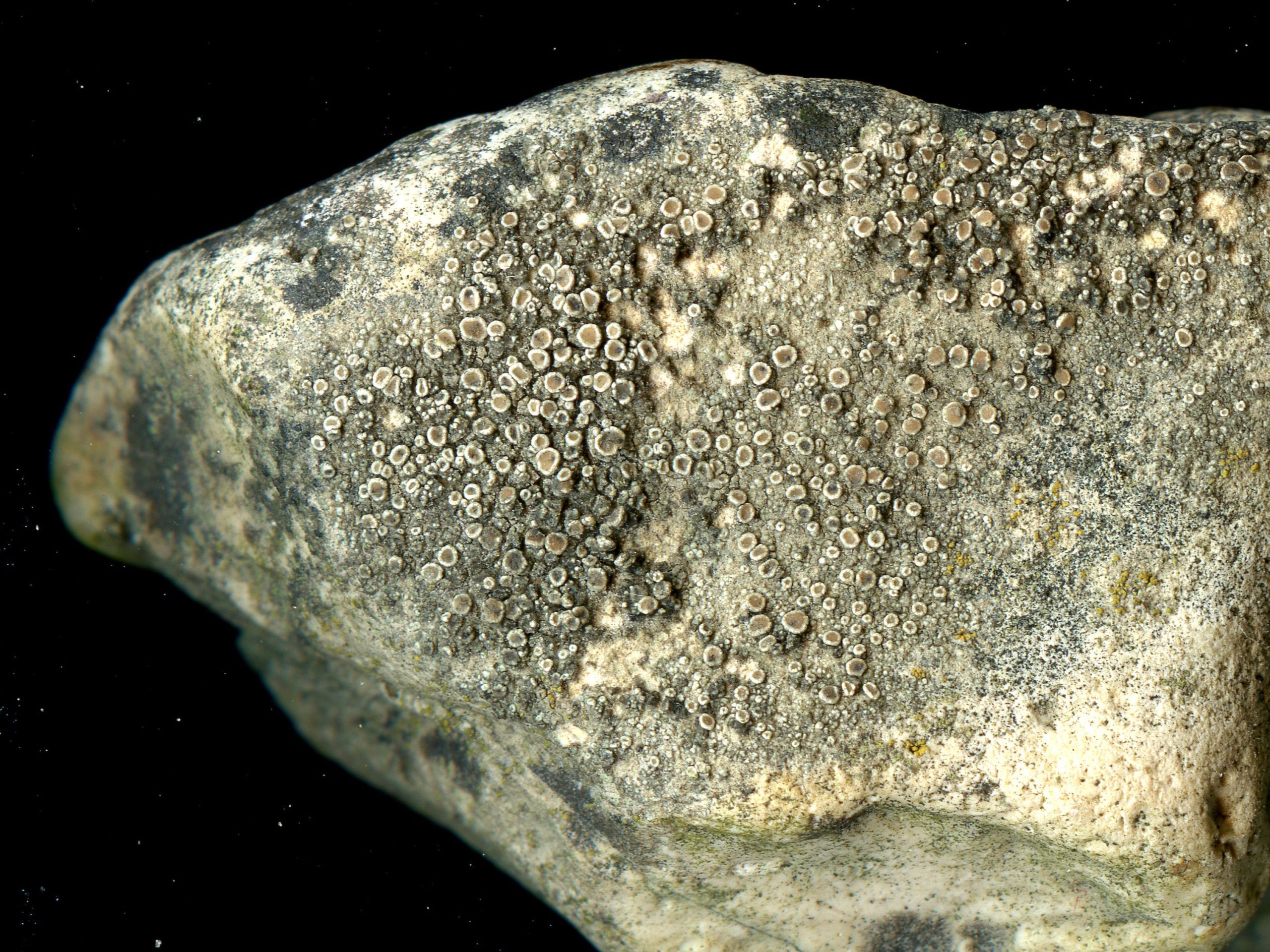
July 18, 2026 at 10:05 pm #46974Anthony These are photos from coastal shingle by the Solent:
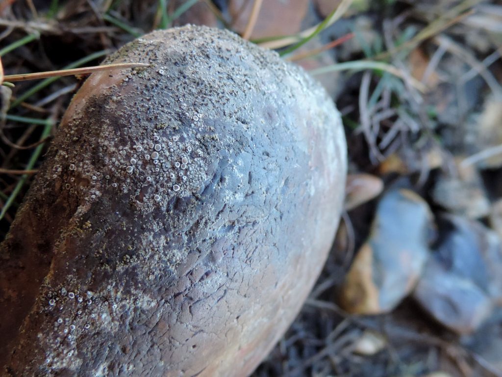
[attachment=2]2013-11-10-03.jpg[/attachment]
Field picture, flint Warren Shore, Hampshire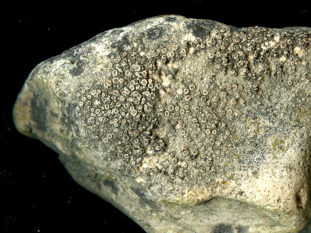
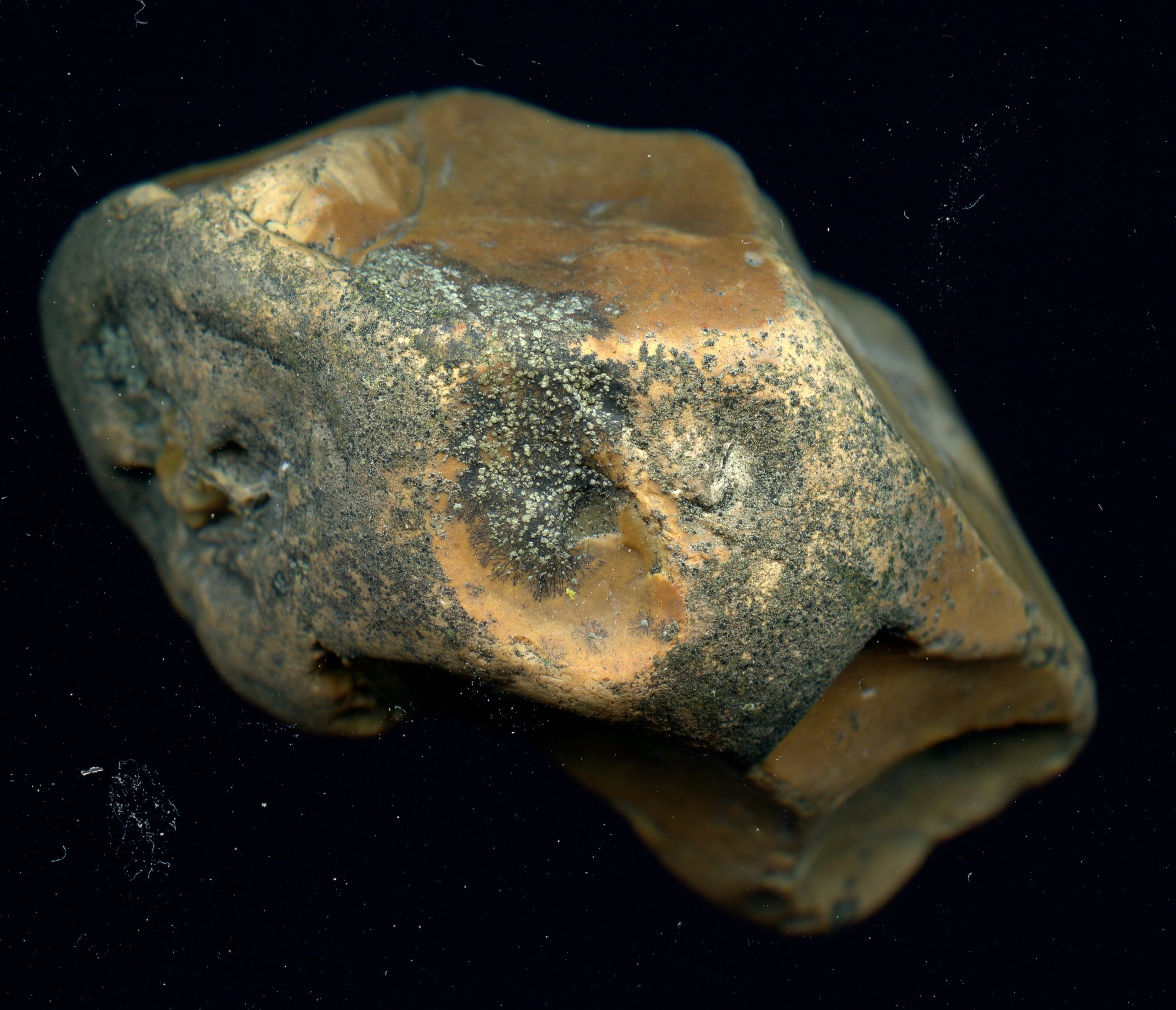
[attachment=1]2013-11-15-06.jpg[/attachment]
Scan of specimen, flint Warren Shore, HampshireI have been identifying it from the thallus composed of rounded granules. Your one is so depurperate I can not realy say any thing about it.
Useful looking at the pictures from that day. This one was labelled
Trapelia obtegens& Amandinea lecideina, but the Trapelia obtegensis clearly Rinodina aspersa, which would be the second Hampshire site, an excellent belated find. 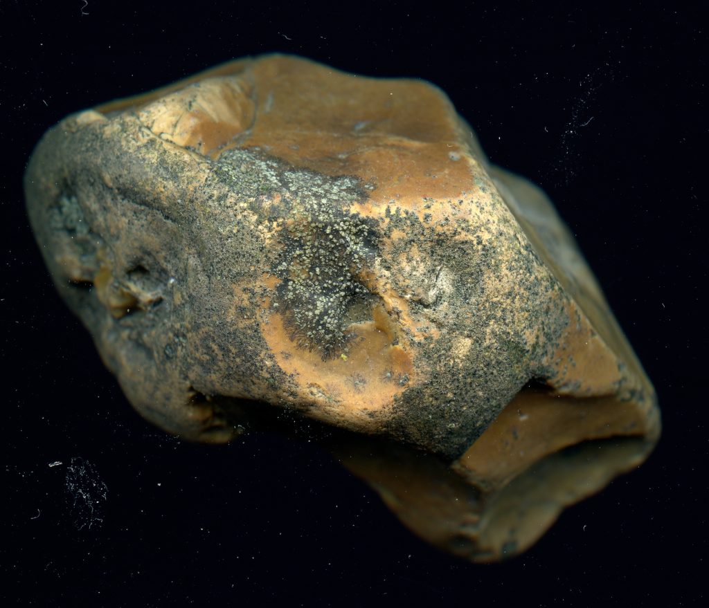
[attachment=0]2013-11-15-05.jpg[/attachment]
Rinodina aspersa(center) & Amandinea lecideinaAll the best
Neil
July 19, 2026 at 6:51 pm #46975Many thanks, Neil — it’s very helpful to see those photographs. Very helpful in more ways than one! Quite apart from
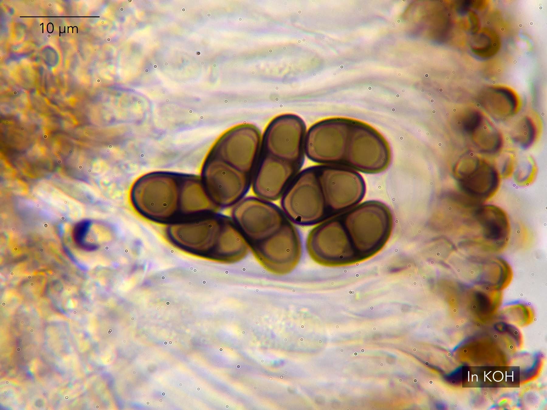
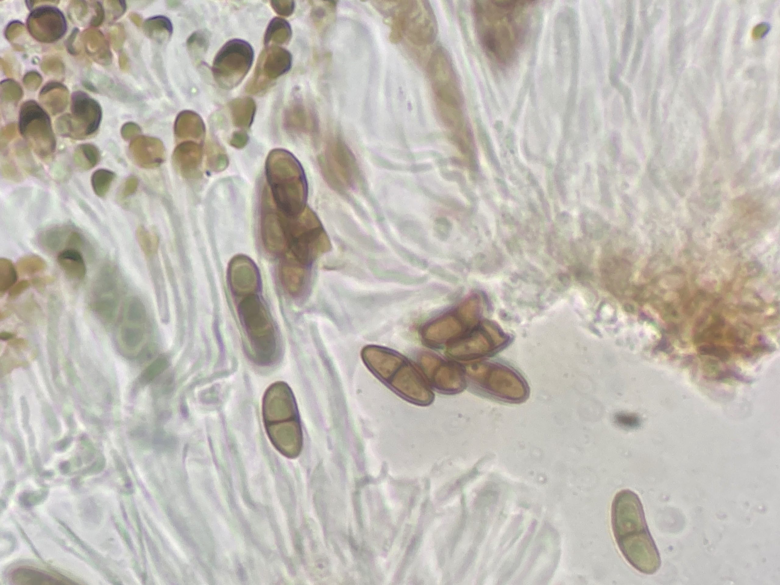
Myriolecis salina, I immediately twigged from your third photo that I might have concluded Amandinea punctatatoo quickly, and that I should have checked more carefully to see whether I had A. pelidna (lecideina)on my pebbles instead. Having looked again at the spores, it seems I do. They appear to have the characteristic ‘girdle’ from median thickening at the septum. Let me know if you disagree.
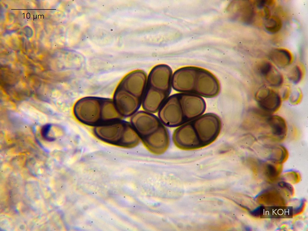
[attachment=0]Amandinea pelidna 20260704-A-7.jpg[/attachment] And as it happens, yesterday I was looking at a lichen on a siliceous gravestone that I couldn’t place, but which looked inexplicably like
Rinodina aspersaand tested C+ red. Thanks to your photo, I now know it’s just a very scattered Trapelia obtegens. Best regards,
Anthony
July 27, 2026 at 3:57 pm #46976Anthony I have not looked at
A. pelidnafor sometime but here is some Amandinea punctataI just looked at for work: 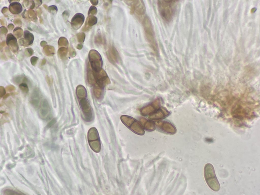
[attachment=0]2026-07-07-02.jpg[/attachment]
Amandinea punctataspores in K, on dead Broom stem Dungeness SO I think your spores are fine for
A. pelidna. All the best
July 29, 2026 at 7:59 pm #46977Oh yes with Rinodina aspersaand Trapelia obtegens, Rinodina aspersahas atranorin, so gives a K/UV(dry)+ bright yellow spot test while Trapelia obtegensdoes not, which is useful. Neil
July 30, 2026 at 9:25 am #46978Many thanks, Neil — very helpful as always. -
AuthorPosts






- You must be logged in to reply to this topic.