
Stigmidium marinum as lichen not LF?
- This topic is empty.
-
AuthorPosts
-
August 29, 2025 at 4:46 pm #33071
Just recently holidaying in Morar, Scotland, where I believe I observed Stigmidium marinumon Wahlenbergiella (Verrucaria) mucosa. But it’s not a taxon with which I’m at all familiar, so I’d appreciate confirmation if possible. Sadly, I didn’t realise what I might have been looking at, so I didn’t attempt to take a sample. My intention was just to photograph
W. mucosaunderwater to show the change in appearance. But later examination, and comparison with photos on the AFL and Lichens Marins websites, put me in mind of S. marinum. Underwater photos below — with apologies that, as the tide was coming in, the water was a bit turbid and some sand was stirred up. So the photos aren’t as sharp and clear as they might otherwise have been in calmer conditions.
The other question I have is much more speculative — though I think Alain Gerault has views on it, and if so I’d be keen to hear them. Andre Aptroot has suggested for some time that
S. marinumis a lichen in its own right, and not (or perhaps not only) an LF parasitising W. mucosa. He describes the S. marinumthallus as thin and filmy, and occuring directly on rock substrate. For his part, Alain suggests on his website that
S. marinumcauses a ‘colour change’ in the W. mucosathallus, but no further description is given there. So I wonder — are the greyer patches on the close-up photo perhaps the thin and filmy thallus of
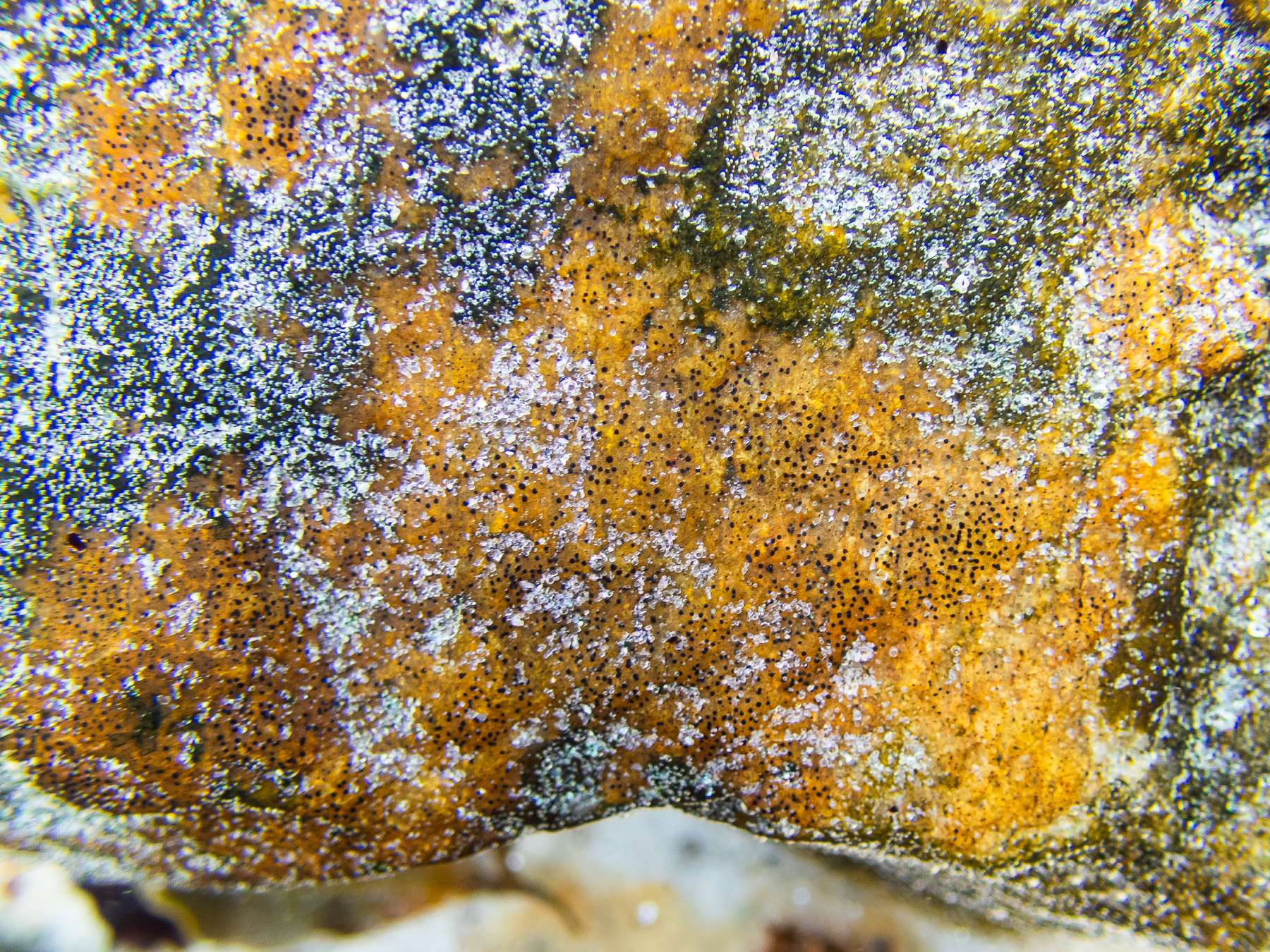
S. marinumoverlaying the greener thallus of (hydrated) W mucosa? They seem to correspond to the potential perithecia of S. marinum, though that’s a first impression. If so, could this mean that S. marinumis growing epiphytically on (or at least not merely parasitising) W. mucosa? And even more speculative still — if the above is plausible, could the black dots on the nearby orange encrusting sponge (possibly
Hymeniacidon perlevis) potentially be S. mariumperithecia, produced from a marginally visible (if I’m not imagining it) thin, filmy and epiphytic thallus? I know nothing of encrusting sponges, but perhaps Alain who understands rocky coasts very well might have seen something similar before. (Again, I was after a photo of the sponge, and didn’t realise what I might have been looking at.)
I certainly understand if there may be nothing further to be said here in the absence of samples to analyse — but I thought it intriguing enough to share even if just for interest. Many thanks in advance for any views!
Best regards,
Anthony

[attachment=2]Wahlenbergiella mucosa 20250820-A.jpg[/attachment] 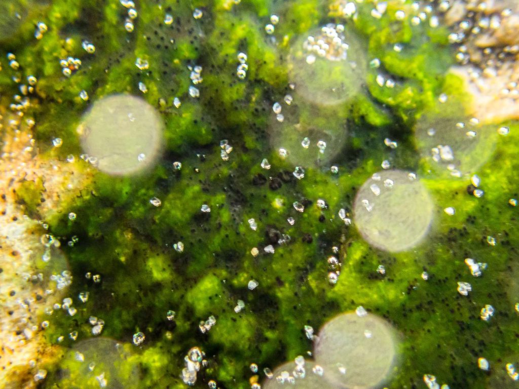
[attachment=1]Stigmidium marinum sp inc 20250820-A-1.jpg[/attachment] 
[attachment=0]Stigmidium marinum sp inc 20250820-A-2.jpg[/attachment] August 31, 2025 at 2:01 pm #46649Anonymous
I don’t recognize Stigmidium marinumand also I don’t recognize Wahlenbergiella mucosawhich doesn’t have such a green and gelatinous thallus. Stigmidium marinumappears in the form of olive-brown then blackish spots bordered with black and never orange. Moreover, the genus Stigmidiumis a “catch-all” genus and the “species” marinum probably includes several taxa, we must expect major upheavals as studies progress. Cordialy.
Alain
September 2, 2025 at 2:46 pm #46650Many thanks, Alain. My apologies — I think I unfortunately created confusion with my speculations. I didn’t mean to suggest that orange body in the third photo is part of
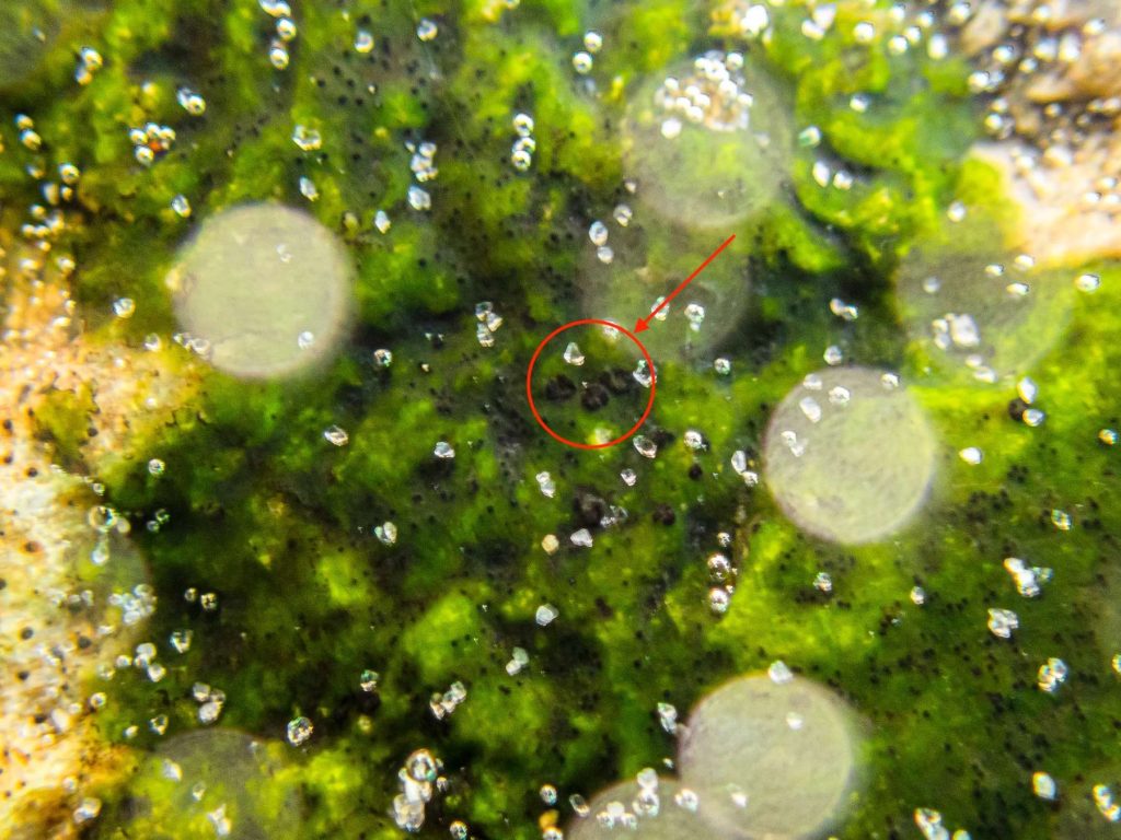
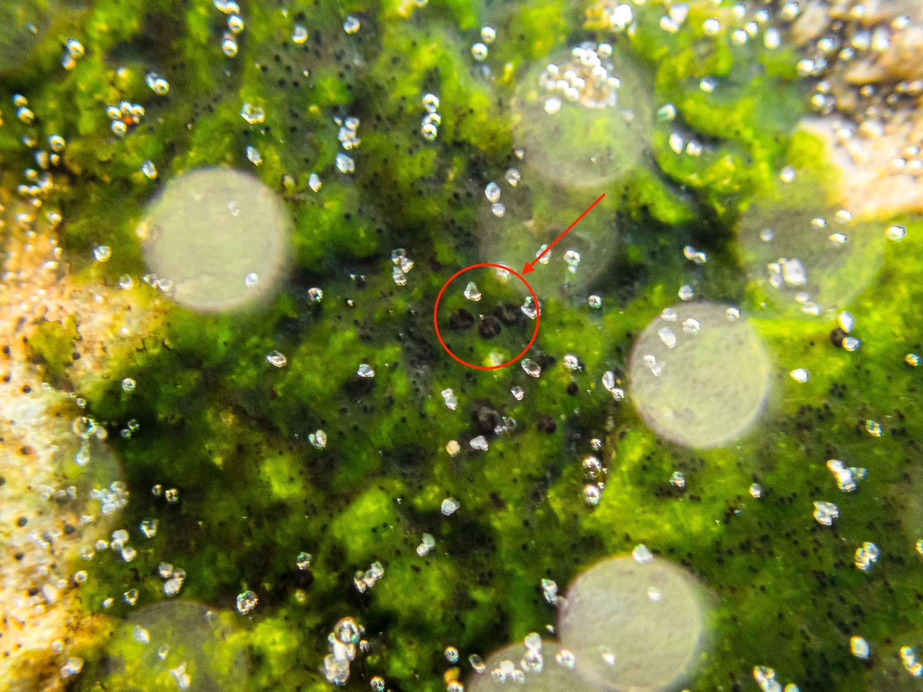
S. marinum. As mentioned, I believe it’s an encrusting sponge. I was only struck by the ‘black dots’ on it that looked at least superficially similar to S. marinum, and that put me in mind of Andre Aptroot’s contention that S. marinumisn’t (or isn’t only) an LF, and that it occurs on various substrates. But it’s perhaps best to leave all that aside. I’ve attached an annotated photo here, encircling what appear to be some of the
S. mariumperithecia on W. mucosa. As you can see from the sand grains in the water, they’re very small. They look very much like some of the structures in your photo above, as well as in the following photos of S. marinum: [list]https://www.afl-lichenologie.fr/Photos_AFL/Photos_AFL_S/Text_S3/Stigmidium_marinum.htm ” class=”bbcode_url”> https://www.afl-lichenologie.fr/Photos_AFL/Photos_AFL_S/Text_S3/Stigmidium_marinum.htm [/list] [list]https://www.researchgate.net/publication/321054505_Ocean_view_A_first_assessment_of_the_littoral_crustose_lichen_biota_of_south_Brazil ” class=”bbcode_url”> https://www.researchgate.net/publication/321054505_Ocean_view_A_first_assessment_of_the_littoral_crustose_lichen_biota_of_south_Brazil [/list] As for the appearance of the
W. mucosa— I should have explained better what I meant by ‘underwater photos’. I took them quite literally underwater, with a submersible camera. The water was in motion as the tide was coming in, creating some softness in the image. The thalli were also in strong light, enhanced by the photo settings, and the thalli were fully hydrated. These factors explain (I think) the unusual appearance. I admit again that there’s unlikely to be any conclusive interpretation of my photos, especially as I have no sample. But I thought it worth posting for interest, and in case anyone’s seen something similar. I know you don’t seem to recognise
S. marinumhere, and it would be really helpful for me to know more precisely what I might be missing given the similarities mentioned above. Thanks again,
Anthony
[attachment=0]Stigmidium marinum sp inc 20250820-A-1.jpg[/attachment] September 3, 2025 at 12:40 pm #46651May be Verrucaria halizoa? Alain
September 3, 2025 at 12:47 pm #46652Many thanks, Alain. Yes — I wondered if it might be V. halizoa, not least because of the colour. But there don’t seem to be any of the fairly abundant, and relatively large and prominent perithecia that seem to characterise that species — and which are shown so well in lichen image banks (including your Lichens Marins). But not having seen V. halizoamyself, I can’t be sure. In any case, it seems it must remain something of a mystery, at least to me! Thanks again for your input.
-
AuthorPosts


- You must be logged in to reply to this topic.