
JennyS

Forum Replies Created
-
AuthorPosts
-
I posted the images on the Facebook ‘Lichenicolous fungi 2.0’ page and have just had a reply there from Valerii Darmostuk:
Quote:Interesting, I’m going to Lichenodiplis poeltii following the key from Tsurykau & Etayo (2017) 10.1017/S0024282916000530. This was described from Australia and is still known only from the type locality (the protologue attached).

[attachment=0]Lichenodiplis poeltii 2.jpg[/attachment] Many thanks Neil – the Arthonia excipienda was from the same 10km square as the pre-2000 record shown on the BLS map but from the other side of the village. Two surprise finds from old photos!
Thanks Jonas – yes I think (finally!) it must just be P. orbicularis Thank Paul, I was starting to suspect ‘undescribed’ might be the case as there was nothing with similar sized spores mentioned in any of the LGBI3 Arthonia descriptions. Despite investigating every possible black dot on Arthonia radiata thallii on that twig other than host pycnidia (which seem to be fairly frequent) there was nothing further.
I’ll be there again in a couple weeks time and will bring more twigs back with me to check!
Hello Paul – yes I realised after posting, very probably either paraphyses or perhaps (if Celothelium ischnobelum) conidia? I didn’t find any mature asci, only what I took to be unsuccessful ones shown in the fourth image, so frustratingly I doubt there’s much hope of getting any further with this!
Hello Di (Chalkie) – just a thought but did you consider a Cornutispora for your ‘windmill-shaped’ conidia? https://lichenportal.org/portal/taxa/index.php?taxauthid=1&taxon=Cornutispora&clid=201 ” class=”bbcode_url”> https://lichenportal.org/portal/taxa/index.php?taxauthid=1&taxon=Cornutispora&clid=201 Hi Neil,there seems to be a green alga over-growing the area round the perithecia and pycnidia, and the same on other parts of the twig too 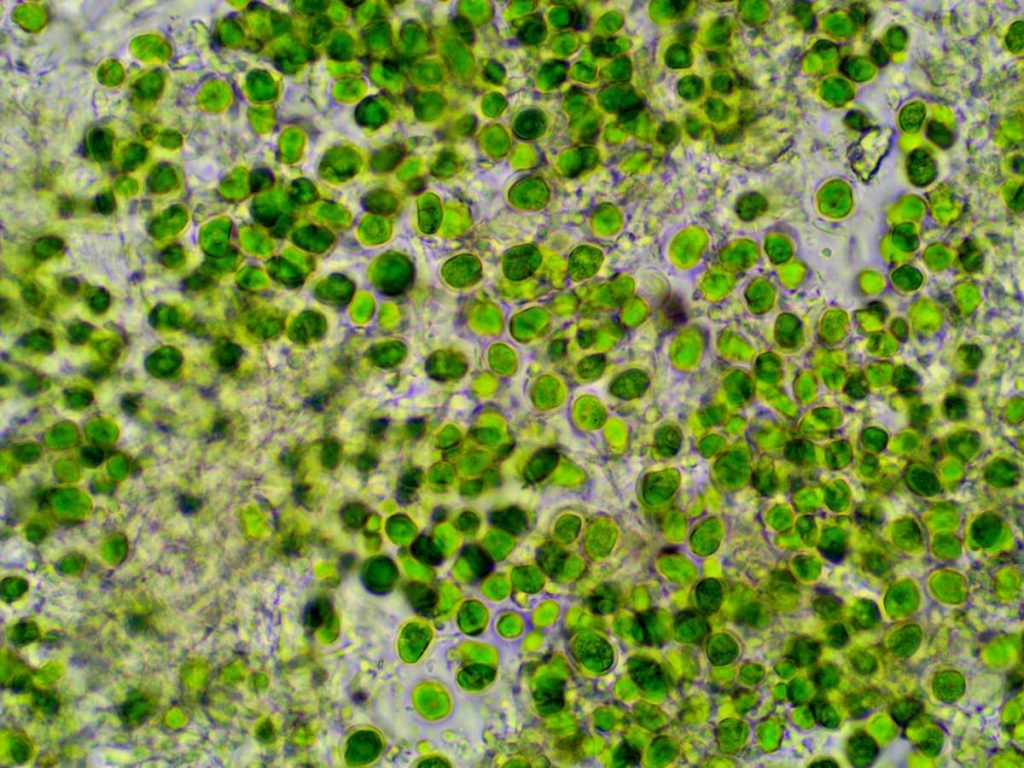
[attachment=0]zz algae An vir thall 240719-1.jpg[/attachment] Thanks for posting the micrographs from your specimen Neil, and might you have any suggestion of what mine might be if its not Anisomeridium viridescens? Photograph below shows where both the first and second group of micrographs were from (the second with both spores and macroconidia).

[attachment=0]zz Anisomeridium viridescens Q 240719 008.jpg[/attachment] Thanks for the confirmation Neil – and very reassuring that the K+ isn’t obvious! (Any chance of adding that to LGBI3?) Thanks for the reply Anthony, and the slides were mounted in K. The lack of an obvious K+ green reaction in my first section concerned me so I was relieved to find both perithecia and pycnidia in the second section as macroconidia aren’t mentioned for Arthopyrenia carneobrunneola.
I checked the LGBI3 descriptions for both Anisomeridium viridescens and Arthopyrenia carneobrunneola and though the K reaction and presence/absence of pycnidia are the most obvious differences there is also a slight difference in the spore shape:
A. viridescens “usually constricted at the septum; the upper cell usually distinctly wider than the lower”
A. carneobrunneola “not or only slightly constricted at the median or somewhat supramedian septum”
Though some of the spores in my sections are a bit intermediate they do compare better with Mark Powells’s illustration of Anisomeridium viridescens here:
https://fungi.myspecies.info/sites/fungi.myspecies.info/files/Anisomerid%20viridescens.jpg ” class=”bbcode_url”> https://fungi.myspecies.info/sites/fungi.myspecies.info/files/Anisomerid%20viridescens.jpg than with the Asco France photographs of Arthopyrenia carneobrunneola here:
http://www.ascofrance.com/recolte/3729/dothideomycetes-pleosporales-arthopyreniaceae-arthopyrenia-carneobrunneola ” class=”bbcode_url”> http://www.ascofrance.com/recolte/3729/dothideomycetes-pleosporales-arthopyreniaceae-arthopyrenia-carneobrunneola Jenny
And from a bit further along the same twig both perithecia and pycnidia present, spores 13-16 x 3.5-4.5 µm, macroconidia 10-12x 2-2.5 µm. 
[attachment=2]w Anisomeridium viridescens 2 240719-10b.jpg[/attachment] Macroconidia

[attachment=1]w Anisomeridium viridescens 2 240719-4 con.jpg[/attachment] 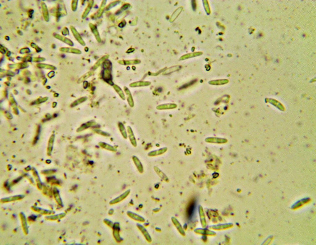
[attachment=0]w Anisomeridium viridescens 2 240719-3 con.jpg[/attachment] Thanks for the suggestion but the underside of the lobes and the rhizines are black which would rule out Phaephyscia nigricans which has Quote:“underside whitish, pinkish or pale brown-white, with sparse, generally pale rhizines”

[attachment=0]Phaeophyscia 240712 007.jpg[/attachment] The conidia with long arms confirmed by David Hawksworth as Psammina filamentosa. Thanks Paul – the arms were 2.5-3.5 µm wide and with septa present, but not clear enough to count how many. I emailed David Hawksworth a few days ago but haven’t had a reply yet. 
[attachment=0]w Psammina filamentosa q 240515-1b.jpg[/attachment] -
AuthorPosts


