JonasRavn

Forum Replies Created
-
AuthorPosts
-
I suspected that it was not a crystal clear case – it rarely is with Verrucaria spp.in my experience, however limited it still is. Thank you for the link, it is a great site that I have used frequently for the high quality microscopy images of quite a large number of taxa. I have considered
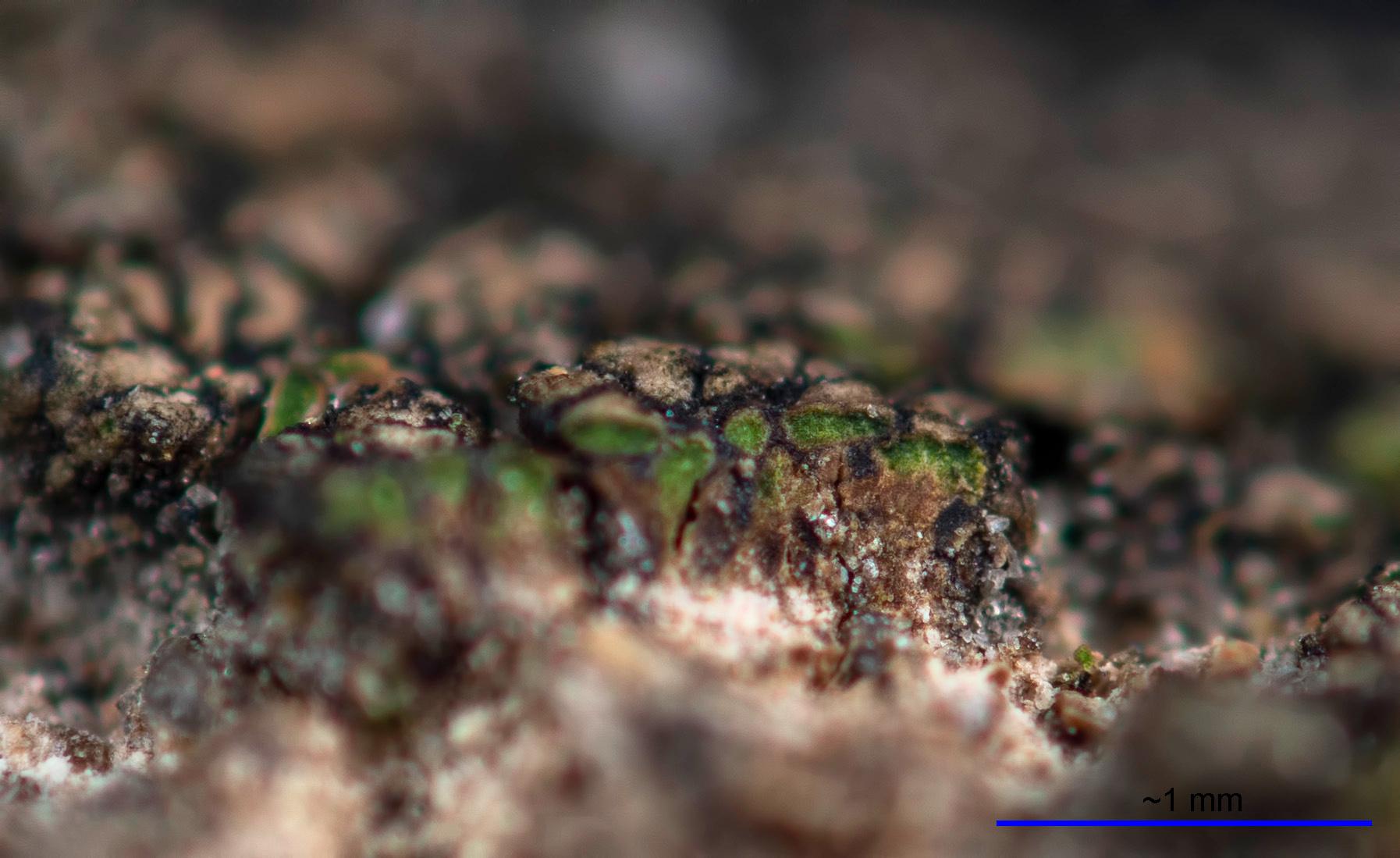
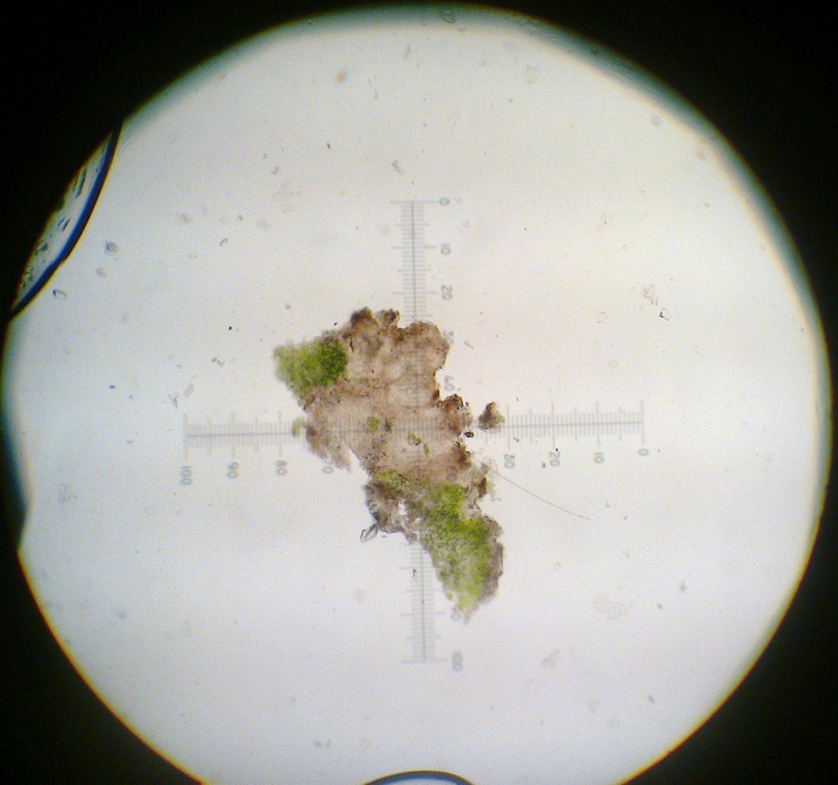
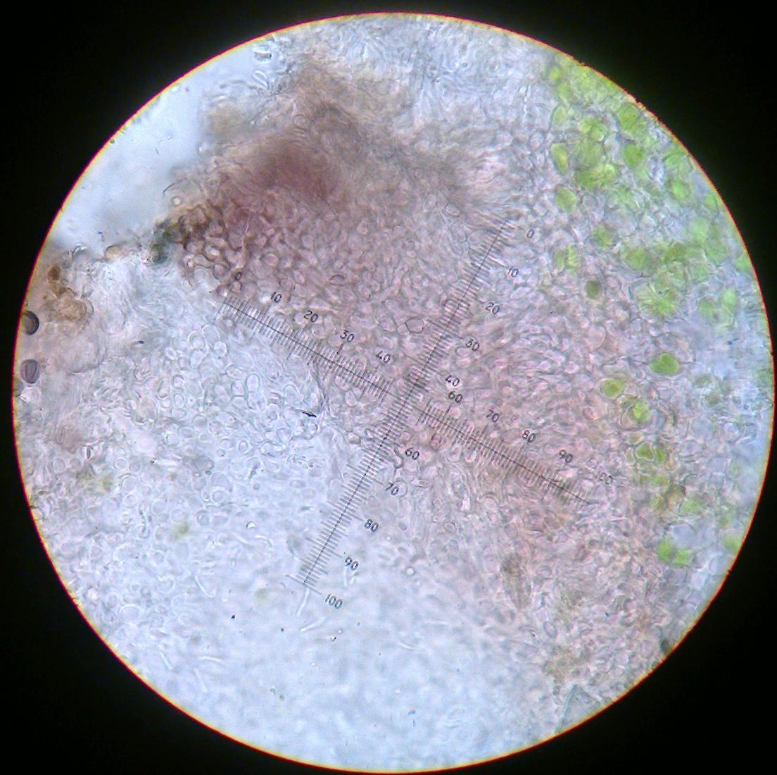
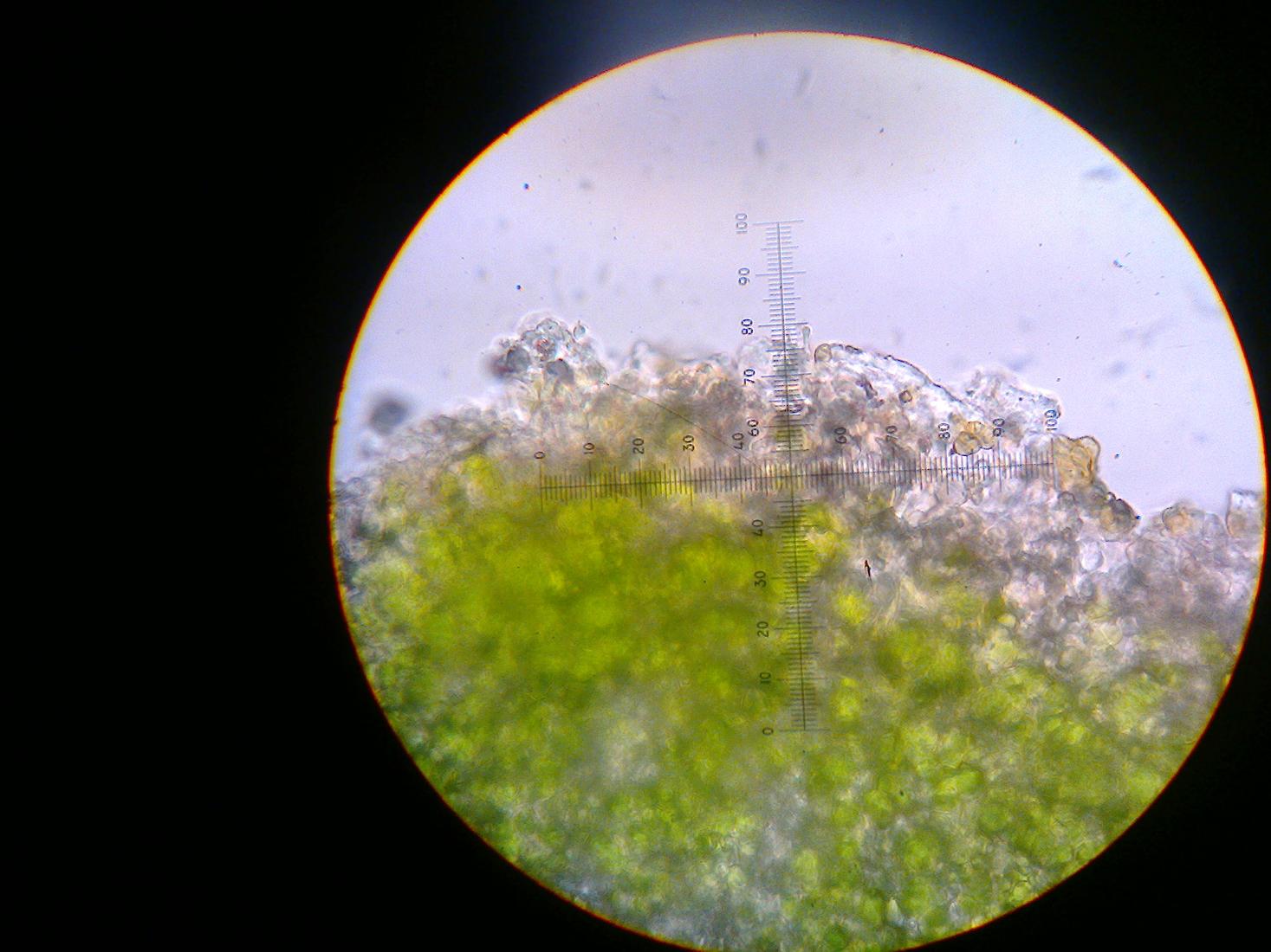
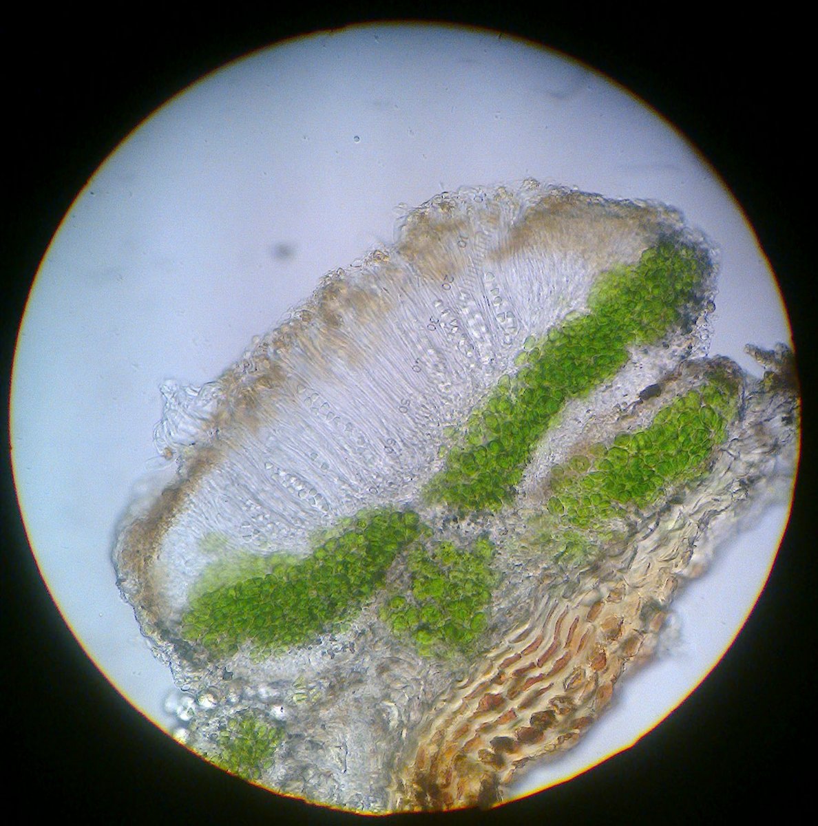
V. elaeinaand it is not a bad fit in terms of spore-size, but I am not sure that the shape of the spores is right. According to LGBI, V. elaeinahas ellipsoid spores whereas the spores in my specimen have a +/- distinct taper towards one end and instead appear +/- sole-shaped. This tapering is mentioned for V. dolosain LGBI, but I am unsure how common this shape is in Verrucariaand if it should be considered as evidence for V. dolosa. What do you think? If it is indeed V. ochrostoma, it would be the first record from Denmark, and I don’t think this sterile specimen is sufficient to document that. I do, however, think that the resemblance is quite striking when I look at photos and descriptions of V. ochrostoma, so I would tend to gravitate towards your option 2 or 3. I have visitied the location again hoping to find a better developed thallus, but so far without luck. It is quite nearby my home, so I will pop by there from time to time and update here if and when I find something conclusive. I did not mention this before, but after cutting some sections, I can see that the thallus is rather thick and thought it would be worth documenting aswell:

[attachment=2]cf. Verrucaria ochrostoma 1-1.jpg[/attachment] I recently spent more time than I would like to admit in trying to identify a lichenicolous species on
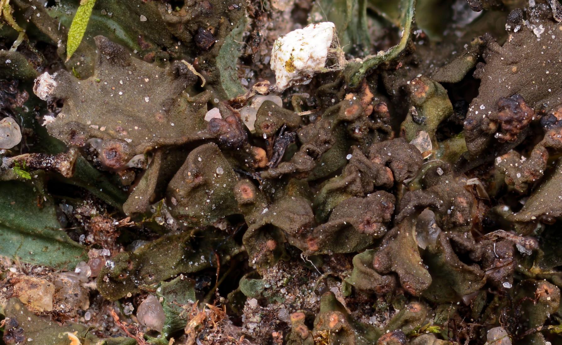
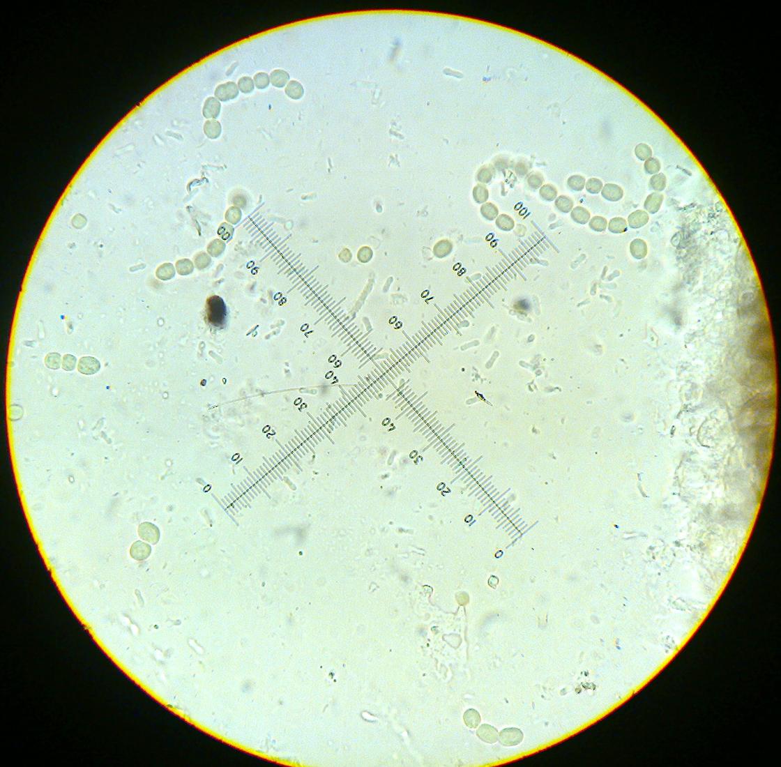
Enchylium cf. tenax, just to realize that it was in fact the “host’s” own pycnidia. I had never seen any Collemataceaewith pycnidia, so I did not even consider that possibility… The conidia are however quite characteristic (almost dumbbell shaped) and fit well with the reported conidia from E. tenax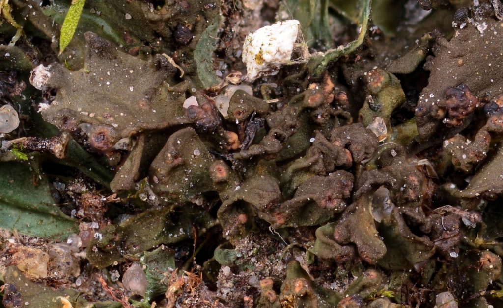
[attachment=3]cf. Enchylium tenax 2-1_01.jpg[/attachment] 
[attachment=0]Billede1890_01.jpg[/attachment] 
[attachment=1]Billede1886_01.jpg[/attachment] Lastly, I would like to add that even though no solid ID may come of it, I much appreciate you taking the time to answer my queries – thank you for that.
– Jonas
Hi again, Mark I like your suggestion, but all the black globular structures I have examined have been what I think is colonies of
Gloeocapsa. The pycnidia are red-brown and immersed in the squamules/areoles. I attach a section through the area you highlighted – as far as I can see it is a cluster of pycnidia. You may be right that the pycnidia are from a lichenicolous fungus – I will attempt to key them as such tomorrow 

[attachment=1]Billede1924.jpg[/attachment] 
[attachment=3]Billede1921.jpg[/attachment] 
[attachment=0]Billede1926.jpg[/attachment] 
[attachment=2]Billede1923.jpg[/attachment] 
[attachment=4]Billede1919.jpg[/attachment] Indeed it has – thank you for your comments. – Jonas
It has been a while and the material from this observation is now lost, I’m afraid. When I am near the locality again, I will attempt to collect another thallus and retry the ID – you don’t think the apothecia here are too yellow (rather than orange)? Thank you for the reply, I enjoyed following your identification process! I may have misinterpreted the paraphyses as simple as I don’t have a good stain for them yet – they are clearly branched in your specimen and I don’t doubt that it is the the same species. I have ordered some ink and will try the ink – acetic acid stain on my next specimen.
My interpretation of the paraphyses as simple led me towards
Biatoraas I first interpreted the K/I reaction as the Biatora-type (and I am still not entirely sure of the difference between Lecidella– and Biatora-type, if I am honest). The picture of B. helvola on ITALIC ( http://dbiodbs.univ.trieste.it/italic/italic07?s=246&us=admin I am interested to hear what you find out regarding the UV-reaction of normal apothecia. I made a very haphazard setup to do a quick test with no overwhelming results. I think I can see a small band of orange UV-reaction in the hymenium that turns yellow and moves “inward” when I add C, but simply replacing my condenser with a UV torch might not be the best way to test this and I suspect that there are some artefacts…

[attachment=2]Billede1858.jpg[/attachment] 
[attachment=0]Billede1881.jpg[/attachment] 
[attachment=1]Billede1876.jpg[/attachment] Hi Neil, Thank you for your reply. You are right that the lower surface is quite thickly tomentose, but I am not sure whether or not this is sufficient to rule out S. dactylophyllum. One of my many challenges with Stereocaulon is the frequent use of terms such are “more or less”, “usually” and the likes in the keys and descriptions. Furthermore the descriptions are not always consistent between different keys. Some sources state, as you mentioned, that S. dactylophyllum should be more or less glabrous, but in “Lichens of Finland” (Stenroos et al.) it described as “more richly tomentose” than S. subcoralloides.
The picture of the lower surface shows the tip of a pseudopodetium, but most of the bases are covered in some sort of “gunk” from the substrate, and evaluating anything on them is quite tricky. I will try to get some fotos of an entire pseudopodetium for comparison, when I get home from a trip in a week’s time and follow up in this thread.
Best regards,
Jonas
Thank you for your reply, Neil, The wall of the perithecia and both the conical and “abberant” pycnidia react K+ green/olive. It seems, however, that the globular “conidia” develop secondarily on the concial pycnidia – and that they occur in “chains”. I found a pycnidium with normal conidia on the inside, but with the grey mass on the outside. While I have no explanation for that phenomenon(a lichenicolous fungus, perhaps?), I am satisfied that the species in question is A. polypori.

[attachment=0]Anisomeridium cf. polypori 2-14.jpg[/attachment]
Thanks again for helping and for welcoming me.Best regards,
Jonas
Hello Paul, Thank you for your response. You bring up a good point about the hypothecium. I have interpreted it as if the algal layer was mor or less continuous, but checking my photos again I am not 100% sure. I will redo the section and double check – and get some better pictures in the process.
Thanks again,
-Jonas
The [img]tag doesn’t seem to like flickr urls. Here are the photos: 
[attachment=1]Athallia sp. 1-1bc.jpg[/attachment] 
[attachment=0]Athallia sp. 1-2.jpg[/attachment] [/img] -
AuthorPosts