
Anisomeridium-confusion
- This topic is empty.
-
AuthorPosts
-
March 25, 2020 at 11:25 am #31626
I am having some trouble with this Anisomeridium collected from the shaded bark of a deciduous tree in an urban woodland, Aarhus, Denmark. It has numerous pycnidia with a rostrum and a white columnar mass of macroconidia (ellipsoid/ovoid 4-4.5 µm x 2.7-3.2 µm) and I have found what appears to be microconidia (bacilliform, ~2.5 µm x ~0.7-0.8 µm) both of which fit with my impression in the field: that this is A. polypori. The confusion arose when I found several pycnidia of a different type; more or less globular or with a very short rostrum and with a distinctly grey amorphous discharge consisting of grey +/- globular conidia (diameter: ~2.5-3.15 µm). I suspect that I might be in the wrong genus, but I can’t seem to find a better alternative. Where have I gone wrong?

First an overview of the thallus:

[attachment=5]Anisomeridium cf. polypori 2-13.jpg[/attachment] Section of a perithecium:

[attachment=3]Anisomeridium cf. polypori (tværsnit, perithecium) 2-8.jpg[/attachment]
Spores (1 div. = 0.96 µm):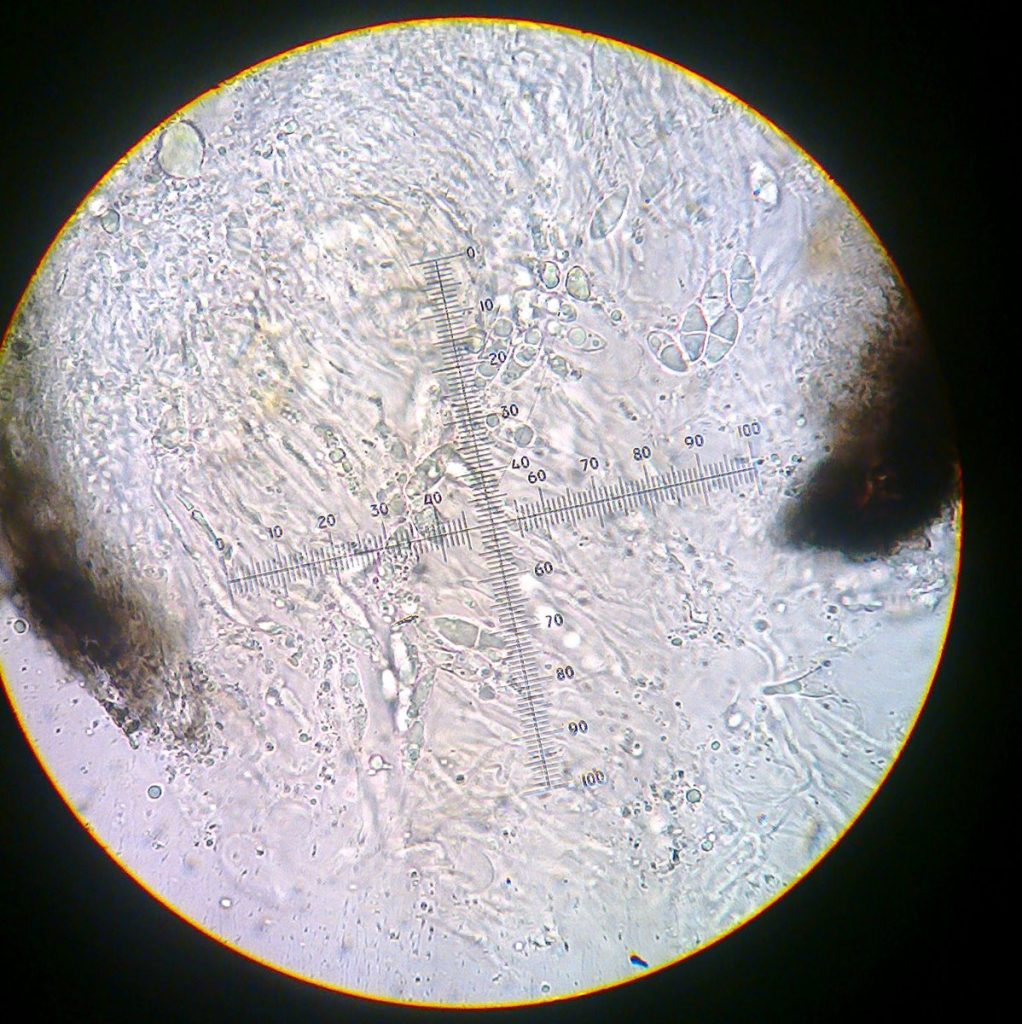
[attachment=9]Anisomeridium cf. polypori (sporer) 2-3.jpg[/attachment] Close-up of pycnidia (the aberrant type marked in the second photo):

[attachment=1]Anisomeridium cf. polypori 2-11.jpg[/attachment] 
[attachment=0]Anisomeridium cf. polypori 2-10.jpg[/attachment] “beaked” pycnidium with columnar macroconidia:

[attachment=2]Anisomeridium cf. polypori (pyknidium, makrokonidier) 2-4.jpg[/attachment]
macroconidia (1 div. = 0.96 µm)
[attachment=6]Anisomeridium cf. polypori (makrokonidier) 2-5.jpg[/attachment] “aberrant” pycnidium with grey mass of conidia:

[attachment=4]Anisomeridium cf. polypori (pyknidium, makrokonidier måske) 2-6.jpg[/attachment]
Close-up of grey globular conidia (1 div. = 0.96 µm):
[attachment=8]Anisomeridium cf. polypori (makrokonidier måske) 2-7.jpg[/attachment] Close up of what I believe to be microconidia, but I am unsure if that is indeed what it is (1 div. = 0.96 µm):

[attachment=7]Anisomeridium cf. polypori (mikrokonidier) 2-2.jpg[/attachment] March 25, 2020 at 12:10 pm #42879Hi Jonas, welcome to the forum. Have you tested the pycnidia walls with K, they should be K + olive green far all
Anisomeridiumspecies, if the typical ones have K + olive-green walls and the odd ones are not then you would have Anisomeridium polyporiwith some pycidia of some other fungi associated. If all are K -, then I have no idea! Neil
March 25, 2020 at 1:18 pm #42880Thank you for your reply, Neil, The wall of the perithecia and both the conical and “abberant” pycnidia react K+ green/olive. It seems, however, that the globular “conidia” develop secondarily on the concial pycnidia – and that they occur in “chains”. I found a pycnidium with normal conidia on the inside, but with the grey mass on the outside. While I have no explanation for that phenomenon(a lichenicolous fungus, perhaps?), I am satisfied that the species in question is A. polypori.

[attachment=0]Anisomeridium cf. polypori 2-14.jpg[/attachment]
Thanks again for helping and for welcoming me.Best regards,
Jonas
-
AuthorPosts


- You must be logged in to reply to this topic.