Di N

Forum Replies Created
-
AuthorPosts
-
Thank you for the tip about watching it under higher magnification. After considerable battling with very awkward samples and many tiny grains of sand, I saw a definite C+ red under the compound microscope in this rather thick and messy section. I videoed it with my phone (in a clamp, which meant I could concentrate on putting the C on the slide), and then took screenshot stills. It took about 6-10 seconds before it started reacting, and lasted another 10 or so seconds before fading. Very satisfying. The only ‘fleeting’ C+ red that I had come across before was with foliose lichens, where I could easily see the reaction under the dissecting microscope, even though it was so quick to disappear. Section in water:
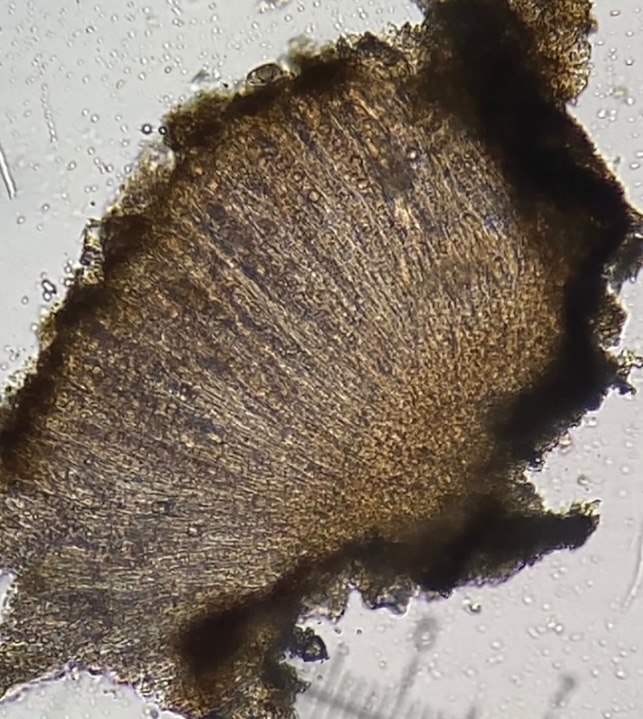
[attachment=2]IMG_5477.jpg[/attachment] Section about 10 seconds after adding a drop of C to the slide: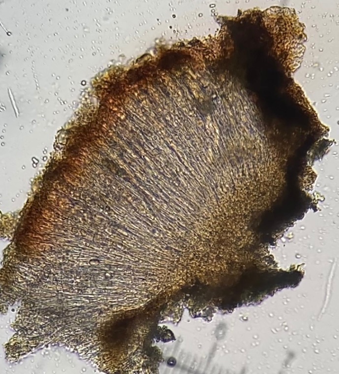
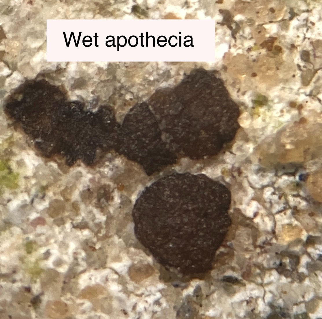
[attachment=1]IMG_5478.jpg[/attachment] After maybe half an hour in water, the apothecia swelled up considerably and changed from black to brown.
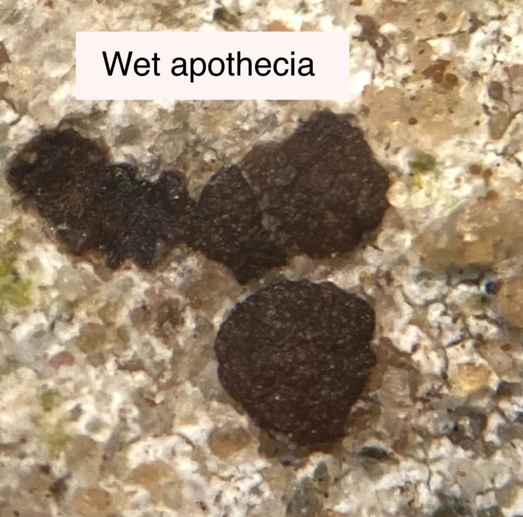
[attachment=0]IMG_5464.jpg[/attachment] Thank you so much for the detailed response. I have tried C again, but cannot get any trace of red reaction with bleach, neither on the thallus nor apothecia (using cocktail stick) nor on apothecial sections (they start reddish, dry or in water, but just bleach out with C). I have tried it four times, and haven’t seen even a fleeting reaction (watching under dissecting microscope). The same bleach gives a strong red reaction with Punctelia this morning, so I don’t think it’s the bleach. No red with KC either. Puzzling. Apologies for photo quality – some are through the dissecting microscope. 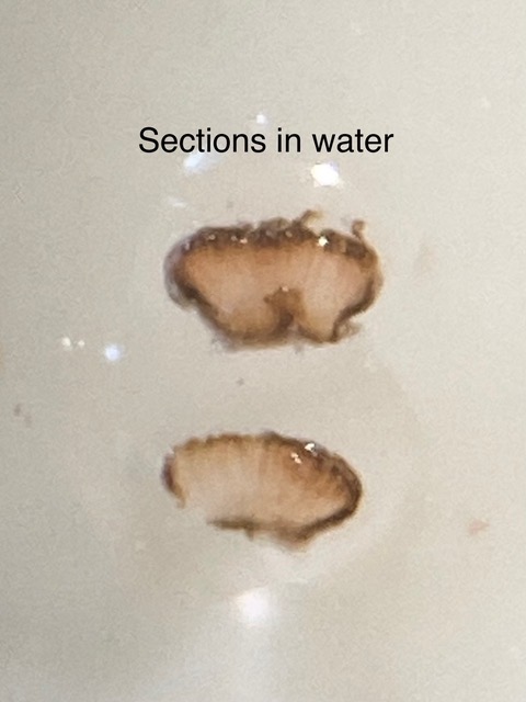
[attachment=6]IMG_5413.jpeg[/attachment] Thick section under x100 compound microscope – it was wetted, and harder to cut.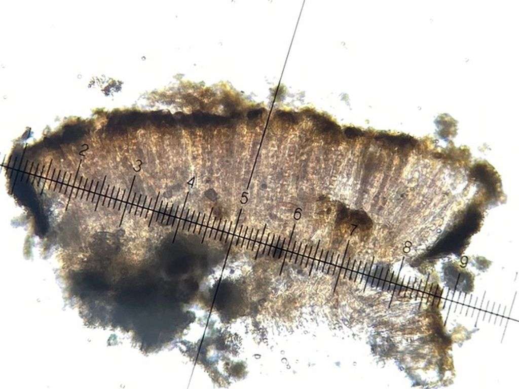
[attachment=2]IMG_5448.jpeg[/attachment] 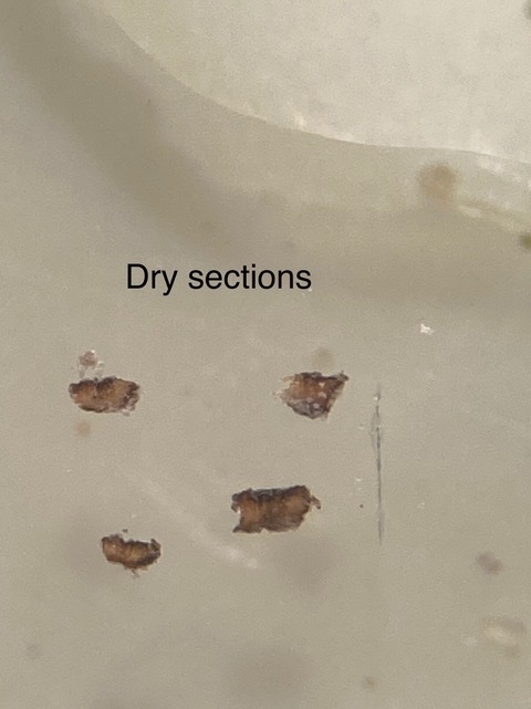
[attachment=5]IMG_5414.jpeg[/attachment] 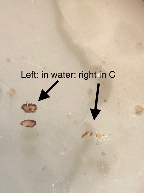
[attachment=4]IMG_5415.jpeg[/attachment] This photo is of an algal blob found in the thallus – it isn’t a discrete algal layer, there just seem to be odd pockets of algae mixed up in the cotton-wool like thallus.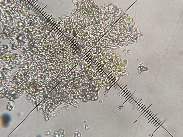
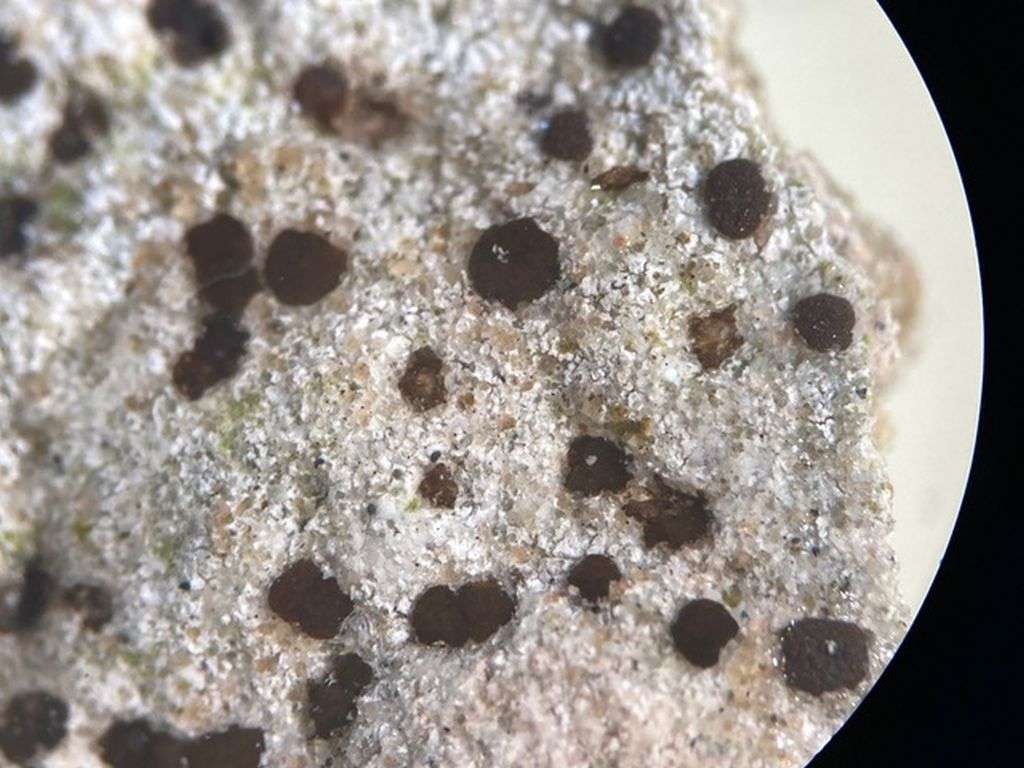
[attachment=3]IMG_5417.jpeg[/attachment] Last two photos are after spraying it with water, and then after leaving a droplet of water on the surface for about ten minutes. Spraying didn’t seem to do much, but after about ten minutes of being submerged, the apothecia have swollen up quite a bit, and look much more reddish-brown rather than black. It is very fragile – the sandstone keeps just rubbing off the back, and the main piece has fractured into several bits. I’m keeping it for future reference. I’ve looked up Trapelia in several places and can see it’s a tricky genus. Happy to leave it at this now.
[attachment=1]IMG_5449.jpeg[/attachment] 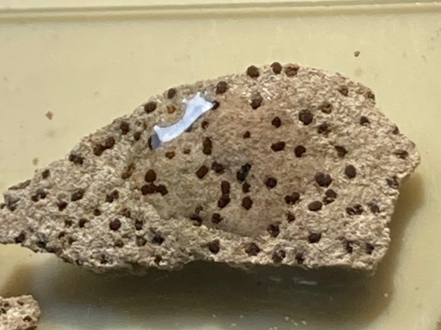
[attachment=0]IMG_5450 2.jpeg[/attachment] I revisited this after about a year of looking at lichens, and learning more. It is a lichen after all, Phlyctis argena. KOH on the thallus goes yellow and then red, and crystals (of norstictic acid) can be seen under the microscope. 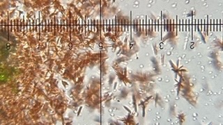
[attachment=0]B5A3198C-E398-4D91-9720-6718C36216F8.jpeg[/attachment] March 18, 2022 at 9:50 pm in reply to: Whitish sorediate crust on woodland twigs and branches #44701Thank you – the third photo was of the same thallus – I think it was only just mature enough for the soredia to have begun to break through the cortex surface in one patch. There were lots of much larger patches on thicker branches, but I just brought back a thin twig. I also tested the K patch with UV, with my new 365 nm torch, and there was no reaction. Thank you – very helpful tips about C vitellina. Beautiful ascus photo too. Thank you again. I did have a look, but I need lots more practise with sectioning bigger things. Just for curiosity, I tried taking a photo with incident light at x100. I wasn’t able to get a better section than the squash before. What appeared to be black spheres at x20 are actually elongated, about 100um wide and 200 um long.
Maybe one day I’ll look again when I’ve learnt a bit more. Thank you.
Thank you (I deleted the first photo as I found a better example of the brown spores). I hadn’t expected such a speedy response. That is all really helpful and encouraging. I had read and re-read your twitter thread about L albescens variations – it helped a lot.
Thank you. -
AuthorPosts