
Whitish sorediate crust on woodland twigs and branches
- This topic is empty.
-
AuthorPosts
-
March 16, 2022 at 6:40 pm #32321
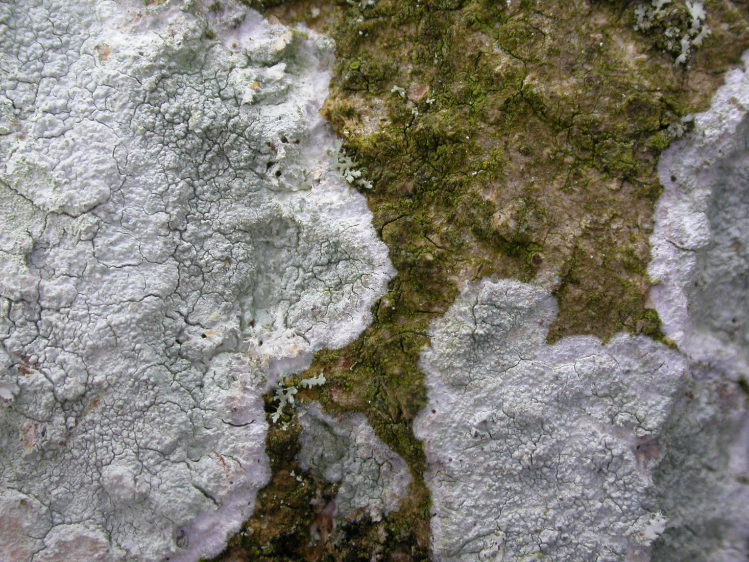
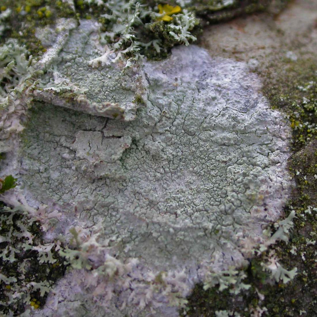
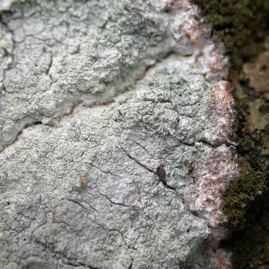
This might be Phlyctis argena, but I could easily be wrong. Lots of bright white to pale greenish mostly circular patches on twigs and branches, with fimbriate white margins, in a woodland in Warwickshire, near a small stream. Some of the sorediate areas were slightly pinkish, but not sure whether they were just unhealthy. 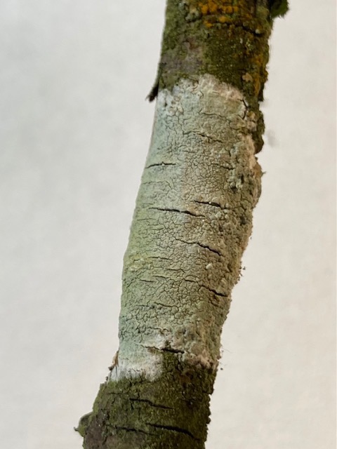
[attachment=3]IMG_4390.jpeg[/attachment] 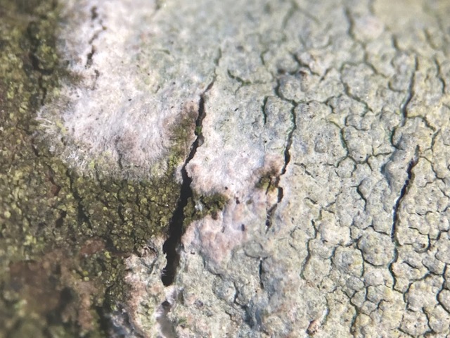
[attachment=2]IMG_4388.jpeg[/attachment] 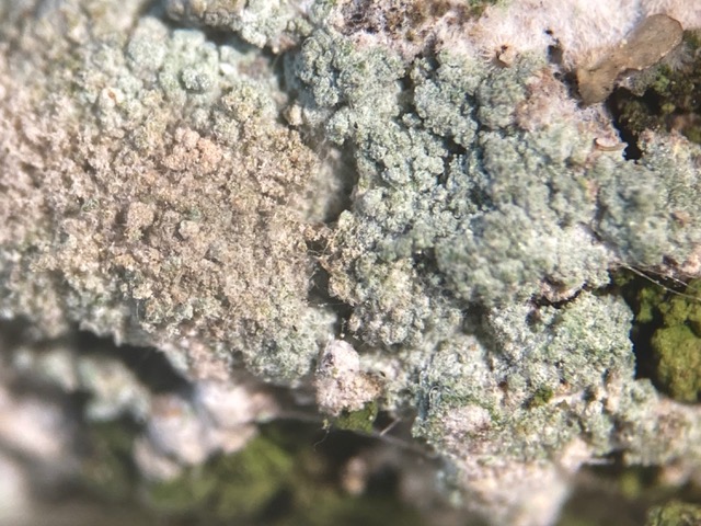
[attachment=1]IMG_4389.jpeg[/attachment] It was C- and K+ y to r (the first attempt using a cocktail stick only went yellow, but a more generous dollop of K gave a distinct orange-red). No UV fluorescence noticed before or after K.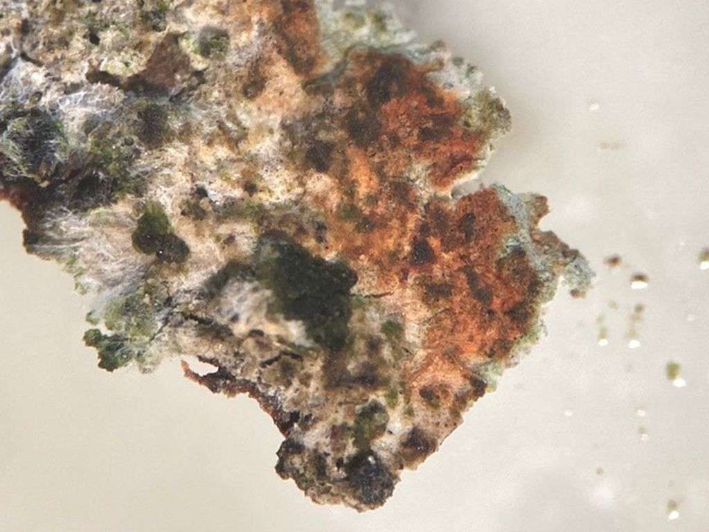
[attachment=0]IMG_4383.jpeg[/attachment] March 18, 2022 at 9:19 am #44700Although I strongly suspect you are correct, I was initially a little disconcerted by an apparent dearth of soredia in your first two photographs. However, when I checked my own photographs of relatively young Phlyctis argena thalli, I notice that they have a very similar appearance to yours. 
[attachment=2]EsHkMLnXIAMqO_-.jpeg[/attachment]
I can’t think of any other common lichen that is pale (whitish to pale greenish grey), sorediate, K+ yellow turning red and has a wispy fimbriate margin.The thallus of P. argena is pale grey and tends to be irregularly warted, cracked and/or lumpy. Soralia initially usually develop from pustules, then the soralia coalesce and the soredia form widespread sheets. The sorediate areas are slightly more creamy white (slightly paler) than the thallus.

[attachment=1]ENigrhkW4AQSFHx.jpeg[/attachment] 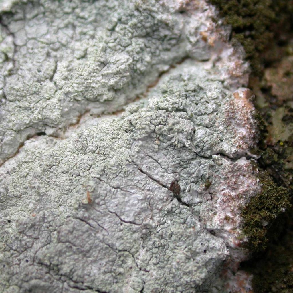
[attachment=0]ENhM7X2VAAIaDuY.jpeg[/attachment]
I have further thoughts and observations about this species which I will post this evening, but for the moment, if the thallus and irregular, pustular soralia development looks right, then taken with the wispy fimbriate margin, the K+ yellow to red reaction and the habitat (on twigs and branches) I think you are fairly safe to assume that you have Phlyctis argena.March 18, 2022 at 9:50 pm #44701Thank you – the third photo was of the same thallus – I think it was only just mature enough for the soredia to have begun to break through the cortex surface in one patch. There were lots of much larger patches on thicker branches, but I just brought back a thin twig. I also tested the K patch with UV, with my new 365 nm torch, and there was no reaction. March 20, 2022 at 9:39 am #44702Until considering your query, I had not been fully aware of how much thallus (as opposed to soredia) is present in Phlyctisargena, especially in young thalli. I suppose we often see what we want to see, in the case of examining P. argenanoting the wispy fimbriate margin and the widespread soredia starting from little pustular convolutions of the thallus, and tend to ignore the relatively unimportant thallus which tends to have little in the way of diagnostic features. As you may know, I am very keen on studying common species and appreciate this fresh prompt to examine P. argena. A previous query about
P. argenaquestioned the Pd reaction stated for this species in LGBI2. I agreed with that previous correspondent that the reaction, even with very strong Pd solution, is not accurately described as “Pd+ orange-red”. 
[attachment=4]Screenshot 2022-03-20 9.15.50 AM.png[/attachment]
Full thread:https://twitter.com/obfuscans3/status/1252713325596721153 As for the K reaction of
P. argena, I usually find that a frugal application of reagent is best but your experience suggests that perhaps one can sometimes be too frugal. The way that the spot reaction of norstictic acid has been cited in the literature has been inconsistent for different species, sometimes causing confusion. 
[attachment=3]Screenshot 2022-03-20 9.24.13 AM.png[/attachment]
Link to thread discussing this inconsistency:https://twitter.com/obfuscans3/status/1213518967739953153 To properly demonstrate the presence of norstictic acid it is best to observe microscopic red crystals of the potassium salt of norstictic acid when mounted in K (however in practise, it is often sufficient just to note K+ yellow turning red taken with morphological characters).

[attachment=2]ENdKDYqX0AAXPNC.jpeg[/attachment] Although primarily a corticolous species,
P. argenadoes also grow on stone and has a liking for the sort of ironstone gravestones that occur in my region. I have checked carefully that this really is P. argenaand not Pertusaria lactescens(which occurs further north in the Midlands, on harder, more siliceous memorials). 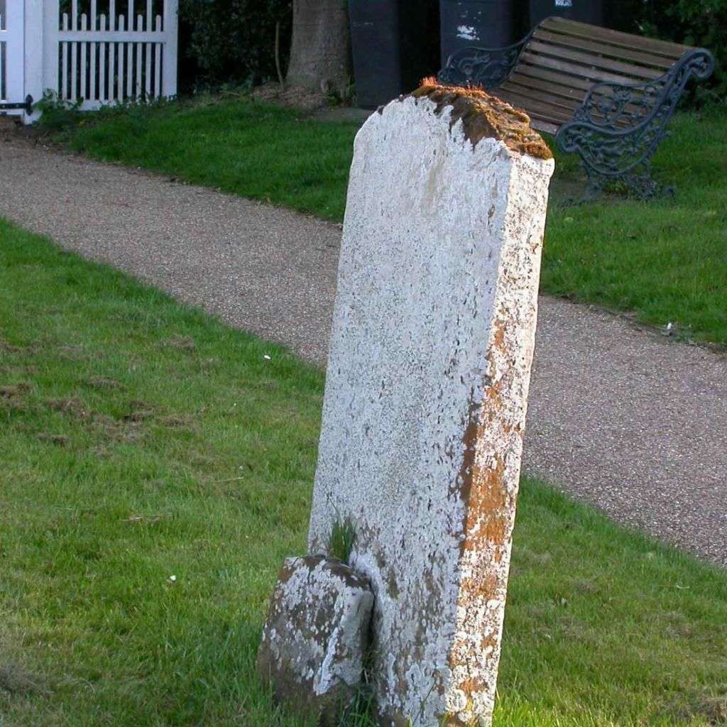
[attachment=1]DV2XDyhX4AAmwVD.jpeg[/attachment] Thanks for taking the trouble to note the (lack of) fluorescence both before and after adding K in
P. argena. As you will no doubt realise, I now shine UV at most spot tests that I perform, just in case there are any other potentially useful tests to add to the K/UV+ intense yellow for atranorin, K/UV+ mauve for confluentic acid and the K/UV+ pale, intense green probably due to arthothelin. 
[attachment=0]Screenshot 2022-03-20 9.39.06 AM.png[/attachment] -
AuthorPosts


- You must be logged in to reply to this topic.