Forums › Forums › Fungi ID requests (post here if you aren’t sure what type of fungus you’ve found) › Small, brown, domed fungus
- This topic is empty.
-
AuthorPosts
-
August 20, 2018 at 7:19 pm #30995
I’d greatly appreciate help with identifying this fungus. Habitat: Path on reclaimed rural industrial site, with willow/birch scrub cut to ground level.
Cap: Brown, domed, slightly slimey, mottled, with flakes near edge; width 2cm; stem 30x3mm;
Gills Grey-brown; Flesh white
Spores: brown/dark ochre; 11.54-13.50 x 7.55-8.90um
Cortinarius have large spores, but there’s no indication of a web.
Hoping you can help.
Nick
August 22, 2018 at 10:03 am #41130Did you look at cystidia? August 22, 2018 at 3:35 pm #41131adampembs wrote:
Did you look at cystidia?
Sorry, no, but I think I’ve found a definite possibility:Redlead Roundhead – Leratiomyces ceres (Cooke & Massee) Spooner & Bridge (prev. Stropharia aurantiaca).
An alien from Australia (first-nature.com), now found on rotting sawdust and woodchips, usually in parks and gardens; autumn.
Cap: Orange-red; convex, becoming flatter; pale veil fragments often cling to cap margins; 1.5 to 6cm across.
Stem: 3 to 9cm tall and 3 to 8mm diameter. Spores: Ellipsoidal, smooth, 11-13 x 7.5um (Phillips).
Key features match, and it’s interesting to note the veil fragments on the cap margin are distinctive.
What do you think?
All the best… Nick
August 22, 2018 at 4:40 pm #41132I must admit when I first saw this my immediate reaction was Strophariaceae, The good image of the spores would immediately rule out Cortinariaceae, whose members have roughened spores, lacking a germ pore, and germ pores are quite obvious here. As regards
Leratiomyces ceres, in my experience that species has much more colourful caps than this one. It would be worth checking (as Adam has suggested) the presence or absence of chrysocystidia. What is the mountant for the spore shot? It makes then look remarkably thick-walled which has thrown me. It maybe the LeratiomycesI will await Andreas’ verdict. For what it’s worth here are images of one of my collections of
L. ceres: 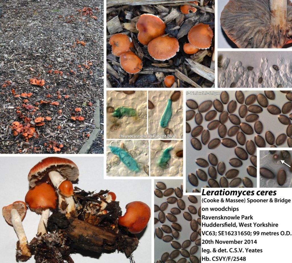
[attachment=0]Leratiomyces ceres 0a.jpg[/attachment] August 22, 2018 at 6:27 pm #41133Many thanks, Chris, for your comments. Andreas sent me the following message, which I read after my last post. I’m sure he won’t mind me copying and pasting it here:
“Hello Nick,
I think it is Leratiomyces percevalii, or may be one of the other large-spored Stropharia species which are now in Leratiomyces.
I have technical problems with the log-in and can’t answer in the forum any more at the moment, therefore as private message ….
best regards, Andreas”
The mountant I used was Melzer’s Reagent. I no longer have the one fungus I took, and the remaining ones have disappeared, so I’m afraid I can’t check the presence or absence of chrysocystidia.
As for the colour of the cap, the photograph doesn’t really do it justice – I remember it being far more vibrant. Even with some manipulation, the image hasn’t captured this.
Thanks, again, for the feedback. I’ll check for any further comments next week when I’m back from a trip.
Nick
August 22, 2018 at 9:06 pm #41134Hello, this is a foto of Stopharia percevalii.
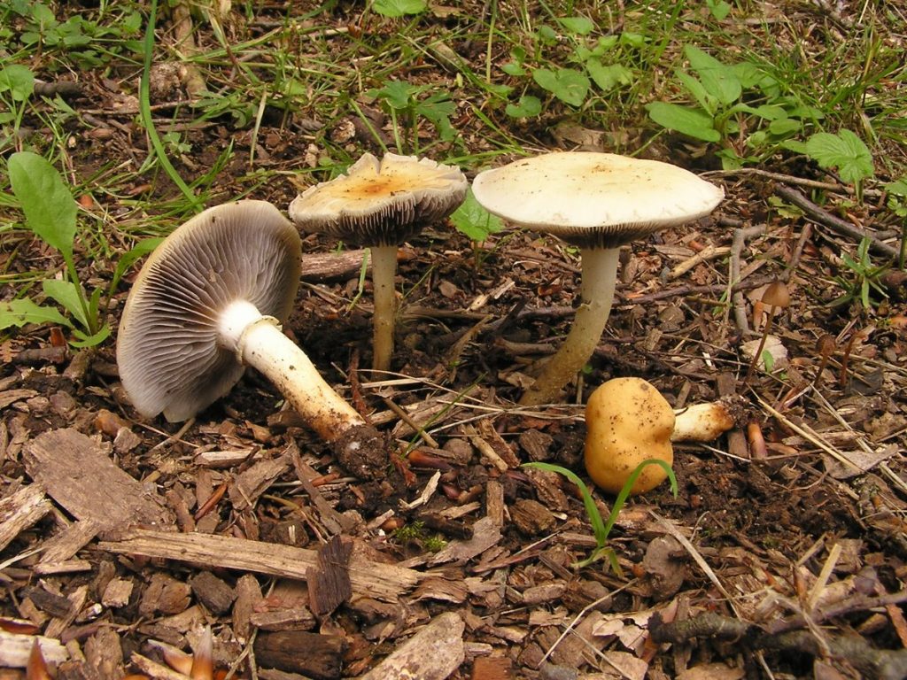
[attachment=0]strophariapercevallii040606Eisenberg 006.jpg[/attachment] I admit that the cap colour doesn’t fit too good to your collection, but L. ceres does neither.
best regards,
Andreas
August 23, 2018 at 8:31 am #41135Interrupting the experts again! My first thought was Psilocybe coprophila. Why would you rule that out? August 23, 2018 at 10:57 am #41136Hello, there are different points that speek against Psilocybe coprophila, but I agree that more microscopical details (especially about the cystidia) would better solve the problem.
– Psilocybe coprophila has gills that are broadly attached
– P. c. is growing on pure dung, not on pure ground or vegetal remnants
– P. c. has a veil consisting of small veils, whereas the veil remnants seen here are more flöoccose and bigger
– P. c. has much veil on the stipe, whereas no trace of veil is seen in the fotos here.
– the overall appearance is a bit too stout in my opinion for P. c.
I agree that these arguments are mostly more or less quantitative, but taken all together I don’t believe that this is Psilocybe coprophila, though I can not exclude it to 100% certainty.
best regards,
Andreas
August 23, 2018 at 1:25 pm #41137What I have called Ps. coprophilain the past has shown – at least in some views – a distinctly “rounded hexagonal” view of the spore outline; from
Funga Nordica: 
[attachment=0]Untitled-1.jpg[/attachment]
I can’t see that here . . .August 24, 2018 at 1:04 pm #41138Very helpful. Thanks both. August 28, 2018 at 10:31 am #41139Many thanks for all the comments and suggestions. Psilocybe coprophila was an interesting interruption, but its features in general and habitat in particular seem to rule it out.
I should have looked at the cystidia (live and learn), but the likelihood is it’s a Leratiomyces sp.
Can we be more specific? Maybe that will have to wait until next year when it re-emerges!
Thanks, again, for your help… Nick
-
AuthorPosts



- You must be logged in to reply to this topic.