Forums › Forums › Fungi ID requests (post here if you aren’t sure what type of fungus you’ve found) › Lepiota or similar
- This topic is empty.
-
AuthorPosts
-
July 17, 2015 at 3:33 pm #29538
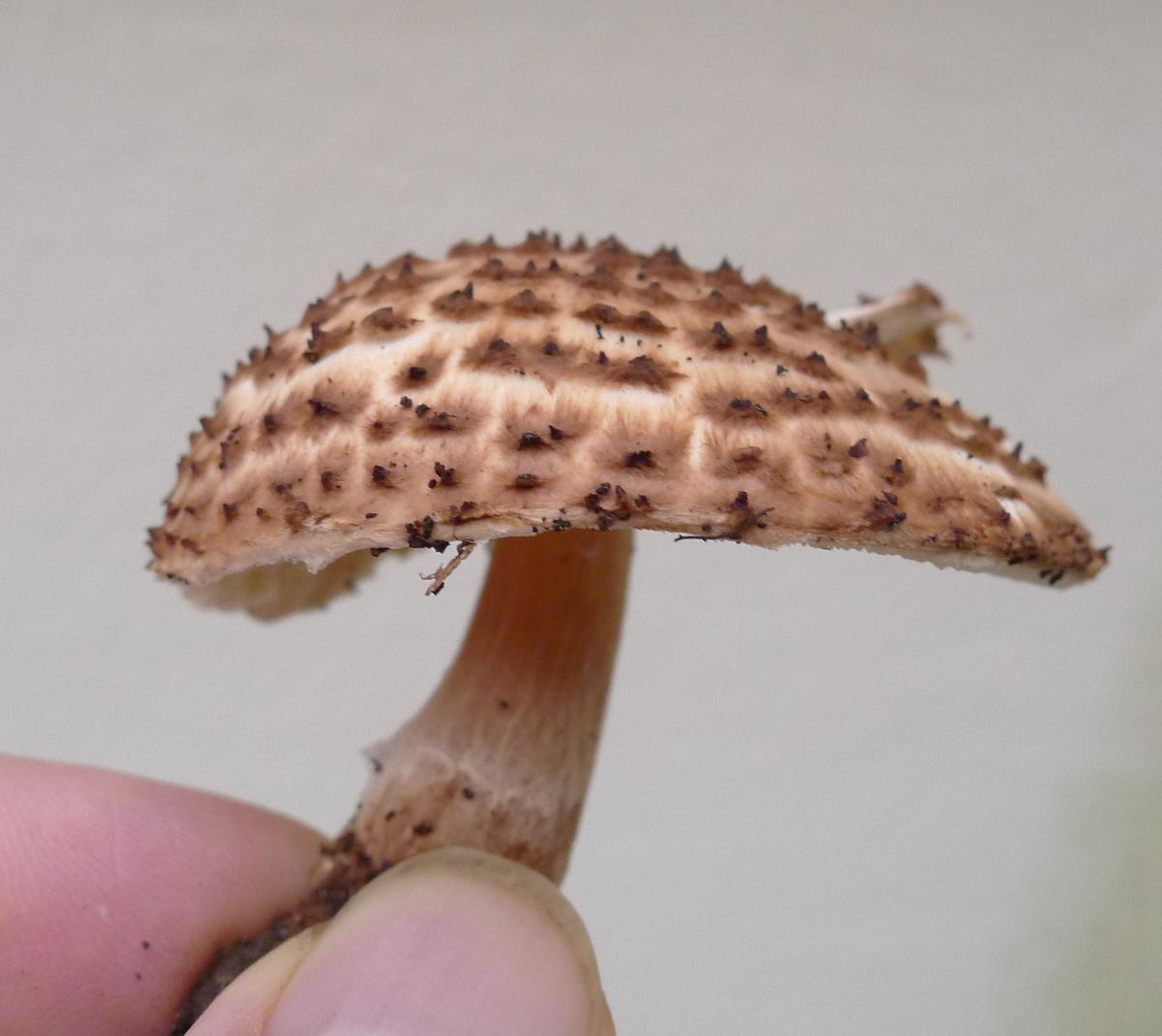
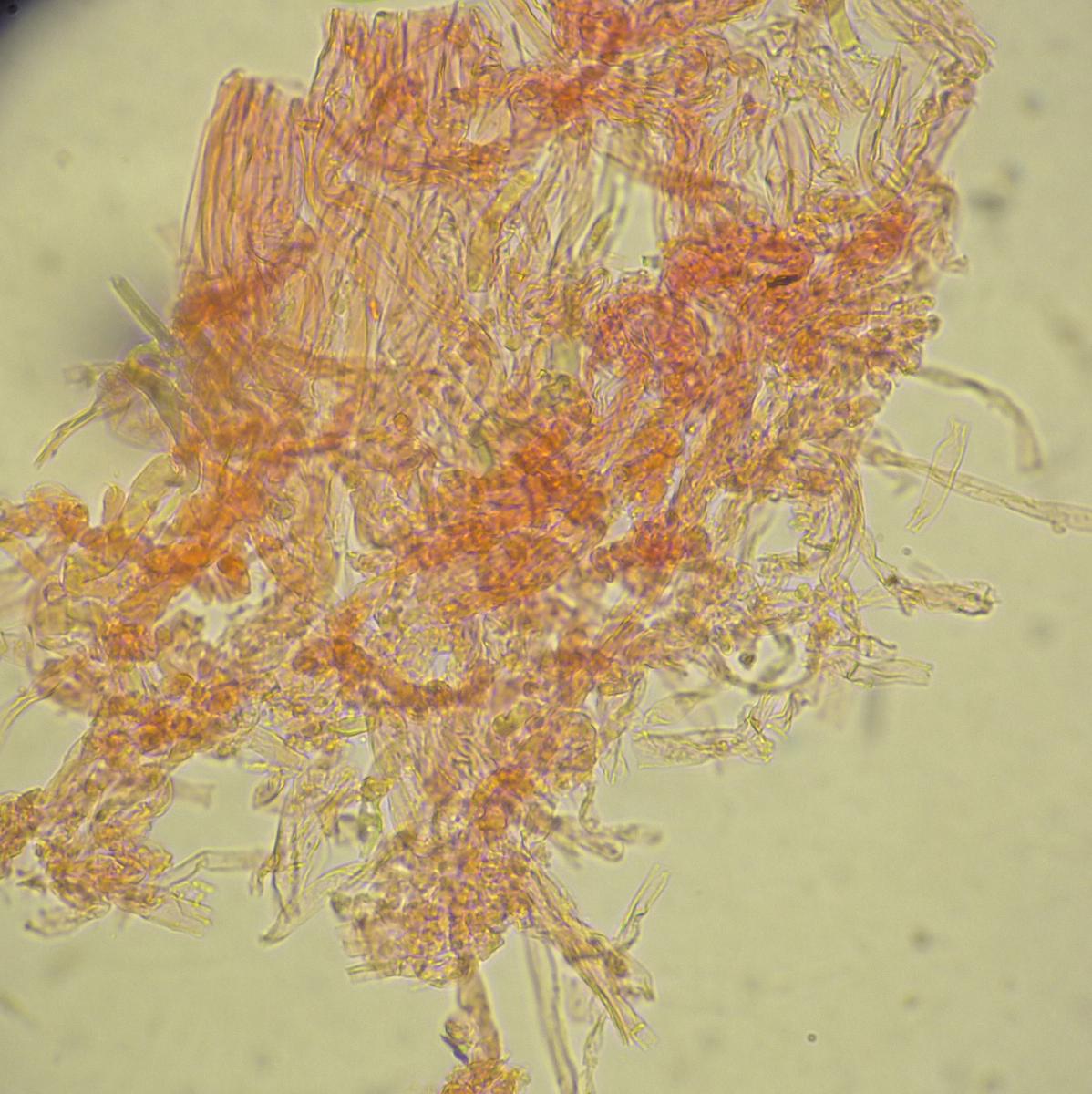
Growing along cyclepath in deciduous woodland. Smell faint, chemical/antiseptic. No ring, just a trace of ring zone low on the stipe. Brown fibrils on the stipe. Some swollen cens looking at a pileipellis section (perhaps these are from the warts/scales as I see no swollen cells in a scalp. I’m keying to Lepiota but struggling to get to species. Any help is appreciated


[attachment=9]P1100634.JPG[/attachment] 
[attachment=8]P1100635.JPG[/attachment] 
[attachment=7]P1100636.JPG[/attachment] 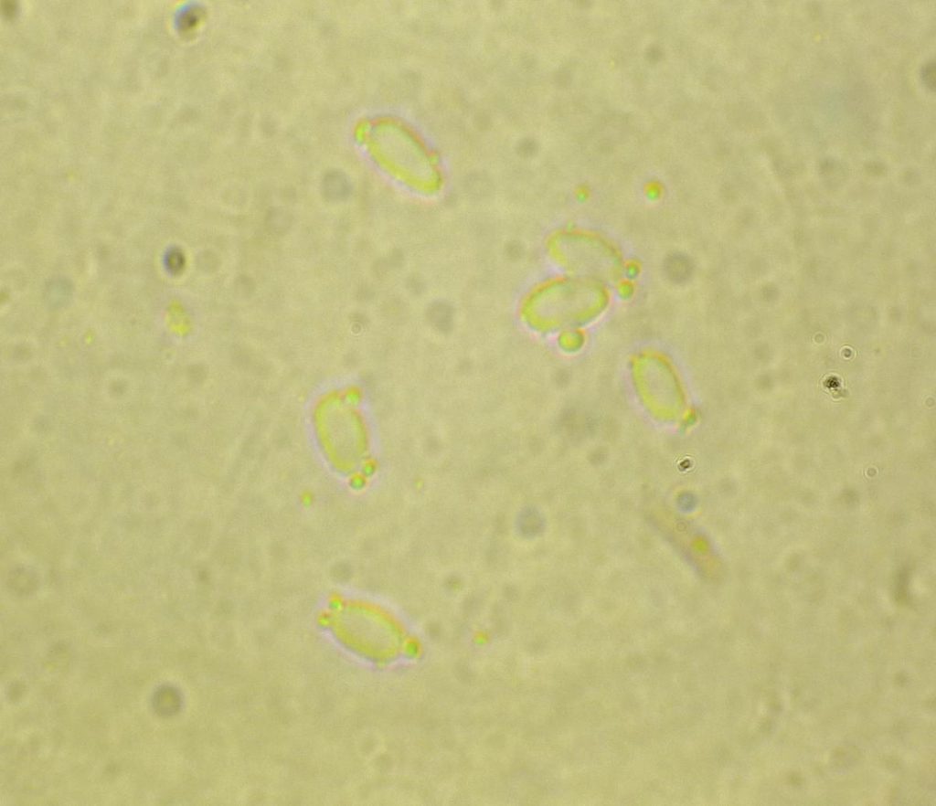
[attachment=6]P1100637.JPG[/attachment] 
[attachment=5]P1100639.JPG[/attachment] 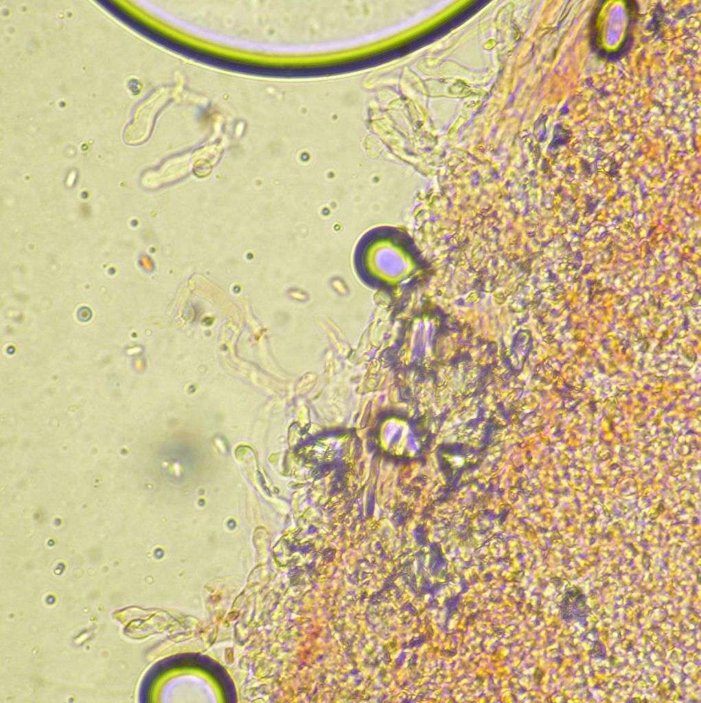
[attachment=4]P1100640.JPG[/attachment] 
[attachment=3]P1100644.JPG[/attachment] 
[attachment=2]P1100646.JPG[/attachment] 
[attachment=1]P1100647.JPG[/attachment] 
[attachment=0]P1010362-001.JPG[/attachment] July 17, 2015 at 4:04 pm #35290Plenty of info. in these images! Important features here are shape, size & dextrinoid aspect of spores, pyramidal warts on cap, gill edge white, clamps present, lack of ring, presence (& shape?) of cheilocystidia. These all point to a group of spp. around Lepiota perplexa(the other possibilities being calcicola & echinacea). Your spore sizes are a tad large but seem to fit the latter 2 better. Unfortunately the key (FAN5) differentiates these two by the shape and size of the apical cells which comprise the pyramidal warts on the cap (there’s always one more thing to measure!). July 17, 2015 at 5:43 pm #35291Do these help? In Funga Nordica, assuming the pileipellis is an epithelium, it takes me to Echinoderma, then as the cheilocystidia are distinct to basidioles, this leads to E.perplexa or E.calcicola. E.calcicola spores are notmetachromatic (will have to look that up!) and below the ring zone should be desnely covered with woolly pointed scales of cap colour, whereas E.perplexa spores aremetachromatic and there is no mention of scales below the ring zone. The specimen has a woolly fibrillose covering below the ring one but not what I would call scales. The black bits in the photo are soil.
July 17, 2015 at 7:49 pm #35292Like ‘Coprinus’, it’s easier to call them Lepiota’s! (can you remember which are now Coprinellus and which Coprinopsis?). But you’re right, they were moved out of Lepiota in the 5th Update of the Checklist (March 2011). I’ve had to look up ‘metachromatic’ also: it’s the reddish-violet reaction of spore walls in Cresyl Blue in water (don’t think I have any of that). It’s mentioned in the descriptions in FAN5 with both
calcicola & echinaceahaving a non-metachromatic reaction. Perplexadoes seem to have fewer warts/scales on the stipe: only at the base in fact. It’s keyed separately on the basis of it’s spores being up to 3µ wide and more “penguin-shaped” (Q=1.8-2.6). The other two have spores of similar length, up to 3.5µ wide, and are more cylindric-oblong (Q=1.35-2.15). Then the terminal cells of the cap warts: in
calcicolathey are ellipsoid-oblong and with echinaceaglobose to ellipsoid: and of similar size. Your spore widths are 3-4µ and on the image they look roughly ellipsoid (which seemed to rule out
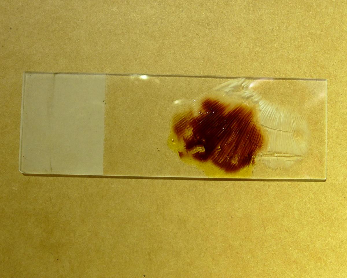
perplexa). There’s not a lot of difference between any of the three really! It’s probably a ‘best fit’ situation between all these factors although some Cresyl Blue would help! July 18, 2015 at 9:18 am #35293Yes, always confusing with name changes, I remember “Lepiota procera” Its NOT metachromatic (at least unless its meant to be heated or something) – here it is in cresyl blue.
I’m now wondering if it is E.aspera (‘Lepiota aspera’) – the cheilocystidia were in tufts along the gill edge (although irregular and unlike basidioles)
July 18, 2015 at 10:30 am #35294Once more into the breach… The ring on
E. asperacan certainly be ‘lost’ (or end up attached to the cap margin), so it’s worth considering. Spores for this sp. are about the same width as the other spp. we’re considering and are longer: FAN5 quotes 6-9(9.5) x 2.5-3.5(4)µ. Also the cystidia are abundant and shown and described as clavate to sphareodunculate and up to 24µ wide. I don’t think this fits you images/description. Ruling out
perlexa(spores metachromatic), the cystidia of the other 2 spp. are much more like your images (both also have them in fascicles or tufts). Time to go for it: the shape of the cystidia and also the apical cells of the cap ‘warts’ look to fit E. calcicolabest. [NB: I notice the coment for
perplexaspores is “… metachromatic in Cresyl Blue, but not very distinctly so.”] July 18, 2015 at 12:44 pm #35295E.calcicola is quite rare! I see you have 2 of the 8 uk records and it’s never been recorded in Wales. 
David might need me to keep a voucher. I’ll dry what’s left of it!
July 18, 2015 at 1:32 pm #35296A timely post and comments – I found a possible ‘Lepiota’ today but will be very cautious about id’ing it further than Genus! July 18, 2015 at 2:26 pm #35297Lepiota s.l. are generally not too bad but as with every Genus, there are pockets of difficult-to-separate species (or species complexes). Like this instance, they may seem to have attributes of more than one species: You would need to find large spores (up to 9µ) to convince me it was
asperum(the lack of ring and cystidia shape do not suggest this species). Looking at FE4 as well as FAN5, the spore size and cystidia shape seems to fit
perplexumbest, but where is the metachromatic (reddish-violet) reaction? The only image of perplexumI have is in BK(FoS)4 where I’ve just notice that the image they use for perplexum(233) is the same image asechinacea(223)!!! The Checklist’s comments for
echinaceum, “Confused with closely related taxa” says it all. My database has 25 asperum, 6 perplexum, 2 calcicolaand 1 echinaceum: but are these all identified correctly? It’s a good idea to keep a dried specimens of ‘cryptic species’ as someone may be able to redetermine them at a later date (several of my collections of perplexum/calcicola/echinaceumhave been sent to Kew). July 18, 2015 at 2:42 pm #35298Afraid I don’t do microscopy so probably best for me to stick with Genus! I am in contact with Kew re ‘Lost and Found Project’ so maybe worth keeping specimen if I can dry it properly. Specimen is probably past it’s best though. I am very much an amateur with fungi but very involved with Glamorgan Fungus Group. Mark
July 18, 2015 at 3:28 pm #35299Ok, measured another 12 spores. This time in Melzers as I can see them better! I have slightly smaller measurements this time. Range 5 – 6.5(7) x 3 – 3.5 µm
Average 5.7 x 3.0µm.
This matches E.calcicola perfectly (and I didn’t check the spore size of E.calcicola until after I made my measurements!)
:geek: Tried again to get a good image of the inflated cells of the pileipellis and below is the best i could get. Really difficult to get a thin enough section even with a new razor.
July 21, 2015 at 7:53 am #35300Hi Adam, Your best two images of the end cells were the two titled ‘wart detail’. These seem to show a mixture of large ellipsoid cells and smaller globose cells. I find the best method is to use tweezers to pull off the tips of the brown warts, then you should get mostly chains of terminal cells.
Another thought; is your Cresyl Blue in water or alcohol? (it’s just that I see the ABFG sell it in alcohol). FAN5 says the metachromatic reaction is with Cresyl Blue in
water. But does it make a difference to the reaction? Much better to see the spore shape in Melzers – they look more the shape of perplexato me. July 21, 2015 at 9:15 am #35301Hi Roy, I always assumed the pileipellis was separate to the warts/scales.
I got the Buyck formula Cresyl Blue from
http://www.micro-science.co.uk/cresyl-blue-buyck-10ml.html It doesn’t mention alcohol but does say it is for metachromatic reactions. Is the sample supposed to be heated?
July 22, 2015 at 1:04 pm #35302Perhaps I’ll have to leave this as an unknown and if I find some more, I will reach a conclusion. -
AuthorPosts






- You must be logged in to reply to this topic.