- This topic is empty.
-
AuthorPosts
-
January 30, 2021 at 1:59 pm #32004
First attempt at ID lichen on living Pear Wood. Brittany France. I’m thinking Physcia aipolia? Thanks for any help.
January 30, 2021 at 5:26 pm #43936You are definitely heading in the right direction. It is a decision between Physcia aipoliaand its close look-alike P. stellaris. The books say that P. stellarislacks the white flecks on the lobes which are present in P. aipolia. In fact it is more a matter of degree; any white spots/flecks in P. stellarisare so inconspicuous that they are often not detected even through a good lens. In your photo there isn’t much lobe visible and I can’t decide whether they are significantly spotted with pale dots/flecks or not. 
[attachment=2]Screenshot 2021-01-30 at 5.12.06 PM.png[/attachment]
The white flecks ofP. aipoliacan be seen in the following image from this website: http://www.lichens.lastdragon.org/Physcia_aipolia.html 
[attachment=1]Screenshot 2021-01-30 at 5.18.13 PM.png[/attachment]
The flecks are sometimes called pseudocypellae but are perhaps better considered to be maculae, pale because the algal layer beneath the surface is disrupted by hyphae rising from the medulla.
[attachment=0]maculae.jpeg[/attachment]
There is a chemical test which distinguishesP. aipoliafrom P. stellarisif you get sufficiently interested to try simple spot tests. The medulla (exposed with a razor blade) gives a yellow reaction with potassium hydroxide solution (K) if P. aipolia. The medulla of P. stellarisgives no reaction. I suspect you are right with your initial identification but I wary of confirming it from the photo due to the potential confusion with
P. stellaris. January 30, 2021 at 9:26 pm #43937Thank you! I am a complete novice, but am also completely fascinated.
I’m glad to know I’m on the right track. Is potassium hydroxide, bleach?
So many lichens here.
Something to really get my teeth into between ‘normal’ fungi and butterflies!
January 30, 2021 at 9:46 pm #43938K is a solution of potassium hydroxide. Small amounts e.g. 10 g of solid potassium hydroxide can often be bought cheaply as samples on sites such as ebay or Amazon. A reasonable substitute is strong caustic soda solution (sodium hydroxide). Caustic soda can usually be bought at hardware stores. Bleach is a source of the other commonly used chemical, sodium (or calcium) hypochlorite (C). I recommend strong types of bleach such as Domestos or Parazone.
January 31, 2021 at 8:11 am #43939Excellent. Thanks again. I have caustic soda to hand. January 31, 2021 at 9:27 am #43940Spot tests are very simple in concept but require a bit of care in practise. It is a case of applying a tiny drop of the reagent on to the tissue to be tested and noting any colour change. Spot tests are most reliably conducted on dry material under a dissecting microscope. Many people, especially beginners, tend to apply far too much reagent. The tip of something like a cocktail stick can be used to apply a tiny spot to exactly the right part of the lichen. If applied to damp specimens, the reagent tends to spread throughout the specimen (not necessarily sticking to the tissue being tested) and the reaction tends to be diluted by the extra moisture present. To distinguish between
P. aipoliaand P. stellaris, it will be best to make an oblique cut through an outer lobe with a razor blade. An oblique (rather than vertical) cut will expose a broader cut face through the medulla. Then a tiny spot of strong reagent is applied to the white medulla. 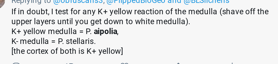
[attachment=1]Screenshot 2021-01-31 at 9.06.44 AM.png[/attachment] With experience, many spot tests can be performed successfully in the field but it might be best initially to collect a small specimen and work on it in more controlled conditions at home. With these K reactions, it is often best to wait until the spot has dried before deciding whether there has been any colour change or not.
In the following image, I have tested
Physcia tenellawhich has K+ yellow upper and lower cortex and K- medulla. The position of the upper and lower cortices are indicated by red lines. In this case I have made vertical sections and this shows how thin the medulla is and why it can be easier to cut an oblique section. 
[attachment=0]tenreac.jpeg[/attachment] January 31, 2021 at 9:58 pm #43941Thanks again! I have been photographing various lichens today and will be sitting down tomorrow to try and make sense of my images! I’m pretty sure I’m going to be posting for more of your advice.
Hope you don’t mind
😀 ! -
AuthorPosts


- You must be logged in to reply to this topic.