- This topic is empty.
-
AuthorPosts
-
February 9, 2020 at 11:59 pm #31598
I’m struggling with this one for some reason. It keys out in Dobson as Parmelia pastillifera but i’m not convinced that is right for a few reasons: 1. The white medulla does not react red with C although it did go a yellow-orange eventually (it also goes yellow-orange with K).

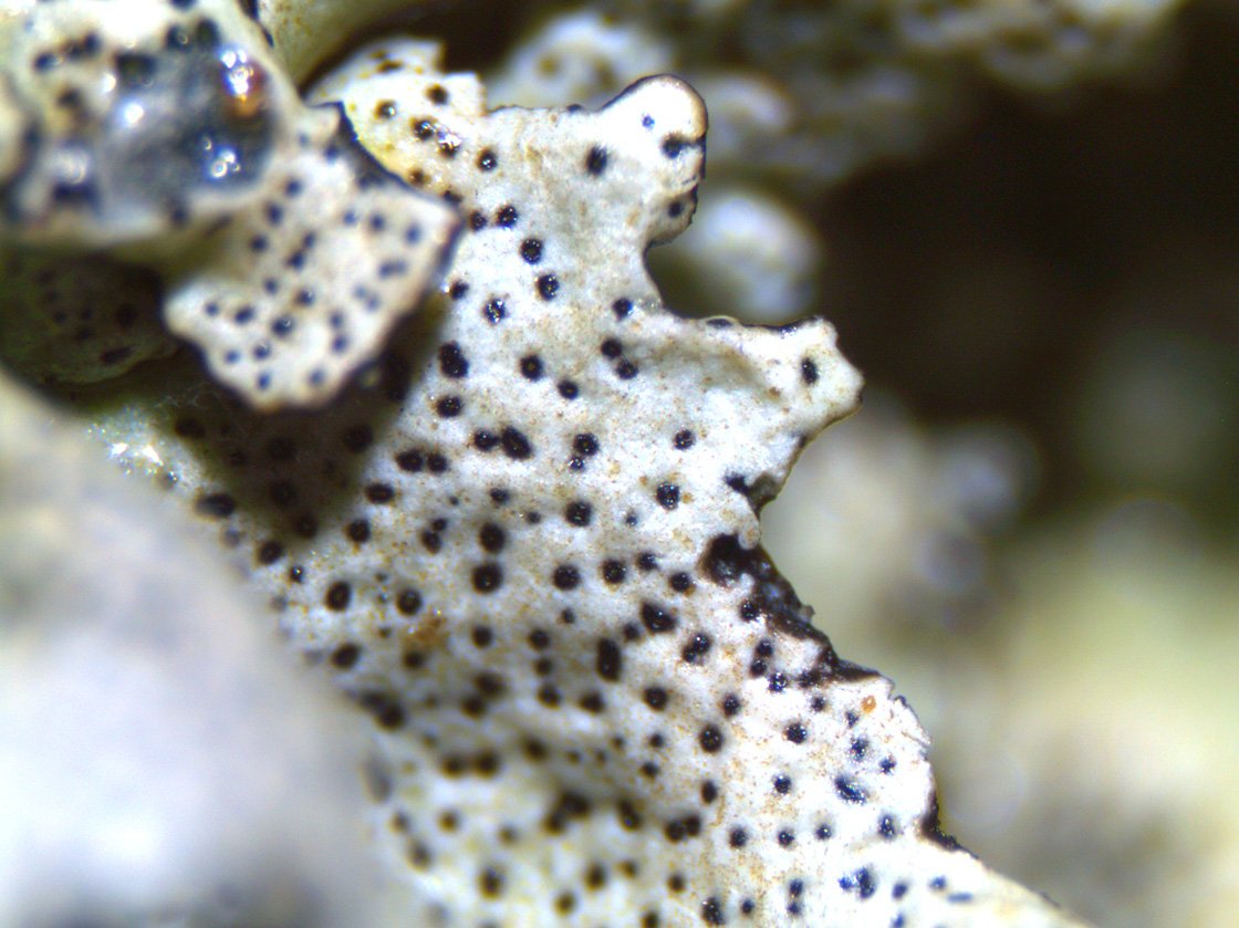
[attachment=0]parmcreactjpg.jpg[/attachment] 2. The normally quite visible small black isidia dusting the surface turn out under the microscope to be what I think are immersed ostioles ? (see pics) and there are rare globose cream to dark greyish-brown isidia on some lobes.
The thallus in general is 7cm in diam on slate stone. The individual lobe heads at the outer margin are 1 mm wide. The outer lobes are a light bluish grey and largely free of the black ostioles while the inner more upright lobes are densely covered in black ostioles giving the inner core of the thallus a darker grey appearance. The lobe surface is smooth and papery (dry) with a slight shine and a black margin. The lower surface of the lobes is generally black (occasionally dark brown at inner thallus) with simple black rhizines extending to the outer edge of the lobes.
I think I am still in Parmelia territory with this one, but cannot pin it down. Any ideas ?
Overall thallus form

[attachment=5]IMG_0790web2.jpg[/attachment] Black ostioles ? And cream to greyish-brown globose isidia
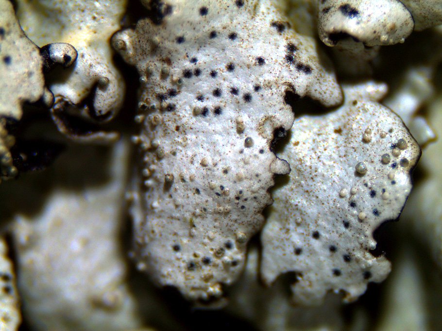
[attachment=4]parmisidia.jpg[/attachment] The simple black rhizines

[attachment=3]parmrhizines.jpg[/attachment] Dense black ostioles ? on inner thallus

[attachment=2]parmostioles1.jpg[/attachment] Detail of ostioles

[attachment=1]parmostioles2.jpg[/attachment] February 10, 2020 at 11:09 am #42794Hi Have you considered Dermatocarpon? Mostly this genus is without rhizines but has immersed perithecia with visible ostioles, as in your pictures. Chemical reactions negative and it doesn’t, in my experience, have adpressed lobes round the edge, as your photos show. The latter lobes do have the look of Parmelina. Could you have two species intermingled? I look forward to someone more knowledgeable commenting!
Caz
February 10, 2020 at 11:45 am #42795Hi Caz Thanks for the comments. It was the images of Dermatocarpon online which looked most like the inner thallus covered in perithecia with ostioles and helped me identify those. I dismissed Dermatocarpon though because I assumed two species could not intermingle in this way, but maybe you are onto something and it would explain why it doesn’t key out right.
Mark
February 10, 2020 at 12:48 pm #42796Mark That does not look like a not a
Dematocarponto me, looks more like a poorly developed Parmelina pastillifera, the “black ostioles” are pycnidia, which are quite frequent on Parmelina pastillifera(see pictures http://fungi.myspecies.info/taxonomy/term/8103/mediahttp://fungi.myspecies.info/taxonomy/term/8103/media” class=”bbcode_url”> Quote:Cortex K+ yellow; medulla C+ carmine-red, K–, KC+ red, Pd–
Scrape away the cortex and carefully apply the reagents to the exposed medulla
I might be something else, but I am not sure what.
All the best
Neil
February 10, 2020 at 1:16 pm #42797Thanks Neil – I did scrape away the cortex to reveal what I think is the white medulla below (beginner so I may be wrong) and applied the chemicals to the white surface. I’ll double check though. So there’s no chance I could have a Dermatocarpon growing on/in middle of a Parmelia that is underneath – is that correct ?
Mark
February 10, 2020 at 2:20 pm #42798Mark Quote:So there’s no chance I could have a
Dermatocarpongrowing on/in middle of a Parmelia that is underneath – is that correct ? No
Dermatocarponhas rounded little divided lobes http://fungi.myspecies.info/taxonomy/term/4783/mediahttp://fungi.myspecies.info/taxonomy/term/4783/media” class=”bbcode_url”> Dermatocarponspecies have no spot tests at all and certainly not C + reactions. I called the black spots pycnidia, but
http://fungi.myspecies.info/taxonomy/term/8103/mediahttp://fungi.myspecies.info/taxonomy/term/8103/media” class=”bbcode_url”> Dermatocarponthe black spots would be fertile perithecia with asci and spores inside. Neil
February 10, 2020 at 5:02 pm #42799I am going to throw another suggestion into the mix, not a definite identification at this stage. What about Xanthoparmelia conspersa? Although I would expect X. conspersato be a yellow-green colour, that does depend on the specimen and the lighting. Mark, when viewed in daylight, does the thallus have any yellowish-green tinge or is it really pure grey? The morphological details seem to fit X. conspersa– the presence of isidia, the often numerous pycnidia, the lower surface mainly black with stubby rhizines. This suggestion would also explain the K reaction. I am not sure what to make of the supposed C reaction – I think we need a retry with that and carefully record what happens immediately on adding strong C to the exposed medulla. If we decide that
Xanthoparmeliais the right direction to go, then we also need a more careful K test just to rule out X. tinctina. February 10, 2020 at 5:20 pm #42800Other Mark Yes it is physical more like
Xanthoparmelia conspersa, the silver-grey colour put me off, rather than the usual yellow-green colour, possibly an usnic acid deficient morph, if the photo is giving the right colour? Neil
February 10, 2020 at 5:58 pm #42801It could be a chemical var. without usnic acid but perhaps more likely to be an individual in which the usnic acid colour is less apparent. Perhaps specimens growing in shade (or other environmental factors) leads to them having lower concentration of usnic acid. I have a nice specimen of X.tinctinawhich appears pure grey and some specimens of Hypotrachyna sinuosaare grey with little apparent hint of yellow-green. One thing I noticed about your spot test is that you appear to have applied quite a drop of reagent. I prefer to apply reagents with e.g. the tip of a cocktail stick. If excess reagent is applied, one of several things might happen. The lichen substances might be dissolved and diluted to the point where they don’t react properly or substances from one structure (such as the medulla) may leach up into the cortex (or vice-versa) and give anomalous results. LGBI2 claims that the cortex of
Parmelia sulcatais ‘K+ yellow>red’. This is presumably due observation of clumsy, profligate testing. The cortex of P. sulcata is K+ yellow and the incorrect stated reaction in the 2009 ‘Flora’ is probably due to salazinic acid from the medulla being flushed up into the cortex and reacting there. In addition, I have seen some good specimens ruined by great splodges of reagent. Best wishes, Mark
The photograph shows the true spot reactions of
Parmelia sulcata, performed carefully with the tip of a cocktail stick. February 10, 2020 at 9:26 pm #42802Thanks for the additional input Mark and Neil Mark – can confirm the thallus is light grey with no hint of yellowish-green. I had been using a pipette before which drops a rather non-specific glob of reagent on the testing area as you noticed. Used the tip of sharp pointed tweezers this time to drop on just the white medulla material. I got C- and K+ orange-yellow in two separate areas.

[attachment=0]test.jpg[/attachment] February 11, 2020 at 9:02 am #42803When I saw the initial post I was tempted to suggest Xanthoparmelia conspersa, but as a novice was rather reluctant to make the suggestion. The comment about using a tiny drop of reagent for the spot test is important, and I would add that the C+ red reaction can be very fugitive with the red colouration dispersing very quickly. These are all things which we learn with experience and I am always appreciative of the comments made by the forum’s team of experts. It is the small things which are not in the books that can often make the difference. I am sure that I am not the only forum member who reads and learns, but rarely contributes. I would hate to place a burden on those who generously give their advice, but perhaps more of the inexperience forum members should post their identification dilemmas so that we may all continue to learn. I would add the provision that these should be genuine conundrums rather than just a simple identification query.
February 11, 2020 at 10:08 am #42804All Quote:I would hate to place a burden on those who generously give their advice, but perhaps more of the inexperience forum members should post their identification dilemmas so that we may all continue to learn
Yes please, if I have time and some idea if will answer if not, someone else will, I hope. I would even welcome
Cladoniapictures!!! Also do not be shy, I missed the
Xanthoparmelia conspersa, as I rarely see it, so was thrown by the colour due to the lack/low level of usnic acid, so everyone can get it wrong, even experts. We only lean by these sorts of discussions. Neil
February 11, 2020 at 10:52 am #42805I wholeheartedly agree with the tone of Chris and Neil’s replies. If anyone has any genuine queries then we can all learn from having them posted on this forum. There are two aspects in particular that I like about airing queries in public. First is the way that so many others can learn from the discussion. Second is that it provides a sort of informal peer review. In the past, most determinations were an interaction between only two people. The forum is a very efficient means of sharing the identification process. There will be instances where online determinations are not possible and a referee will have to handle the specimen. However, in most cases the determination can be discussed online which helps people to help themselves (much more so than a name scribbled on to a returned packet).
The best lichenologists are usually the most willing to accept that mistakes are made (and Neil is one of the very best). Sometimes a rush to help leads to insufficient consideration, other times an aberrant form throws us off guard, and we all have our weak spots. The collective wisdom of the community on a forum can often lead towards a satisfactory resolution.
Mark – thanks for performing the medullary reactions again and letting us know. I think we will provisionally consider your lichen to be a grey individual of
Xanthoparmelia conspersa(fairly high probability). I am a strong advocate of examining things side by side and in this case, if you come across more typical X. conspersa, this could be used as comparison. February 11, 2020 at 1:45 pm #42806Thanks once again Mark/Neil for your input on this and the advice on chemical testing procedures. Will compare a good X. conspersawhen I see one I can only echo everyones comments about how useful it is to have a forum discussion about ID or other aspects of lichen. It really helps with all of the common pitfalls and mistakes that we make with identification as beginners.
-
AuthorPosts




- You must be logged in to reply to this topic.