- This topic is empty.
-
AuthorPosts
-
November 11, 2023 at 10:51 am #32714
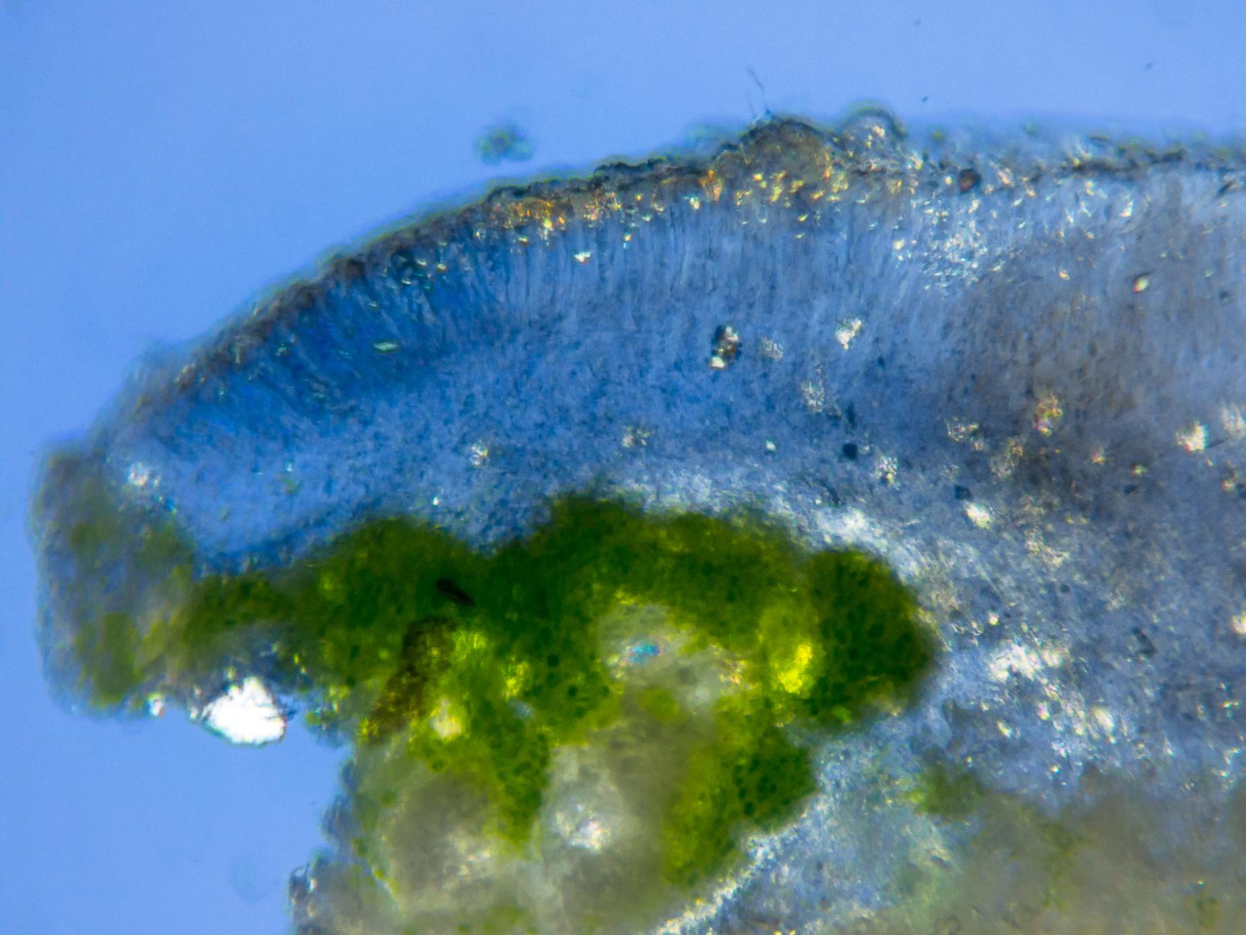
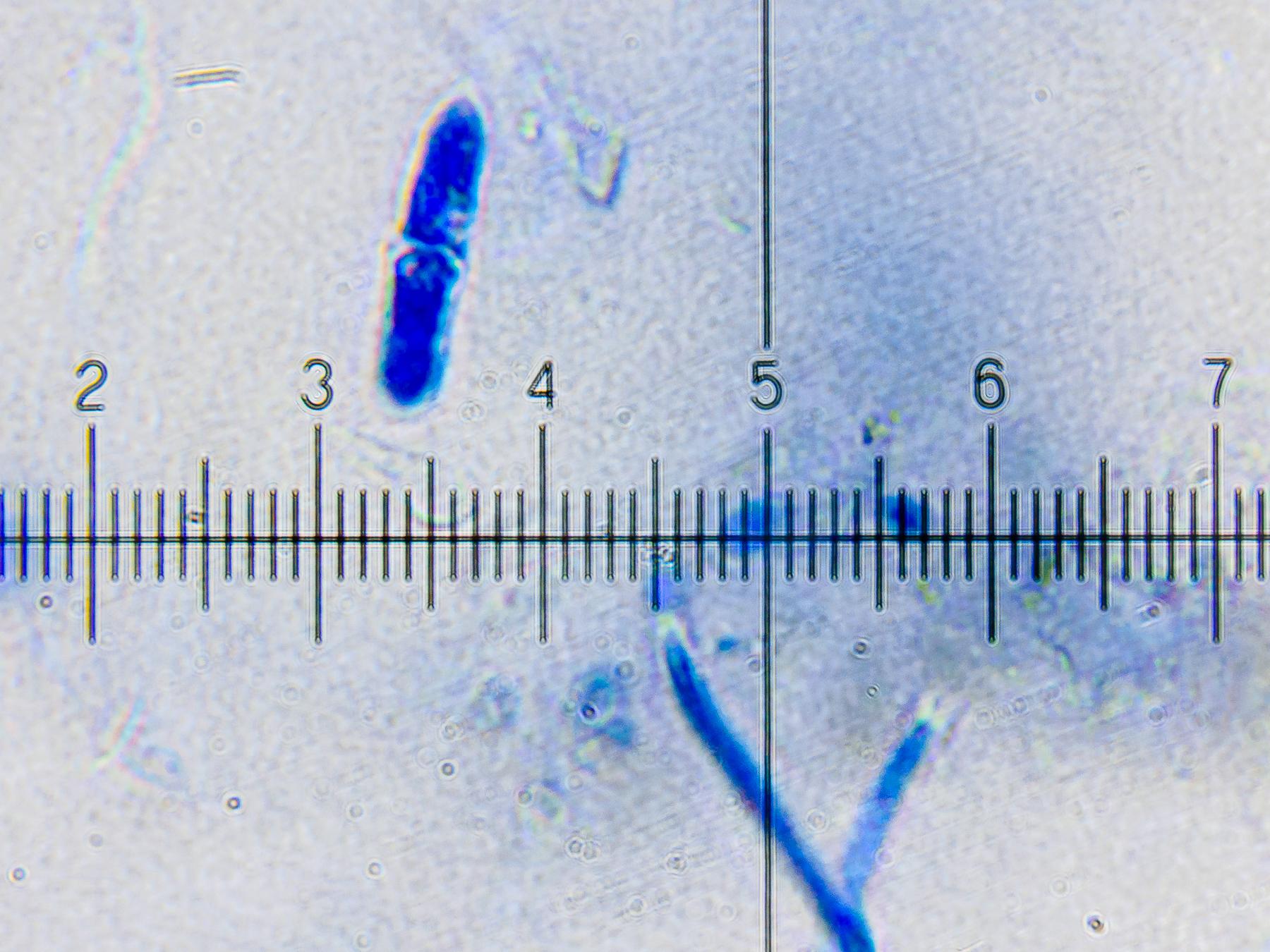
When I first came across this fungus in 2020 I had no way of knowing whether it was obligately lichenicolous or a general saprobe. I didn’t share details of it beyond posting a thread about it on twitter. Recently, two further occurrences, suspected to be the same organism, have been reported on L. hybocarpa. In the field, infections are quite conspicuous due to the darkened discs of infected apothecia (but note that other fungi may cause darkening of similar appearance). Sections of affected apothecia reveal that the hymenial tissue of the host is very degraded, largely replaced by hyphae belonging to the LF (lichenicolous fungus). The infection is confined to the host’s hymenium and does not extend into the thalline margin. Colourless, bacilliform conidia are produced at the surface of the infected hymenium. The conidia are 1-septate when mature and up to about 4 x 18 µm. The following images will provide a good impression of the fungus. Note that in the line drawing the host is named as
L. chlaroterabut it has been subsequently redetermined as L. hybocarpa. Does anyone recognise this fungus? I anticipate that further details of other specimens will be posted in response to this post. 
[attachment=6]EOASymzXUAE_Wo5.jpeg[/attachment] 
[attachment=5]EOATjlJX4AMAUy5.jpeg[/attachment] 
[attachment=4]EOAURJDWsAINUZs.jpeg[/attachment] 
[attachment=3]EOAUwmRX0AEDLuf.jpeg[/attachment] 
[attachment=2]EOAUwmRWsAINCiI.jpeg[/attachment] 
[attachment=1]EOAVb3oXUAAL-tf (1).jpeg[/attachment] 
[attachment=0]EOAV0cCXsAAfyD4 (1).jpeg[/attachment] November 11, 2023 at 12:18 pm #45728Hello Mark I haven’t seen this before. There are precious few characters to go on, but it would be identified as an Acremonium, I suppose, many of which are anamorphs of Hypocreales. The anamorph of Pronectria anisospora (Acremonium pedatum) fits in terms of spore size, but I can’t find any record of a Pronectria or similar associated with Lecanora.
Sorry, that’s the best I can do…
Paul
November 11, 2023 at 1:31 pm #45729Many thanks for starting this forum topic, Mark. Last month I found ostensibly the same mould-like anamorphic conidial fungus in a specimen of
Lecanora hybocarpafrom near Lavenham, Suffolk (VC26/TL921498). The thallus is on I think a Corylustwig — the twig was broken and could have come from another tree, but it was well caught in a hazel at the edge of an arable field, and most of the surrounding trees seemed to be hazel. As with Mark’s specimen, the LF shows up as a red staining and ‘rotting’ in the apothecia. It doesn’t seem to affect the thalline margins or the thallus more generally.

[attachment=9]20231005-PA050262.jpg[/attachment] Even apothecia that seem relatively unblemished are affected. In this section, mounted in water in cross-polarised light, a reddish stain appears within the hymenium. The LF seems to have inhibited or degraded the normal development of the apothecium. The descending POL+ epithecial granules characteristic of
L. hybocarpaare missing, except in the ‘healthy’ part of the apothecium. Asci with spores are visible, but again only in the healthier part. Where the reddish stain is present, there seem to be no asci at all. The LF seems to have inhibited or destroyed them as well. 
[attachment=8]20231005-PA050265.jpg[/attachment] An overview section of a visibly fully parasitised apothecium, again mounted in water, shows that the reddish stain of the LF dominates the fruiting body. No asci are seen at all (nor POL+ epithecial granules, not shown here due to the limit on the number of photos per post).
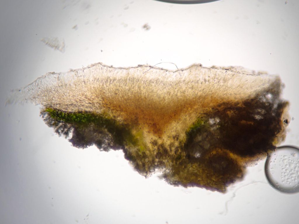
[attachment=7]20231005-PA050268.jpg[/attachment] However, the LF’s hyaline conidia can be made out peeping about the surface of the apothecium (a different apothecium than the one above). As with Mark’s specimen, the conidia are 1-septate (1 div = 1 µm).

[attachment=6]20231110-PB100790.jpg[/attachment] Clearing and staining in K-ink-vinegar brings out the LF’s hyphal network. Again, the LF appears confined to the hymenium of host apothecia, seemingly drawing its nutrients from the hymenial tissue, not the photobiont. Clearing and staining also make obvious that the host’s asci and paraphyses are nearly entirely inhibited or degraded or destroyed. Essentially only the LF’s own hyphae remain (1 div = 10 µm).

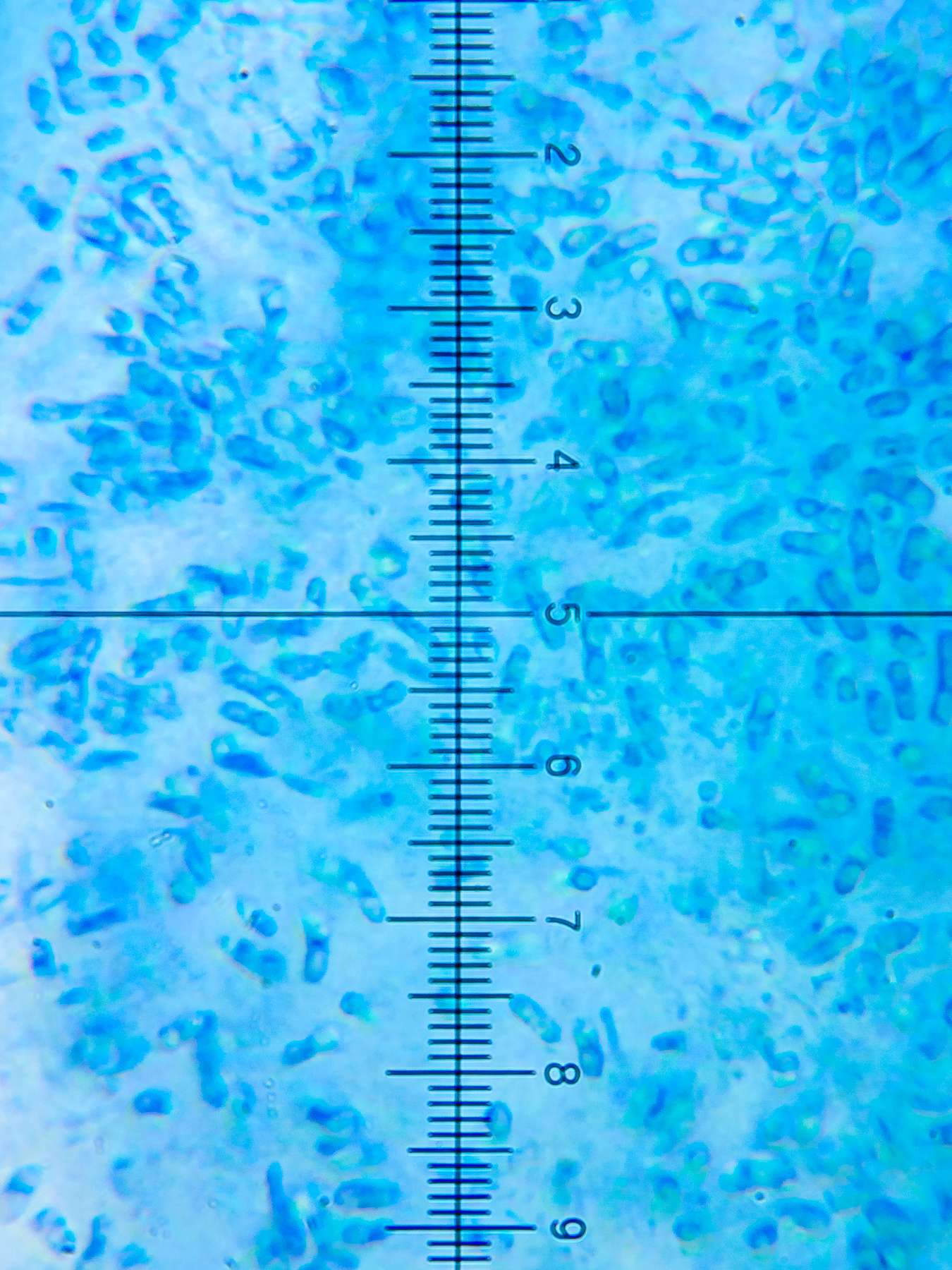
[attachment=5]20231110-PB100793.jpg[/attachment] The LF’s hyphae appear mostly to be trending towards the surface of the host apothecium to sporulate. Developing conidia and perhaps also phialides are visible near the 5 to 8 marks on the scale (1 div = 2.5 µm).

[attachment=4]20231110-PB100797.jpg[/attachment] The hyphae themselves are hyaline as the unstained images above show. They seem to be roughly 3-4 µm wide, including a roughly 0.5 µm cell wall. It’s hard to get a sense of the length of each hyphal cell, as the hyphae seem to twist. They don’t seem to branch too much, though (1 div = 1 µm).

[attachment=3]20231110-PB100800.jpg[/attachment] A close look at free and developing conidia shows that fully mature spores are roughly 13-16 µm in length and 4-5 µm in width, including a roughly 0.5 µm cell wall — in line with the size of the conidia in Mark’s specimen (1 div = 1 µm).

[attachment=2]20231110-PB100803.jpg[/attachment] 
[attachment=1]20231110-PB100806.jpg[/attachment] 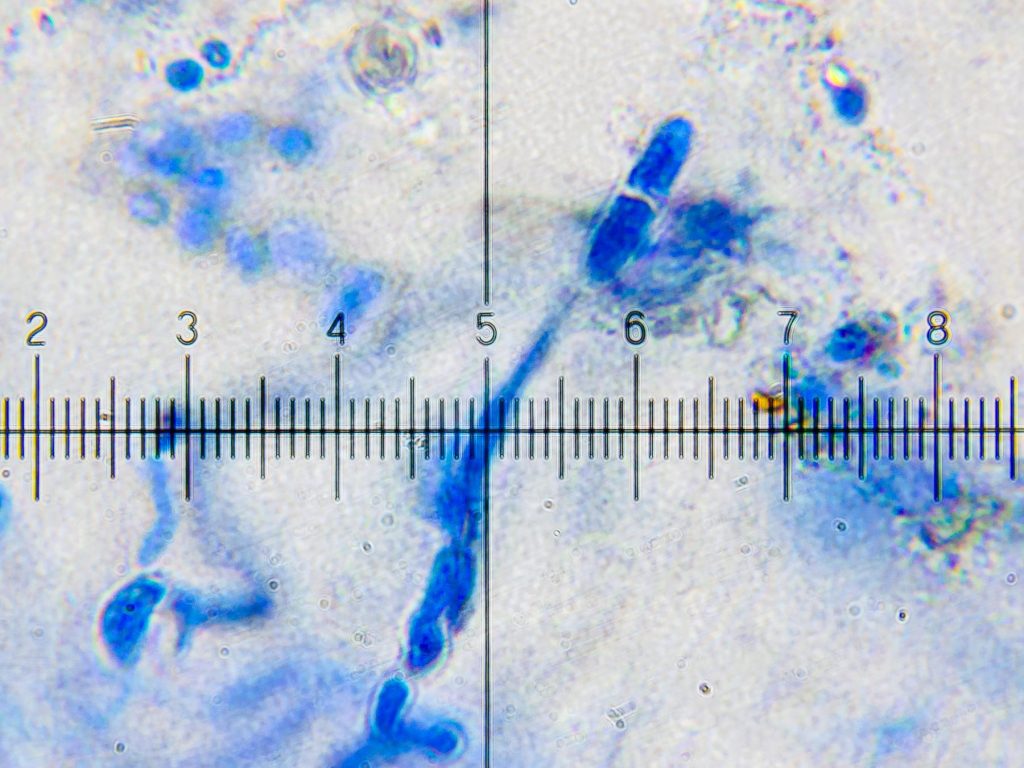
[attachment=0]20231110-PB100809.jpg[/attachment] Apart from this thallus, there are other
L. hybocarpathalli from the same twig that appear to be similarly infected. I also have a more recently collected L. hybocarpaspecimen from near Glandford, Norfolk (VC27/TG037408) that I think might also host this LF. Many thanks, Paul, for your suggestion of
Acremonium pedatum/ Pronectria anisospora. Acremoniumhas come up in the Twitter discussion, which has also brought forward additional finds that others may wish to post here. Grateful for any further advice about next steps towards identifying or describing this LF.
November 11, 2023 at 3:11 pm #45730Following up on Paul’s suggestion of Acremonium/Pronectria, I’ve found that there has indeed been at least one Pronectriaspecies very recently described on Lecanora. Valerii Darmostuk (2021) describes Pronectria gromakovaeas new to science from an occurrence on Lecanora populicolain the region of Kharkiv, Ukraine. I’m not sure how strongly this bears on the LF described here, though. The details are available open-access from Lindbergia— https://doi.org/10.25227/linbg.01141https://doi.org/10.25227/linbg.01141” class=”bbcode_url”> November 11, 2023 at 3:40 pm #45731Following the above posts on Twitter, I had a look at some reddened Lecanora hybocarpa apothecia on a Prunus spinosa twig that I’d already been looking at for other lichenicolous fungi. The first reddened apothecium that I sectioned had similar 1-septate spores with conspicuous oil droplets and a small bump on the end where they had been attached.Free conidia were 15-18 x 3.5-4.5 μm. Others still attached to the conidiogenous cells in the hymenium were shorter. January 12, 2024 at 10:04 pm #45732I received an email from Brian C. yesterday regarding this LF on Lecanora hybocarpa. He writes: “When you sent the message & link below, I thought “I know that thing”, but don’t seem to have made a specimen. I picked up a cherry branch in the local park the other day, and sure enough I found it, so will make a specimen of it this time. Similar, moribund L. ‘hybocarpa’ also frequently has the asteroconidia of Tremella macrobasidiata.
As for the oblong conidia on L. hybocarpa, I suggest recording it for the BLS Database as Acremonium [sp.], with a note on conidia size/septation. Hopefully it won’t be too long before we have a real name for it.”
January 13, 2024 at 12:16 pm #45733Many thanks, Mark, for bringing this LF to the attention of Brian C. It’s exciting to hear that he recognises having seen it before, and that he was easily able to find a likely specimen. It’s also good to know that our finds can be more publicly disseminated via the BLS database as Acremonium sp, at least until a proper name is found. I’ve emailed Brian, and copied you, bringing some further micrographs to his attention. These micrographs relate to the Norfolk specimens that I found, one of which fairly clearly seemed to be our hyphomycete LF, and the other of which may or may not be. Perhaps the additional evidence will help with identifying the LF — and at the very least, Brian may discount it as relating to a different LF, which would be equally valuable.
I sent him the micrographs by way of a link to the presentation about this LF that I recently gave to the BLS Lichen Chat and Improvement Group. Having published that link on Twitter some time ago, it occurred to me that I should just as well have done so here! —
https://drive.google.com/file/d/1bTuHFgsKoJo_XnTPlgr-qm7Kz1vs23tf/view January 13, 2024 at 3:19 pm #45734I have found and confirmed two further occurrences of our Acremonium on L. hybocarpa in the past couple of weeks (North Bedfordshire). It seems to be easy to find it in an identifiable state at the moment so if anyone is interested in finding their own specimens, this is a good time to look. Some lichenicolous fungi seem to be seasonal, others periodic in a less predictable way and this Acremonium may be an example of the latter. Candidates are easily noticed in the field as the dark reddish-brown slightly degraded discs mean that the infections form conspicuous patches. Microscopy is required for confirmation. I keep hoping to find the Tremella macrobasidiata which Brian mentions and which supposedly causes similar discolouration of the discs, but no luck so far. July 25, 2024 at 11:45 am #45735I have just been looking at another sample of Lecanora hybocarpa with reddened apothecia, on an Ash trunk: 
[attachment=5]IMG_9969.jpeg[/attachment] This heavily affected apothecium had Acremonium-type conidia as described above, and swollen hyphae within the hymenium.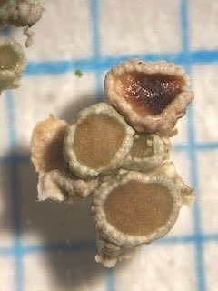
[attachment=4]IMG_9970.jpeg[/attachment] 
[attachment=1]IMG_9989.jpeg[/attachment] I then sectioned a second apothecium (to check that they were L hybocarpa, using polarised light, as the first one had no epithecial crystals at all). This one only had a limited reddened patch. The non-reddened part had descending epithecial crystals, but the reddened part did not, nor did it have healthy host asci in that part.

[attachment=3]IMG_9973.jpeg[/attachment] 
[attachment=0]IMG_9981.jpeg[/attachment]
In this apothecium, I didn’t find any conidia like the above, but there were two windmill-like conidia (?), each with three long arms, c 25 x 2 um. Thought it might be worth adding the photo here, to see whether anyone else has seen anything similar. I haven’t yet managed to find any information about what they might be. Maybe the reddening is not related to these ‘windmills’.
[attachment=2]IMG_9997.jpeg[/attachment] July 31, 2024 at 1:34 pm #45736Thanks, Di. It’s great to see that you’ve observed Acremonium-type conidia once again in the same sort of reddened L. hybocarpa auct. br.apothecia. My own observations were in October last year, and I was waiting until the autumn to resume investigations in case the sporulation was seasonal. Even so, I’ve seen the reddened apothecia numerous times so far this year — though I suppose the same discolouration can be caused by other LFs as well. Like you, I’ve observed partial degradation of infected apothecia, with POL+ granules remaining in the healthy tissue. (See my post of 23/11/23 above for a photo.)
But I’ve never seen the windmill-shaped conidia that you’ve observed. Could they be the asteroconidia of
Tremella macrobasidiatathat Mark Powell cited Brian Coppins as having mentioned, in his post of 12/01/2024 above? Or of a related Tremellaspecies? It seems that Tremellaalso causes discolouration of the apothecial disc. I believe T. macrobastidiataasteroconidia might have four arms rather than three, but perhaps there’s some variation. The protologue for
T. macrobastidiatais here: https://www.cambridge.org/core/journals/lichenologist/article/abs/tremella-macrobasidiata-basidiomycota-tremellales-a-new-lichenicolous-fungus-from-the-iberian-peninsula/D1F9462B30AAB676E913E82B70415AF4 ” class=”bbcode_url”> https://www.cambridge.org/core/journals/lichenologist/article/abs/tremella-macrobasidiata-basidiomycota-tremellales-a-new-lichenicolous-fungus-from-the-iberian-peninsula/D1F9462B30AAB676E913E82B70415AF4 Although I’ve never seen asteroconidia of any sort, I happen to have seen ‘dumbell-shaped’ conidia in reddened
L. hybocarpa auct. br.apothecia. Perhaps they come from yet another LF in addition to the putative Acremoniumand the T. macrobastidiata. Some micrographs below (ignore any ‘ Acremonium‘ label on the image files if you can see it, as for now I keep the images in the same photo folder as micrographs of our putative Acremonium). 
[attachment=2]Acremonium sp 20231027B-14 Glandford.jpg[/attachment] 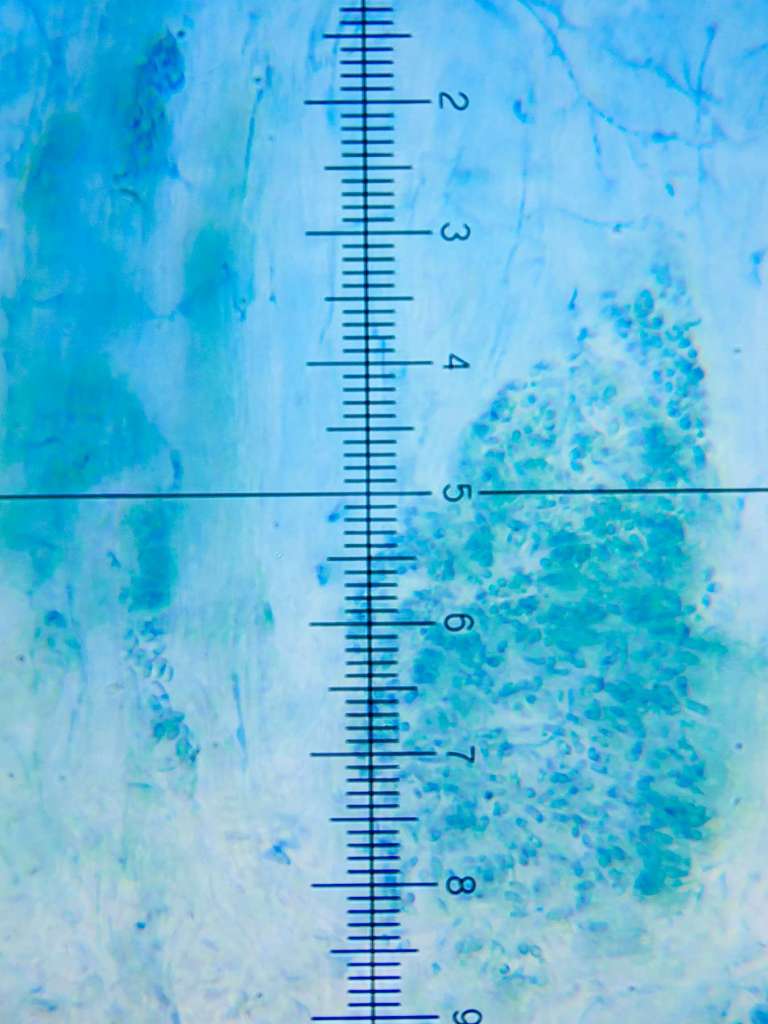
[attachment=1]Acremonium sp 20231027B-15 Glandford.jpg[/attachment] 
[attachment=0]Acremonium sp 20231027B-16 Glandford.jpg[/attachment] Best,
Anthony
August 5, 2024 at 1:14 pm #45737Hello Di (Chalkie) – just a thought but did you consider a Cornutispora for your ‘windmill-shaped’ conidia? https://lichenportal.org/portal/taxa/index.php?taxauthid=1&taxon=Cornutispora&clid=201 ” class=”bbcode_url”> https://lichenportal.org/portal/taxa/index.php?taxauthid=1&taxon=Cornutispora&clid=201 -
AuthorPosts







- You must be logged in to reply to this topic.