
Yellow fungus, with spore photo!
Forums › Forums › Fungi ID requests (post here if you aren’t sure what type of fungus you’ve found) › Yellow fungus, with spore photo!
- This topic is empty.
-
AuthorPosts
-
July 27, 2015 at 10:52 am #29576
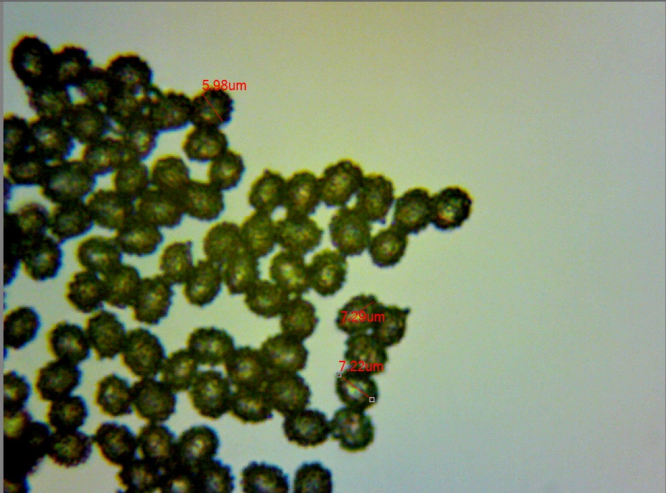
Found on the lawn, near Oak, Rhododendron, Weeping Willow, Beech. My first ever spore photo:
[attachment=1]Site002 – Fungus001 – Spores002 – measured- 27th July 2015.jpg[/attachment] 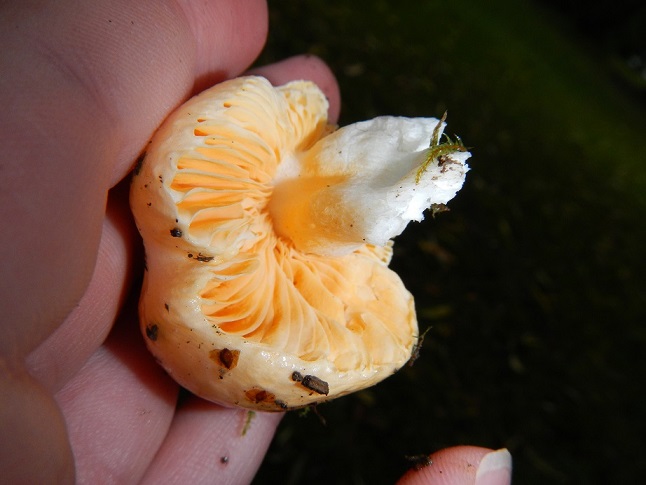
[attachment=0]DSCN5518 r.jpg[/attachment] July 27, 2015 at 12:01 pm #35448Hi Jimmy, You always need to provide a good sharp photo of the top of the cap as well.
PS: you say it was yellow? – The colour looks decidedly orange on my screen (but it looks right – i.e. it doesn’t look like a colour cast on the photo or trick of the light etc.) can you confirm if the colour was definitely yellow?
Regards,
Mike.
July 27, 2015 at 2:52 pm #35449Hi Mike, Sorry, I was rushing! The cap and gills were a mix between both yellow and orange and the spore mass was orange.
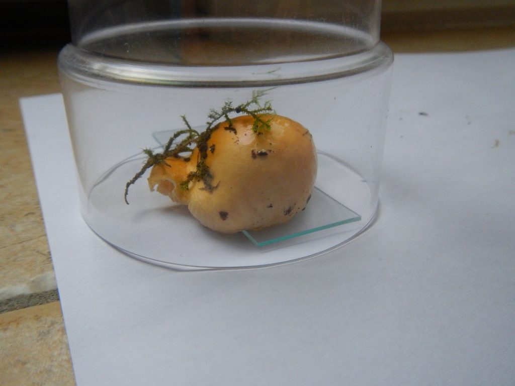
[attachment=0]DSCN5522 r.jpg[/attachment] July 27, 2015 at 3:04 pm #35450Hi Jimmy, I’ve got a feeling it will turn out to be something obvious – but I’m stumped.
I’m assuming that the cap was quite viscid/sticky with the debris that’s stuck to it.
Those spores look very distinctively warty, and with an orange spore print, that must narrow things down.
Regards,
Mike.
July 27, 2015 at 3:06 pm #35451Hi Mike, The cap was very slimy and sticky and the spores did strike me as being very warty (but I don’t see spores that often anyway!).
James
P.S. Do you have any idea what group it is? That might help me help to identify it!July 27, 2015 at 3:56 pm #35452The Genus is Russula. July 27, 2015 at 4:00 pm #35453Thanks Roy, I’ll get searching! July 27, 2015 at 4:26 pm #35454You need to check how much the cap cuticle peels and whether the taste (of the gills) is mild or hot. Spore colour narrows the field considerably (which is good as there are well over 100 British Russula spp…. and counting). All Russula spores are warty to some extent: the size of spores and height of spines/warts is useful info. and whether the spines are isolated or joined (either in short ridges or more extensively, even forming a network). Then there are the reactions of the stipe to Iron Sulphate and Guaiac…… and so on. With the limited info. I’m guessing, but it could be around
risigallina/acetolens. July 27, 2015 at 6:04 pm #35455Russulas are also known as “brittlegills” – they are mostly quite chunky and have white or whitish gills and stipe and the gills are often brittle. You can recognise them instantly after a while. Looking at the spores is best done in Melzers reagent, and try to mix them up a bit with a needle so you get separate spores.
Photos through glass jars are not ideal. A top shot in its habitat is preferable.
Having said all this, Russulas are quite tricky but with the aid of Kibby’s British Russulas, it could be a way to start as the microscopy only really involves looking at the spores and the cap cuticle, although you will need special stains to see the cap cystidia really clearly.
cheers, Adam
July 28, 2015 at 10:59 am #35456The taste is mild, and I’ll measure the height of the spines/warts this afternoon. July 29, 2015 at 7:42 am #35457The height of the warts/spines was around 0.6 µm. -
AuthorPosts




- You must be logged in to reply to this topic.