
Xerocomus species?
Forums › Forums › Fungi ID requests (post here if you aren’t sure what type of fungus you’ve found) › Xerocomus species?
- This topic is empty.
-
AuthorPosts
-
October 3, 2025 at 5:50 pm #33092
Another request for help. I photographed these fungi yesterday in the suburbs of Rugby. They were within a few metres of one and other in an area of mown grassland (verge) between the pavement and a laurel hedge. I initially assumed all three were the same species due to their close proximity but now suspect it may involve two species. I tentatively identified the first image as Xerocomus rubellus. This was a group of five very close fungus. The cap diameter of the majority was about 4-5cms but as the image shows the largest was about 10cms. All had a reddish stipe and yellowish pores. The second image shows two effectively merged together. The stipe is pale reddish, yellowing at the top as it merges with the pores. The diameter of the cap is less obvious due to the merging but again probably 5cms. Although the cap was not cracked I wondered on reflection whether this may be Xerocomus chrysenteron? The final image was the largest fungus with a cap diameter about 12-14cms and I suspect is a more mature version of the species shown in image one. As always I would really appreciate any comments and thoughts. 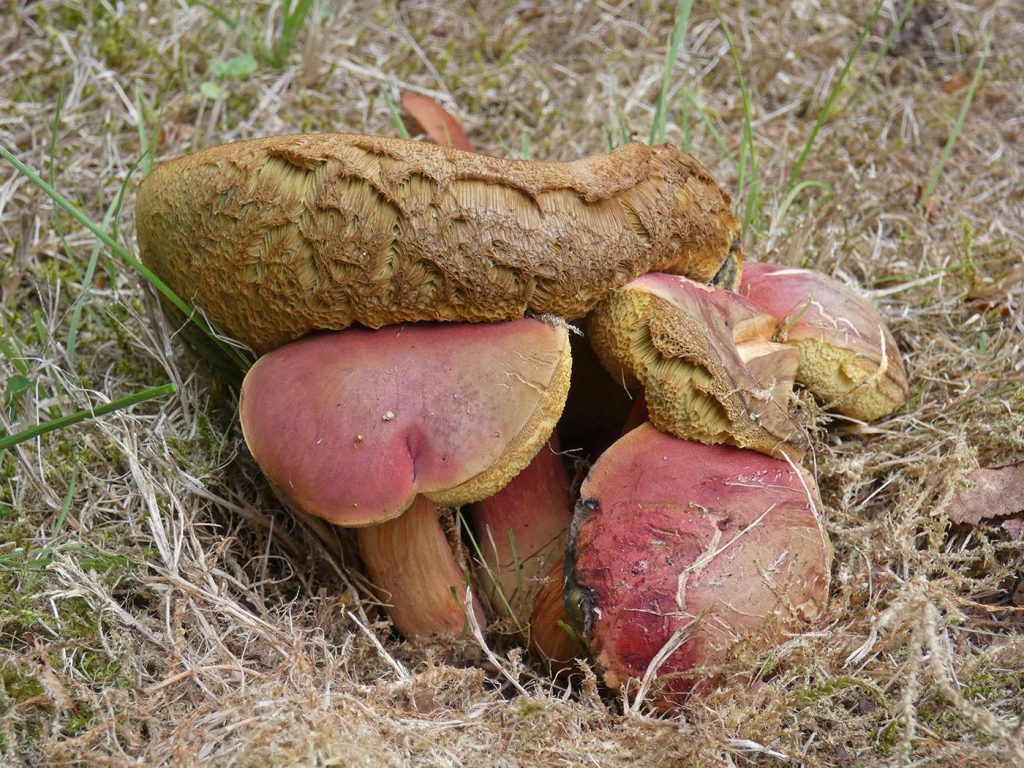
[attachment=2]1Xerocomus rubellus(1),Rugby.jpg[/attachment] 
[attachment=1]1P1910498.jpg[/attachment] 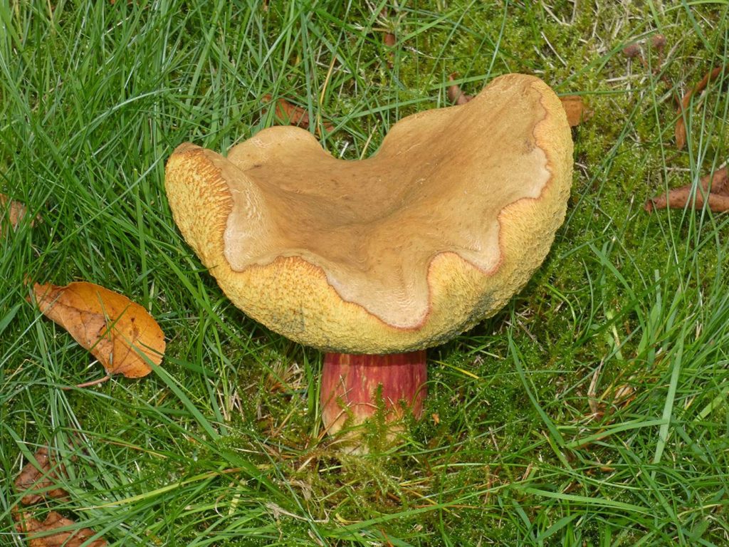
[attachment=0]1P1910519.jpg[/attachment] October 4, 2025 at 11:40 am #46696Hi Bob, I think you are probably correct with your ID of the first as
Xerocomus rubellus. (Now Hortiboletus rubellus). I don’t think you have
X.chrysenteronfor the second. – I think that one is showing more of the macro characteristics of Xerocomellus porosporus. The cap crazing showing in your photo is very typical of that species (and will become more properly “cracked” as the caps age).
There seems to be some differences of opinion (within ID guide books etc.) as to the extent of the reddish area found on the stipes of
X. porosporus. Some literature suggests that the red area will be a fine band near the apex, some will indicate quite an extensive area of red in the top half of the stipe, and some will indicate that the stipe below the red areas will be sepia toned.
All references seem to agree about the thin yellow band that will be seen above the red area, at the very apex of the stipes.
I’m not sure about the final one. I don’t think the cap is showing what would be expected of a fully mature
X.rubellus, but (for me) there aren’t enough macro characteristics showing to hazard a guess. As with almost all boletes, cutting the examples in half through the cap and down the stem can give some good indicators such as what the inner flesh looks like, any internal red areas etc, and whether the sample “blues” or not, which areas, if any, do turn blue, and how long it takes to turn blue, etc. etc.
And, as I do with most ID reply suggestions, I’d add the usual caveat that microscopy would give the most certainty when macro characteristics alone leave some doubt.
Regards,
Mike.
October 4, 2025 at 12:23 pm #46697Many thanks for your detailed reply and caveats Mike, it is much appreciated. As a complete novice I love the spectacle that fungi bring, but am somewhat overwhelmed by the regularity with which they are reclassified as they are further studied. Thanks again. Regards,
Bob
-
AuthorPosts


- You must be logged in to reply to this topic.