
Vocabulary
Forums › Forums › General fungi discussion › Vocabulary
- This topic is empty.
-
AuthorPosts
-
June 9, 2015 at 6:23 am #29452
As you probably will have noticed, I’m new in this field of natural history! Here’s some vocabulary I would quite like to learn the meaning of: 1. Basidia
2. Cystidia
3. The ‘sections’, which you put under the microscope. For example, are these cross sections? Of the stem, cap, gills etc.?
4. Reagents
5. ‘light’ microscopy
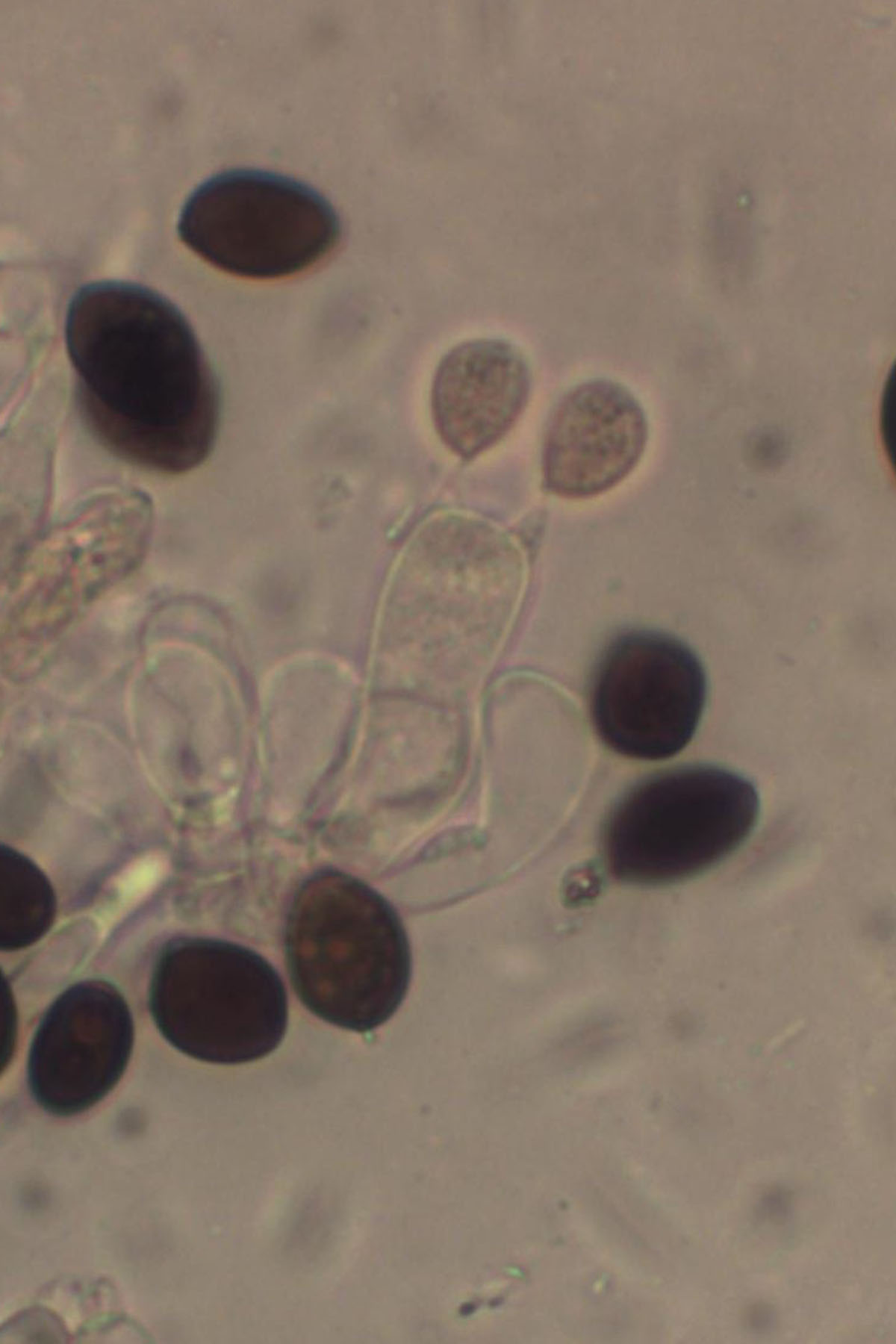
June 9, 2015 at 9:04 am #34895Hi The basidia is the structure that the spores grow on in the Basidiomycetes (most of the “mushrooms”) that we talk about and they look like a hand with usually four fingers not the best photo but it shows the spores
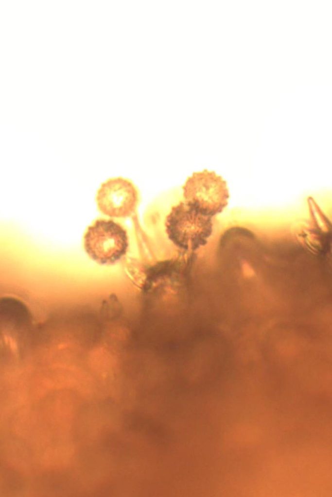
[attachment=4]Basidia (2)a.jpg[/attachment]
They can also have 1 2 or 3 fingers and this can be an aid in identifying some species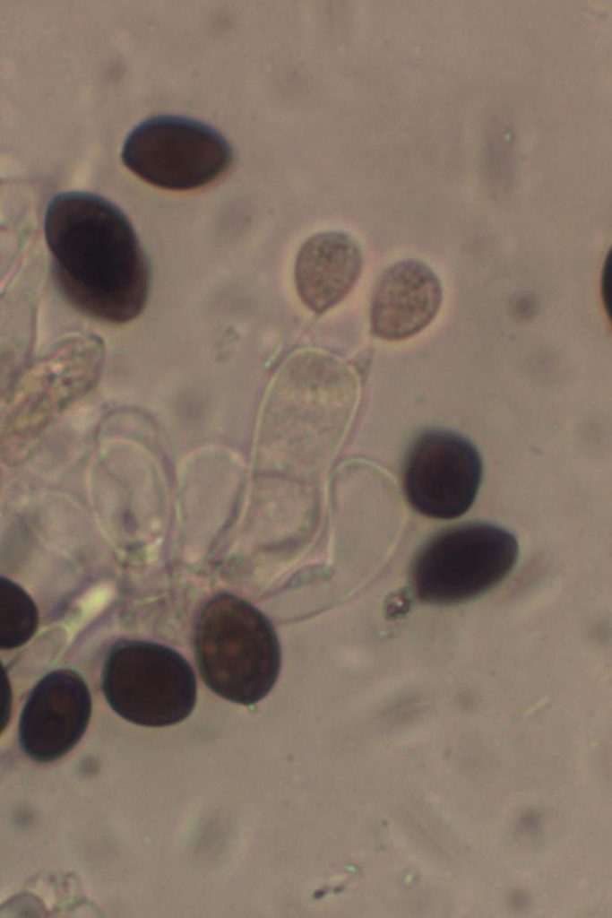
[attachment=3]basidia 2.jpg[/attachment]
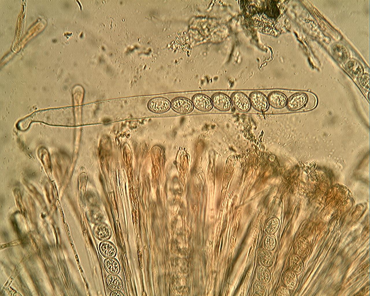
The other big group collected are the Ascomycetes and they tend to be much smaller and include groups such as Peziza and Scutellinia and they grow their spores in an tube like structure called an Ascus usually with 8 spores to each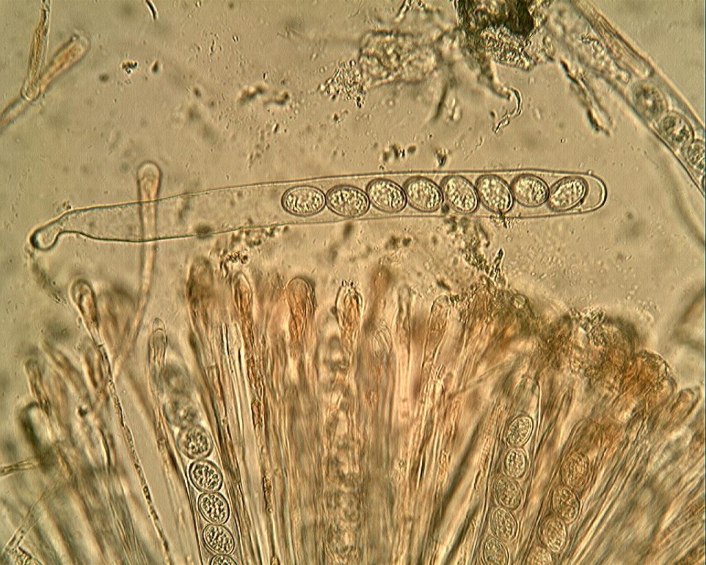
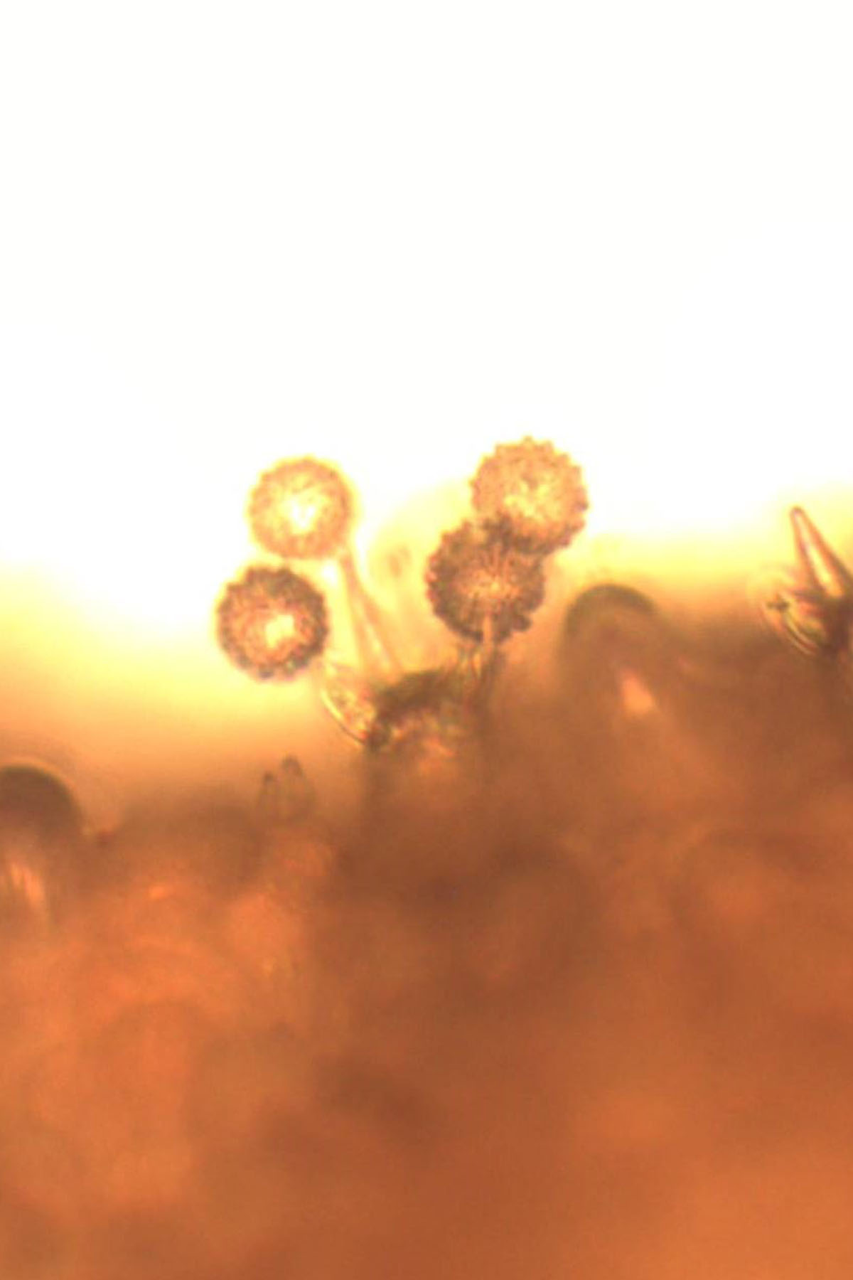
[attachment=2]ascus2.jpg[/attachment] The cystidia is part of the microscopic make up of the Basidiomycetes. They can sometimes be found on the edge of the gills (cheilocystidia) on the face of the gills (pleurocystidia) on the cap (pileocystidia) and on the stem (caulocystidia). Whether they are present and their shape and size is again an important feature in some group such as Mycena.
The sections can be of any or all parts of the fungus depending on the family. In Mycena the cheilocystida is one of the main methods of splitting some species. They can be all sorts of shapes two of which are shown hear
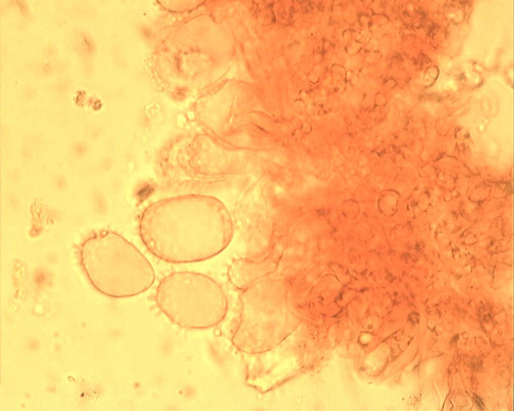
[attachment=1]cheilocystidia.jpg[/attachment] [attachment=0]cheilocystidia.jpg[/attachment]
The stains, reagents and microscopy are discussed elsewhere in the forum.That’s your starter for 10.
There are books out there giving much more information on how examine fungi with a microscope but you then need the mycology books that give you the microscopic details of each fungus.
Mal
June 9, 2015 at 9:44 am #34896Excellent post, Mal. I’ve also posted a sticky on literature and resources on general fungi, not covered in a sub-forum. No doubt it needs adding to! June 9, 2015 at 1:29 pm #34897I would again refer you to the microscopy primer/tutorial mentioned here: – 
http://www.fungi.org.uk/viewtopic.php?f=5&t=108 You will find that it contains the answers to your questions here, and a great deal more information too!

Regards,
Mike.
June 10, 2015 at 6:34 pm #34898Excellent information. Details like this encourage new starters into the world of fungi. -
AuthorPosts




- You must be logged in to reply to this topic.