
Trochila ilicina (was Microscope for spore examination)
Forums › Forums › Ascomycetes › Trochila ilicina (was Microscope for spore examination)
- This topic is empty.
-
AuthorPosts
-
July 25, 2015 at 3:33 pm #29574
Here’s a photo of Trochila ilicina, taken with the eye cam on ToupView, using an IKEA desk lamp as the illumination: 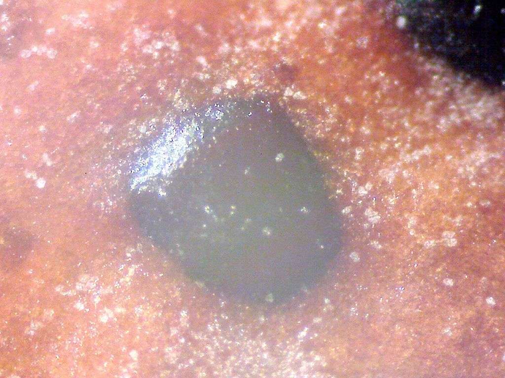
[attachment=0]Trochila ilicina (Holly Speckle) 25th July 2015 t3.jpg[/attachment]
It’s a terrible photo, but that’s probably because it’s my first!!July 25, 2015 at 8:10 pm #35422Hi That doesn’t look quite right for
Trochila ilicina; more likely a ‘coelomycete’ perhaps a Coleophoma. Checking the ‘innards’ would help . . . . regards
Chris
PS
Trochila ilicinaportrait here: http://www.ascofrance.com/search_recolte/3594http://www.ascofrance.com/search_recolte/3594” class=”bbcode_url”> July 26, 2015 at 7:12 am #35423That’s strange… Have a look here:
http://www.ispotnature.org/node/704000 July 26, 2015 at 7:46 am #35424Check this page by Chris Johnson (a respected mycologist) – you can see distinct “eye-shaped” apothecia. To be honest, it looks different to both your fungus and the plate by Chris  July 26, 2015 at 1:07 pm #35425adampembs wrote:
Check this page by Chris Johnson (a respected mycologist) – you can see distinct “eye-shaped” apothecia. To be honest, it looks different to both your fungus and the plate by Chris
http://www.outerhebridesfungi.co.uk/species.php?id=363
OK . . . .now I have seen all three photo’s I agree, this looks like
the very early stageof Trochila ilicinaquite a while before the “lids” open; seeing a single apothecium misled me. I must have a word with Chris J.

Phacidium multivalve, another very common species on Ilex, probably the anamorph: 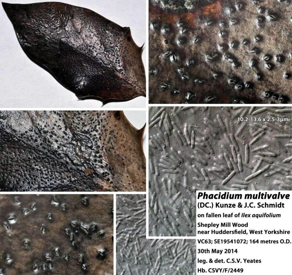
[attachment=0]Phacidium multivalve.jpg[/attachment] (note the rod-shaped conidia)Cheers
Chris
PSThis is not, of course, in any way intended to impugn Chris Johnson’s excellent website, which has many splendid images of Hebridean fungi, including many species without similar images on the Web. The portraits of mosses are of extremely high quality as well. July 26, 2015 at 4:51 pm #35426Hi Guys Just been tipped-off by Chris Y about your new forum – news filters through very slowly to the Outer Hebrides. I hope it’s a great success and wish all those involved the best of luck.
Thanks for the nice comments regarding my website – pity it’s centred around getting one wrong
😳 Out of action for 3 months while work is carried out on extending my study area but hope to be active again by mid-August.
Regards, Chris
July 26, 2015 at 6:04 pm #35427Welcome to the forum Chris, sorry word didn’t get to you about the forum. I guess you weren’t on Lancashire Lad’s list! I had no idea your ID was wrong, you never know with these plant diseases if it is a certain stage, or morph!
July 26, 2015 at 9:51 pm #35428Hi Chris, Good to see that you’ve found the forum!
As Adam has suggested, When WAB went kaput, I didn’t have any contact details for you, (along with quite a few others), so was unable to get the word out to everyone.
Regards,
Mike.
July 27, 2015 at 6:54 am #35429Hi Mike Much as I thought. Didn’t think I’d been black-listed (yet).
Regards, Chris
July 27, 2015 at 8:01 am #35430posts moved here as subject has changed… July 27, 2015 at 7:56 pm #35431Rather than people having to move to an external website here is the ‘portrait’ (fungi.org.uk gives a bigger image in any case – toggle F11 and it gets even bigger 😳 ):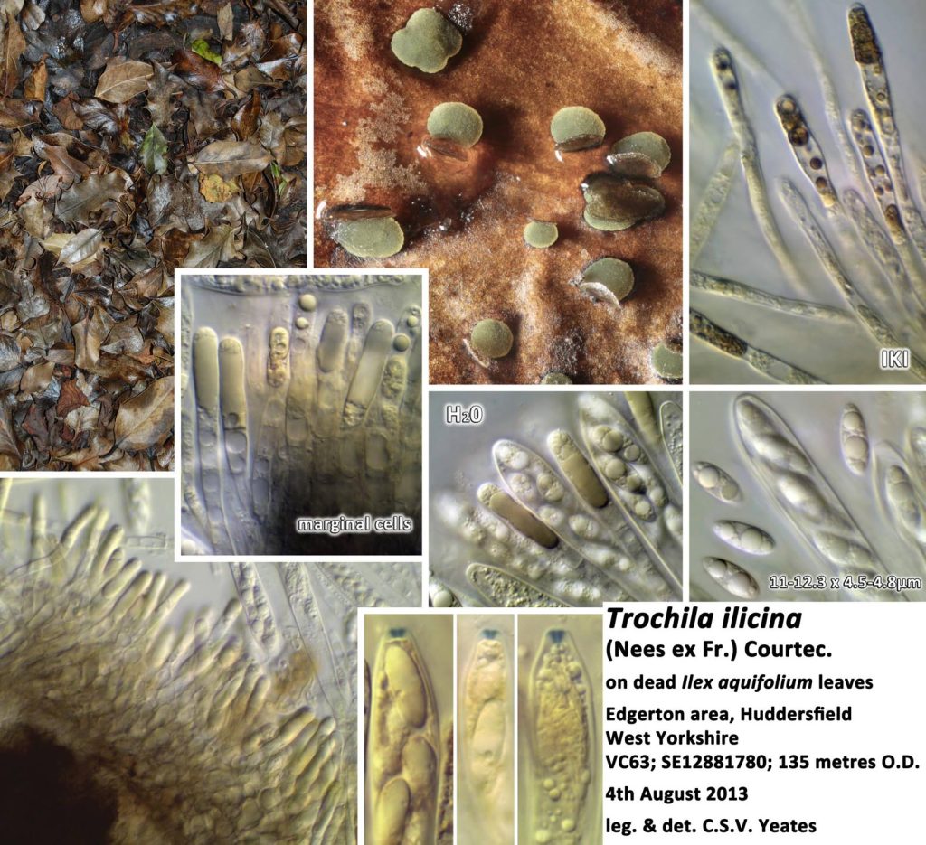
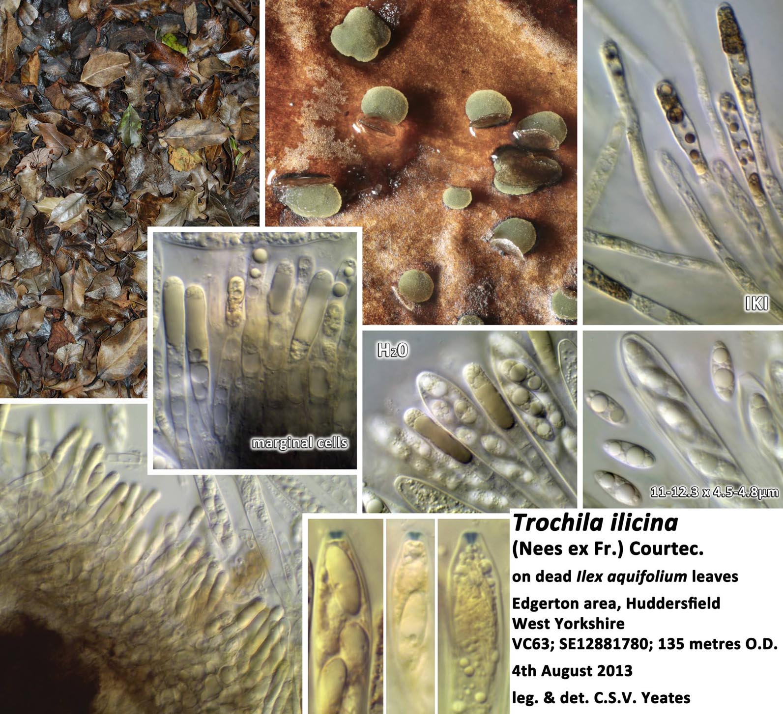
[attachment=0]Trochila ilicina.jpg[/attachment] note the pale olive-green contents of the paraphyses and marginal cells which give the exposed apothecia their characteristic colour. Also note how mounting in IKI (Lugol’s Iodine), while useful for showing positive or negative ascus tip reaction, destroys those paraphysis contents and also loses the distinctive guttules of the spores. This is why it is important, whenever one can, to first examine living material in water – Zotto Baral’s “Vital Taxonomy“: http://www.gbif-mycology.de/HostedSites/Baral/http://www.gbif-mycology.de/HostedSites/Baral/” class=”bbcode_url”> Chris
-
AuthorPosts


- You must be logged in to reply to this topic.