Resupinate (was "Pink, crusty Lichen")
Forums › Forums › Crust fungi, Bracket Fungi etc › Resupinate (was "Pink, crusty Lichen")
- This topic is empty.
-
AuthorPosts
-
August 7, 2015 at 5:24 pm #29612
Found on a Weeping Willow branch, quite high up. 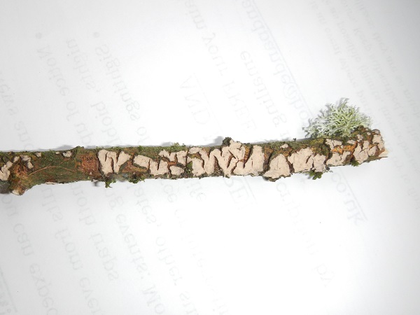
[attachment=0]RSCN6372.jpg[/attachment] August 7, 2015 at 6:50 pm #35619Hi Jimmy The image isn’t too clear but you might want to consider a resupinate rather than a lichen.
Regards, Chris
August 8, 2015 at 10:18 am #35620Is a resupinate a crust fungus? August 8, 2015 at 11:12 am #35621jimmymac2 wrote:Is a resupinate a crust fungus?
Yes, resupinates are part of the Corticioid fungi group.
I don’t think yours is too far from
Peniophora incarnatabut some microscopy would be require to be certain. August 8, 2015 at 11:39 am #35622What would I need to look at under the microscope and how should I prepare the specimen? August 8, 2015 at 1:08 pm #35623jimmymac2 wrote:What would I need to look at under the microscope and how should I prepare the specimen?
First you need to make a spore print. Take a piece of the twig and place it over a glass slide raised on two matchsticks (or something similar), cover it to retain moisture and leave it over night.
This group belongs to the basidiomycetes so you are looking for basidia, type of hyphae, clamps, cystidia in their various forms, crystals, etc.
Moisten a piece of the fungus in water for a few minutes (or longer depending on how hard and dry it is) then take a tiny piece with tweezers and place on a slide with water. The other method requires some skill, only acquired with practice. That it taking a very thin traverse section with a razor-blade.
Good luck, these are not the best fungi to begin with.
Chris
August 8, 2015 at 1:15 pm #35624Thanks Chris, I’ll give it a go. Don’t expect great results though!!  August 8, 2015 at 1:25 pm #35625jimmymac2 wrote:
Thanks Chris, I’ll give it a go. Don’t expect great results though!!
It will be good practice.
 August 8, 2015 at 2:06 pm #35626
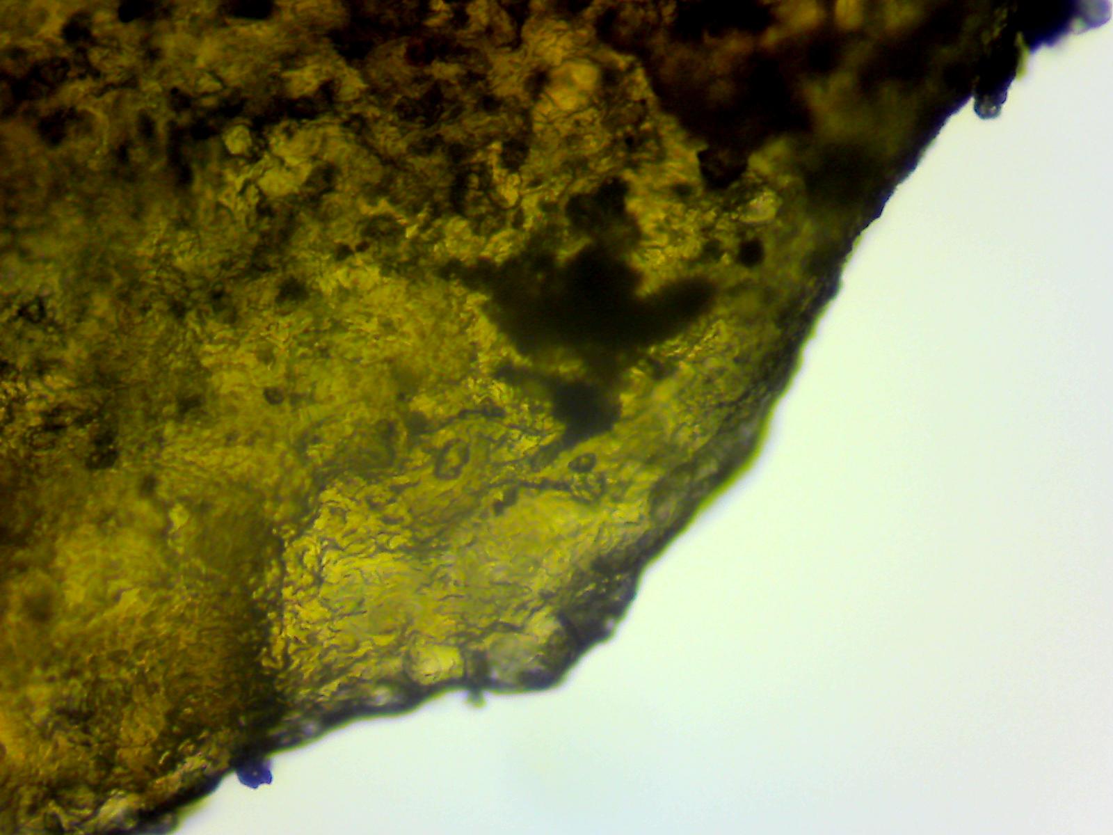
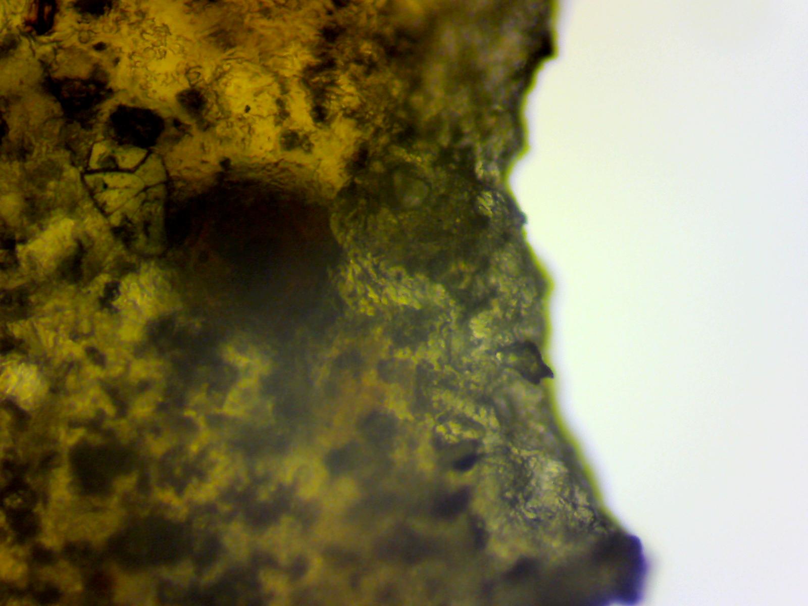
Can you make out anything from these photos? I couldn’t pick out any basidia or cystidia, but maybe you can see something else? If not I can have another go. As you said, it’s good practice! 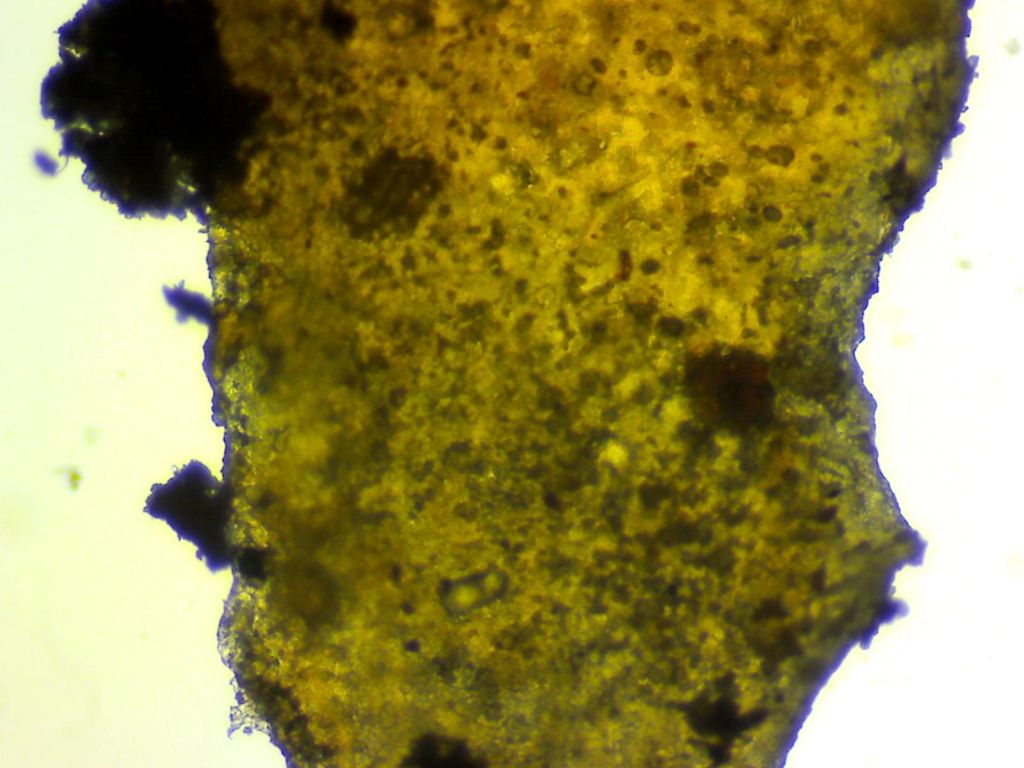
[attachment=5]Site001 – Fungus003 – 8th August 2015 (2).jpg[/attachment] 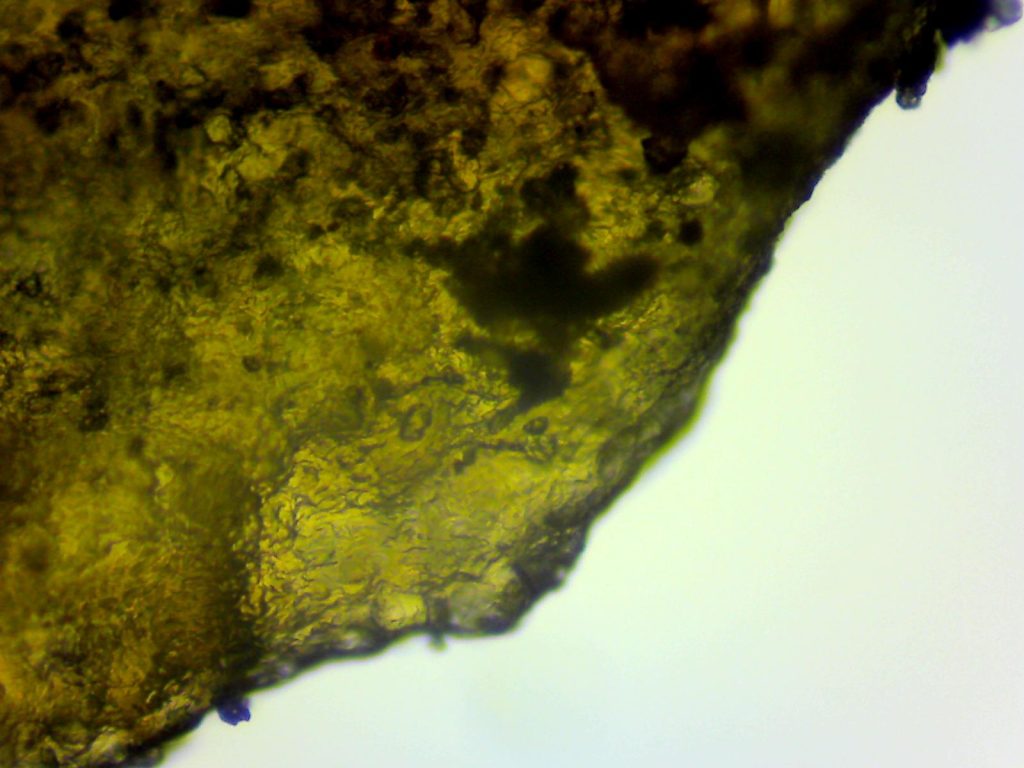
[attachment=4]Site001 – Fungus003 – 8th August 2015.jpg[/attachment] 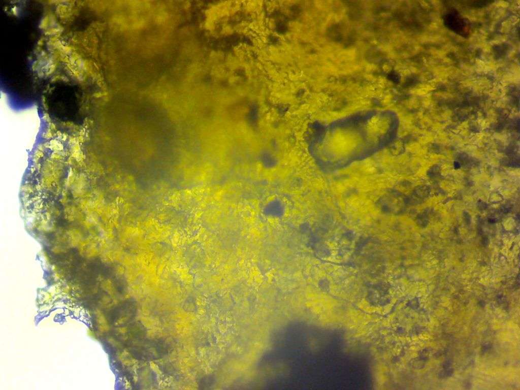
[attachment=3]Site001 – Fungus003 – 8th August 2015.jpg (3).jpg[/attachment] 
[attachment=2]Site001 – Fungus003 – 8th August 2015.jpg (4).jpg[/attachment] 
[attachment=1]Site001 – Fungus003 – 8th August 2015.jpg (5).jpg[/attachment] 
[attachment=0]Site001 – Fungus003 – 8th August 2015.jpg (6).jpg[/attachment] August 8, 2015 at 2:08 pm #35627August 8, 2015 at 4:26 pm #35628jimmymac2 wrote:Can you make out anything from these photos?
No, the sample is too think. You only need a minute piece at a time, barely visible with the naked eye.
August 8, 2015 at 4:34 pm #35629jimmymac2 wrote:Chris Johnson wrote:You could raise this in the Change Requests forum:
http://www.fungi.org.uk/viewtopic.php?f=2&t=249 Done.
 August 8, 2015 at 4:41 pm #35630Chris Johnson wrote:jimmymac2 wrote:
Can you make out anything from these photos?
No, the sample is too think. You only need a minute piece at a time, barely visible with the naked eye.
Awwww…. Tomorrow thenQuote:Take a piece of the twig and place it over a glass slide raised on two matchsticks (or something similar)
Is this what you had in mind? Is it too high?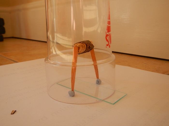
[attachment=0]DSCN6472.jpg[/attachment] August 8, 2015 at 6:18 pm #35631jimmymac2 wrote:. . . . Is this what you had in mind? Is it too high? . . . .
You only need to raise the sample fungus sufficiently so that there is a tiny gap between it and the glass-slide. (A couple of mm at most).Matchsticks, cocktail sticks, or even bits of folded paper, laid across the width of the slide, would give you more than enough of a gap.
To be honest, with fungi samples on small twigs and the like, I generally just rest the twig directly on the slide, as there is usually plenty of gap left by the curvature of the twig, or differences in thickness of the fungus, to allow more than enough spores to drop.
Likewise, with gilled fungi, (unless there is something atypical going on), I just cut the stem off, and rest the cap, (or part cap), directly onto a slide, and it always gives a good solid spore print.
What you’ve done in the photo will work, but the spore drop will be more diffused, because the spores will be able to float down over a far wider area than if the fungus were much nearer to the glass-slide.
Think of it this way. – Say for example, a spore is 10µ in diameter. It would have to travel 100 times its own diameter to fall just 1mm.
If you raise the fungus up by about 50mm as here, the spore has to fall 5000 times its own diameter before it hits the glass-slide.
Even when the sample & slide are enclosed to retain moisture and stop draughts, spores will still be able to float about quite considerably during a fall of 50mm.
So, when taking spore prints/samples, the nearer you have the fungus to the glass, the better.
Regards,
Mike.
August 8, 2015 at 7:29 pm #35632Thanks Mike! I’ll leave it for the night and see if an adequate amount of spores drop onto the slide. AuthorPosts- You must be logged in to reply to this topic.