
Odd Alyxoria(?) from Denmark
- This topic is empty.
-
AuthorPosts
-
July 15, 2020 at 2:23 pm #31701
Last week I found this yellowish Alyxoria growing on wood inside a small hollow in a living Oak near Tibirke, Denmark. I have tried keying it with LGBI and I end up in a tentative ID as Alyxoria varia. There are, however, some deviations from the desciption that leave me doubtful: The thallus and the yellowish pruina reacts orange with K, some of the spores have a rather large (sometimes “flaring”) perispore (not very visible in water, becoming very clear with K and then after a while in K they just appear as a “halo”), the ascomata are very contorted (reminiscent of O. viridis(?)) and there are several spores with seven to eight septae.
The spores are rather variable in length ranging from 22-32 µm x 5-6 µm, mature spores predominatly 6-septate. I have checked many candidates for pycnidia, and the closest candidate are some red-brown “blisters” that should be visible in the first photo. When squashed under the microscope they release a few (not a hole mess of conidia as I am used to) oblong objects that might be conidia.
Any suggestions as to how to proceed from here? I have checked against A. viridisorediata, but the spores are much too large and the septation does not match.

[attachment=9]Alyxoria sp. 1-1.jpg[/attachment] 
[attachment=8]Alyxoria sp. 1-2.jpg[/attachment] 
[attachment=7]Billede1162.jpg[/attachment] 
[attachment=5]Alyxoria sp. 1-4 (Ascus. vand).jpg[/attachment] 
[attachment=2]Alyxoria sp. 1-5 (spore, vand).jpg[/attachment]
[attachment=3]Alyxoria sp. 1-5 (spore, K, lige tilsat).jpg[/attachment] 
[attachment=4]Alyxoria sp. 1-5 (spore, K, efter noget tid).jpg[/attachment] 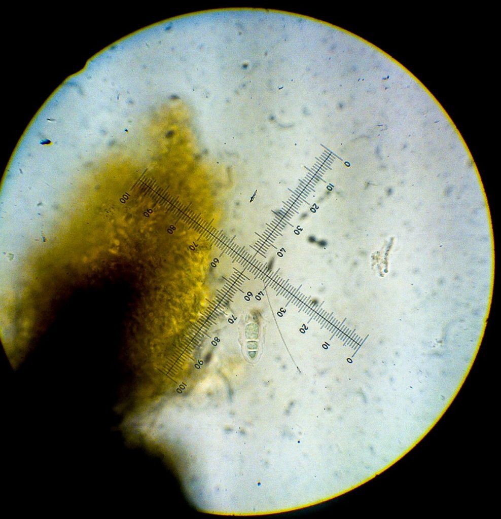
[attachment=6]Alyxoria sp. 1-3 (Mrkelig perispore).jpg[/attachment] 
[attachment=1]Alyxoria sp. 1-6 (konidia, måske).jpg[/attachment] 
[attachment=0]Alyxoria sp. 1-7 (konidia, måske).jpg[/attachment] July 19, 2020 at 7:54 pm #43059Coincidentally, I have been puzzling over an Alyxoria from a friable limestone headstone. I got as far as couplet 27 in the Opegrapha key in the 2009 Flora but there I faltered, unable to decide between mougeotii and saxicolous varia. 
[attachment=1]habitus_20200719_103921_7190042X.jpg[/attachment] I was particularly confused by the “enlarged central cells” of the spores of mougeotii, whereas couplet 28 indicates that varia does _not_ have an enlarged central cell. However the text entry for varia on p 645 says “middle cells somewhat enlarged”. My spores are more like the illustrations of varia on p639 than of mougeotii. I think I found bacilliform and ellipsoid conidia of the right dimensions, which should be a clincher for mougeotii, but I also think that the ends of the apothecia are rounded which rules it out. The conidia are mostly at least 5 microns, whereas O varia conidia dimensions are given as 3-5 microns.
Knowing that his various comments and corrections to the Flora were on the BLS website, including the Opegrapha section,
https://www.britishlichensociety.org.uk/resources/downloads ” class=”bbcode_url”> https://www.britishlichensociety.org.uk/resources/downloads With his permission, here is his response:
“I think that the drawings of O. mougeotii and O. varia ascospores in the Flora are misleading. I have compared the spores of O. mougeotii and ‘O. varia’ and don’t see such a difference. Of course we have the additional problem that O. varia looks to be more than one taxon. Also, since LGBI2, O. viridipruinosa was described as new (2011) and this has shorter conidia (3 microns or so). In the past O. viridipruinosa was clumped in with O. varia and that may explain why the range of conidial length in the LGBI2 is 3 to 5.One of the more conspicuous differences between O. varia and O. mougeotii is the K+ yellow-dissolving of any pruina on the disc and the distinct K+ yellowish of the epithecium. (However, I haven’t checked whether all of the forms of corticolous ‘O. varia’ show this K+ yellow and I need to check more specimens before stating too categorically that there is no K+ yellow in O. mougeotii). This is not a character that is mentioned in the Flora (where the epithecium is said to be K- in both O. varia and O. mougeotii).
Another thought that has crossed my mind is that O. mougeotii may simply be a saxicolous form of one of the ‘O. varia’ taxa.”
The K+yellow pruina rang bells with this posting. An odd Alyxoria from Denmark, on lignin this time, that was tentatively O varia but with K+ orange pruina. It was from inside a tree hollow, and I’ve noticed that you often get exceptionally good pruina when sheltered from rain – maybe dense enough to look orange with K! It is something to bear in mind, anyway.
My saxicolous Alyxoria has a slight K+ yellow bleed of the pruina, so I’m going to record it as Alyxoria cf varia for now and retain the specimen.
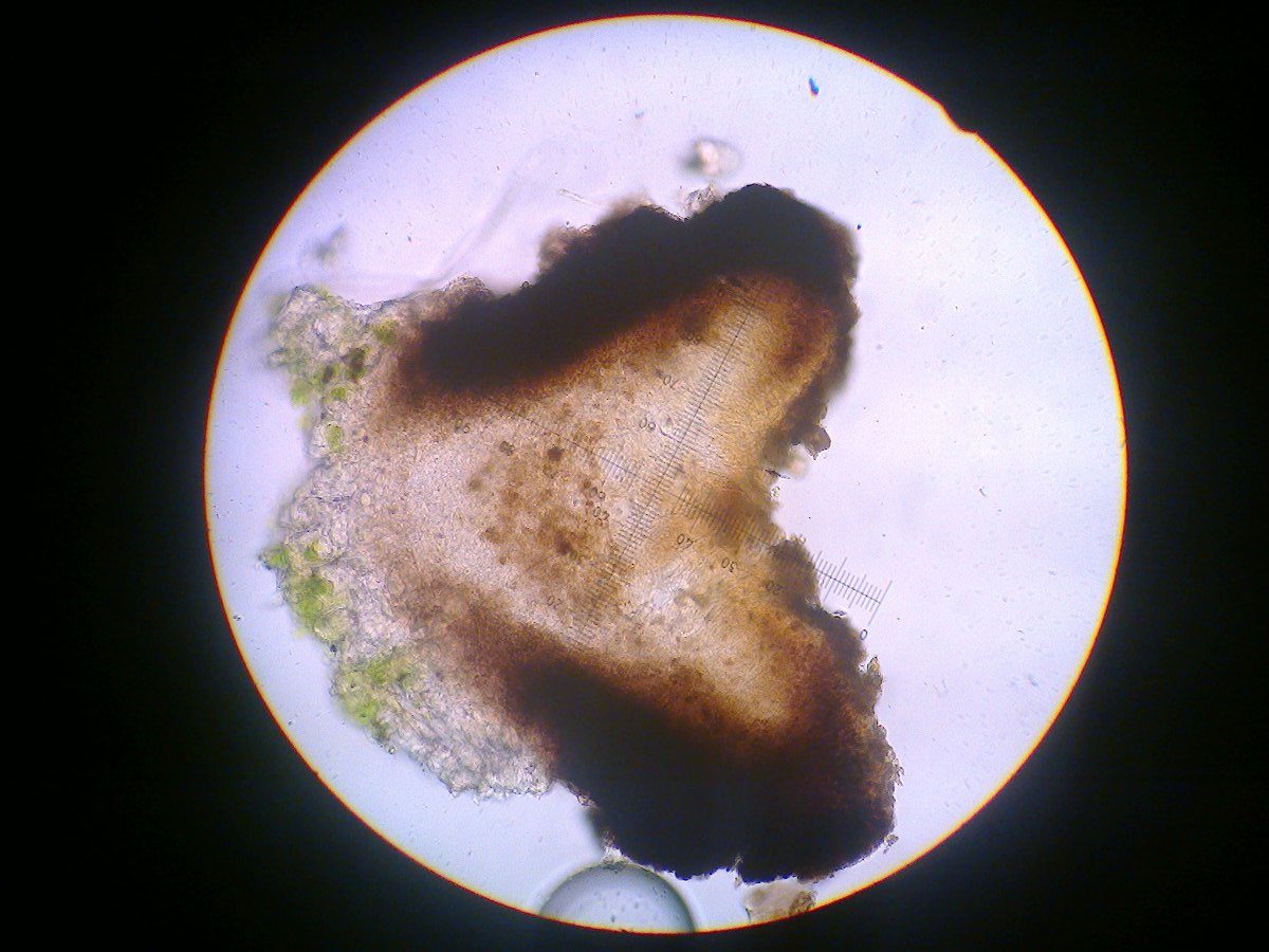
Here are some more photos of my specimen.
Juliet
-
AuthorPosts




- You must be logged in to reply to this topic.