
Mystery soriediate lichen
- This topic is empty.
-
AuthorPosts
-
December 10, 2023 at 12:00 pm #32737
Hi, I’m hoping this is only a mystery to me, but I’ve found this lichen in two churchyards in Sussex and I am really struggling to ID it. There are several photos of it below. I have found it on a sandstone church wall (east), gravestone, and on the top of a brick wall. It is C – and K+ dark brown quickly. The most surprising find was that under UV the soredia were strongly pink/red. The photo shows the UV directed from the side and doesn’t really show the colour quite right, it was a lovely dark pink. The thallus on brick does also have a black prothallus, but this was not as clear or perhaps not present on the sandstone. I was wondering if it was a fertile Lecanora orosthea (there was a lot of this in the churchyard) due to the K test. At the moment I haven’t got access to a microscope, so sorry no spore photos.
Thank you
Sylvia
December 10, 2023 at 5:38 pm #45823When I first examined your really good photos, I couldn’t bring any likely species to mind. However, with a bit of mulling over, I think I might have a suggestion. First however, I very much doubt that most of your images show Lecanora orosthea, They just doesn’t look right for various reasons. In L. orosthea the coherent non-sorediate thallus is the same colour as the soredia, a pale green-grey with a hint of yellow. In most of your photos, the yellowish soralia seem to contrast in colour with the purer grey thallus. Also, on the rare occasions that I have seen L. orosthea fertile, the discs have been predominantly similar in colour to the thallus, often varying in colour with some pinkish brown and dark blue-green but not the consistent pale pink seen in most of your photos. The two following photos are of non-fruiting L. orosthea. 
[attachment=3]FJo2ZPWWYAAGZDY.jpeg[/attachment] 
[attachment=2]FJovV_3X0AES_n8.png[/attachment] Before making my suggestion, I will make a comment about the UV+ pink/red fluorescence you observed in the soredia. It is possible, especially if the lichen was somewhat damp, that the fluorescence is due to the chlorophyll in the algal partner. When I was getting to know Bacidia fuscoviridis (which your lichen is certainly not), I thought I could discern two different forms of it. I seemed to find a form with fluffier soralia and these soralia were UV+ orange in contrast to what I thought was a different form in which the soralia were UV-. It took me a while to realise that my ‘fluffy’ form was merely what B. fuscoviridis is like when damp and in this state the chlorophyll of the algal partner made the soralia seem UV+ orange. I can’t say for sure but I suspect that you are seeing a similar phenomenon.
My tentative suggestion (not confirmed) is that you have Trapelia placodioides which is fairly common on acid/siliceous substrata in churchyards. There are some problems with reconciling my suggestion with some of your observations. T. placodioides should be C+ red and K-. The C reaction may be difficult to detect in the field, especially if the conditions are damp. If you go back when it is very dry, or better still collect a small portion and conduct a C reaction with strong bleach (e.g. Domestos or Parazone) on thoroughly dry material, you might find it is C+ red. If so, that would add weight to my suggestion. The ‘K+ dark brown’ might possibly due to the reagent making the thallus translucent and allowing the colour of the substratum to show through. T. placodioides is rarely fertile but it does sometimes fruit in churchyards. The apothecia in your material are particularly spectacular and they may be plumped up in damp conditions (something that happens particularly markedly with Trapelia fruits).
The following photo is from the dorsetnature website:
https://www.dorsetnature.co.uk/pages-lichen/lch-378.html 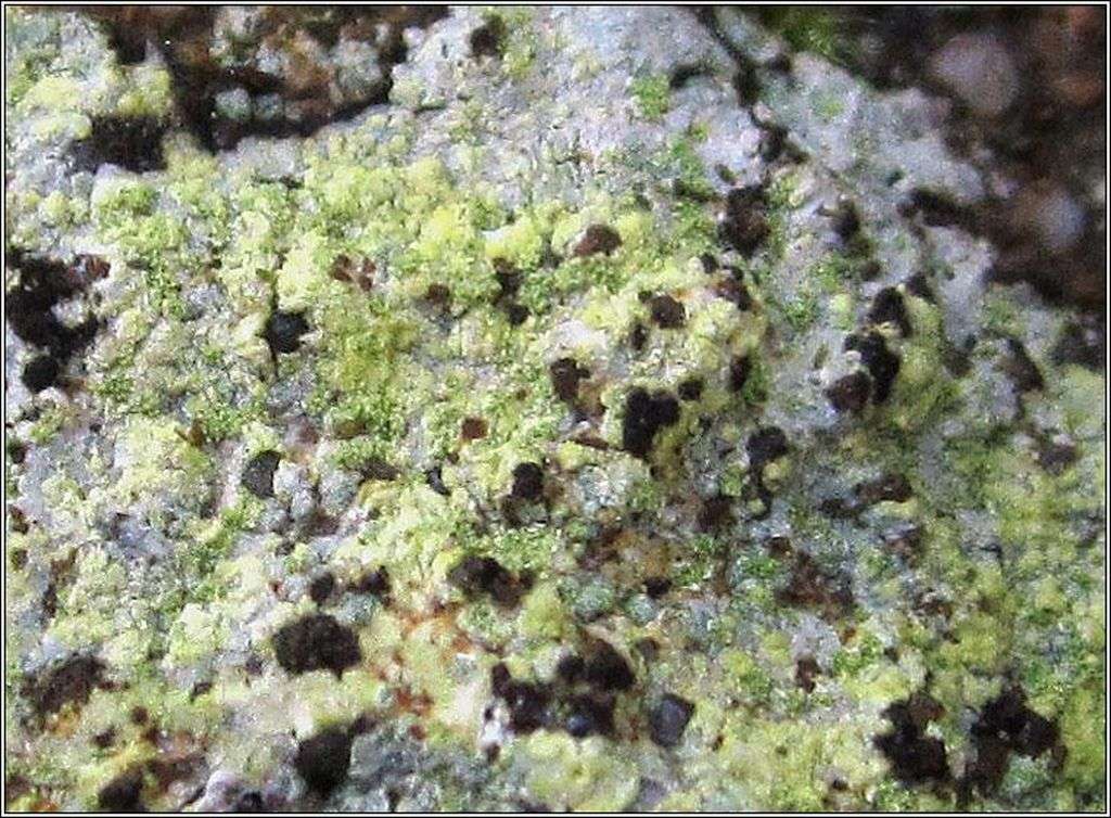
[attachment=1]lch-378a8.jpg[/attachment] You will see how similar the colour and form of the soralia are to your lichen.
The next photo is some sparingly fertile (and thoroughly dry) T. placodiodes which I found in a Buckinghamshire churchyard.

[attachment=0]Trapelia placHamb2.jpg[/attachment] The apothecia of T. placodioides should have a pale thalline rim around them but perhaps this feature is not apparent due to the lichen being damp.
I think I have seen a slight dark prothallus around individuals of T. placodioides even though the literature suggests that it is supposed to whitish. However, I have never seen such a well-developed dark prothallus as shown in your last photo. In fact, I wonder whether your last photo is something different and indeed that one might well be L. orosthea.
December 10, 2023 at 8:19 pm #45824Hi Mark, Thanks so much for all the information. As you can imagine, the rain has been incessant here and it was still very very wet in the churchyard and the first thallus I found was on a shady east wall, which probably won’t dry out until spring. But the final photo is of the one on the top of the wall, so that may well dry out sooner. I’ve added another photo here of a very similar lichen found in September in another Sussex churchyard that I was puzzling over. My notes say K+ yellow and possible C yellow, but uncertain and the UV was not pink/red. So not sure what’s going on there. The soriedia are there but not as distinctive a colour in this one. Although it was in partial shade of a Yew, it had been dry for some time and it was a very hot sunny day.
I need to go back and get samples for each of these and do the tests again. This is proving fascinating.
Best wishes,
Sylvia
December 10, 2023 at 8:26 pm #45825Hi Mark, I just examined one of the photos, the one from the top of the wall and notice down in the right hand corner what could be an apothecia which resembles more closely a Trapelia placodioides apothecia. I’ve cropped the photo and attached it.
December 10, 2023 at 9:32 pm #45826Sylvia I can not help with the IDs. but agree with Mark that the reddish UV fluorescence is most likely activated chloroplasts, which fluoresce red. To see this well, shine a UV torch on damp bryophytes. Most lichens shield their algae, so you do not see this so much in lichens, but it is obvious in
Micarea prasinas. lat. group sorediate and isidiate species when damp. This is especially irritating with Micarea xanthonica, which fluoresces orange from xanothones when dry but goes bright red when wet, which it often is as an inhabitant of oceanic woods. Neil
December 10, 2023 at 10:28 pm #45827As for the further lichen, it is one or other of very fertile Lecanora orosthea, or L. sulphurea growing isolated (as it sometimes does) rather than on its usual Tephromela atra host. I have sometimes found such occurrences and not been able to decide which it is. If proper sorediate areas are present then that indicates L. orosthea but I am not sure there are any ‘proper’ soredia in this case. There isn’t any easy/reliable way to tell between these two as far as I know, in cases in which L. orosthea is so fertile that soredia production has been effectively suppressed. If it is growing in the vicinity of T. atra I tend to assume L. sulphurea, if any convincing soredia are present I tend to assume L. orosthea, but there are some specimens which I leave unnamed. Note that the apothecia have a sort of molten wax appearance and are pale brown with some darker patches, rather than the pale pink or reddish pink typical of Trapelia. The difference in colour is subtle but yours certainly has a pale brown colour rather than pink in my opinion. To confirm which one I have been discussing in this paragraph, I will repost your photo of it immediately below. 
[attachment=2]Unknown.JPG[/attachment] As for that little reddish rimmed speck, the thing is that it occurs on the thallus which also has well-developed apothecia. I don’t know what those little specks are (there is at least one other on the thallus). I am still tending towards fertile L. orosthea for this one (the photos reposted immediately below). The apothecia of this lichen are probably plumped up and looking more transparent than usual due to the damp conditions. Most lichens are much more difficult to identify when wet than when dry.

[attachment=1]PC080574_A.JPG[/attachment] 
[attachment=0]PC080574.JPG[/attachment] December 10, 2023 at 10:51 pm #45828The following two photos are of Lecanora sulphurea. Note that (rather confusingly) the edges of the areoles sometimes become raised and almost break up into irregular granules, which could be confused with soralia. 
[attachment=3]ELQoa4kXsAASdjK.jpeg[/attachment] 
[attachment=2]Screenshot 2023-12-10 10.38.38 PM.png[/attachment] As for conducting chemical tests for our suggested identities, Trapelia will be C+ red, Lecanora orosthea/sulphurea will be K+ dull yellow, KC+ brighter yellow. For the C test, just apply strong bleach to the thallus, and include the soralia as the reaction may be strongest there.
For the usnic acid test for those Lecanora species:

[attachment=1]Screenshot 2023-12-10 10.36.52 PM.png[/attachment] 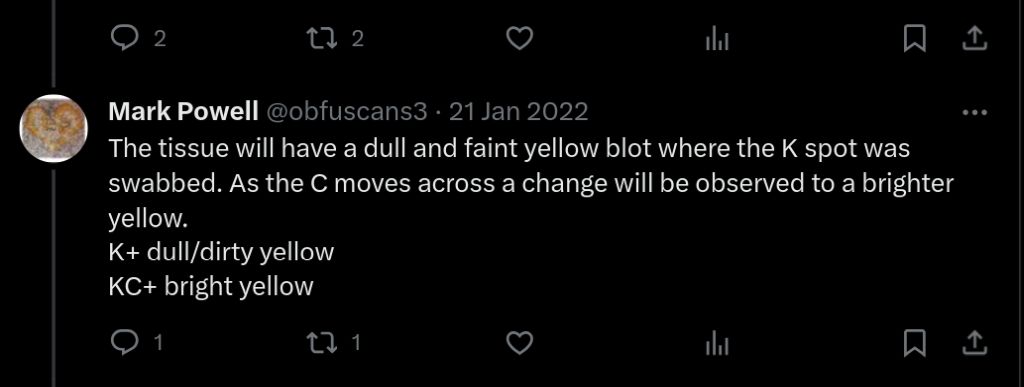
[attachment=0]Screenshot 2023-12-10 10.37.29 PM.png[/attachment]
The KC test is rather subtle but also rather pleasing when you see the dull faint yellow spot on the tissue turn a slightly but noticeably brighter yellow. As with all spot tests, it is important to be rather frugal. I rarely apply drops straight from a dropper bottle, but rather prefer to transfer a smaller drop with a cocktail stick or similar (dry grass stem in the field if nothing else to hand). I use a cocktail stick at home too. Many nice specimens have been ruined when an embarrassing splurge of reagent has accidentally flown out of the dropper.December 11, 2023 at 9:26 am #45829Thanks Mark, that’s really helpful. There are some very subtle differences in shades of pink between the last one I posted and the one in the original post. The latter ones do have the waxy appearance more of Lecanora sulphurea, but the first are more dusted with icing sugar. Your comments have really cleared up a lot of confusion I had over Lecanora sulphurea. It is very common in churchyards here and I realise now that it does have a remarkable number of guises, including looking very different if away from T atra and looking like it has some type of soredia. Well off to get some samples in the next few weeks, so the tests can be done dry.
Really good to know about the UV tests as well, Neil.
Thanks once again
Sylvia
January 28, 2024 at 3:35 pm #45830I have received a specimen relevant to this topic. From the photos above I still think that we are dealing with two separate species and I have been sent just one of them. Anyway, the specimen sent to me is a Lecanora which contains usnic acid, and taken with the photo re-posted below allows us to be confident that it is a lovely example of fertile L. orosthea. I took some micrographs while looking at the specimen which I will post here. 
[attachment=9]PC080574 (1).JPG[/attachment]
The specimen was in the form of some crumbs attached to sellotape. Although this method of collecting is better than nothing, I very much favour my own method in which a scraping ends up the right way up glued to card, retaining more of the look of the lichen and much easier to work with while sectioning. The first thing I did was to remove the best ‘crumbs’ and glue them to a bit of card. The collector is now aware of a better way of collecting useful scrapings from stone. Note that there is one healthy apothecium and some blackened ones.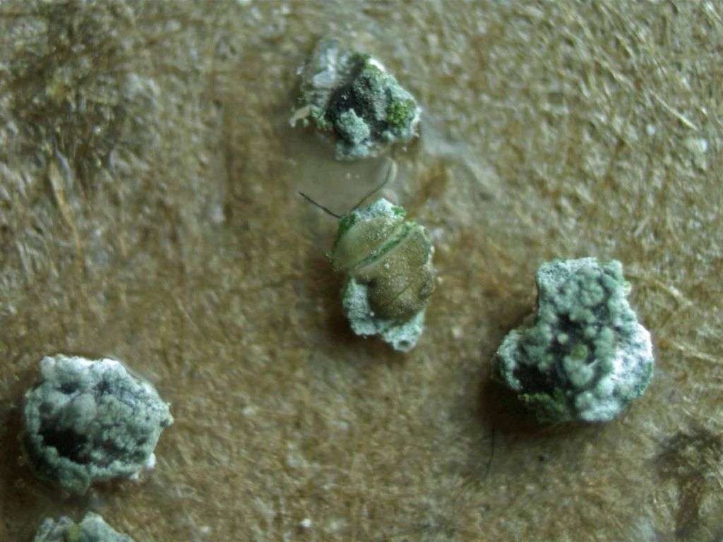
[attachment=8]Lecoros11.jpg[/attachment]
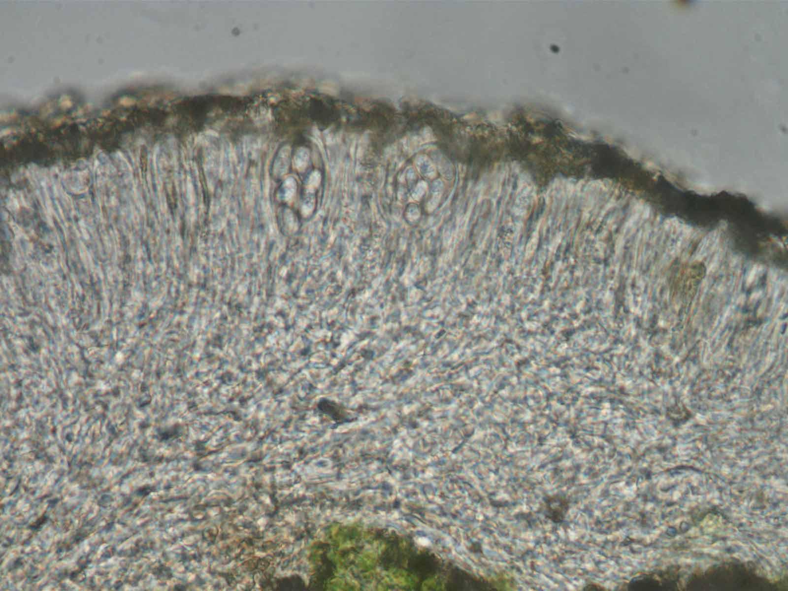
I managed to detect usnic acid by using the KC reaction described above. This has ruled out Trapelia as the identity of the received crumbs and already makes L. orosthea seem very likely.Let’s have a look at some apothecial sections in water. At low power we see that the hypothecium is pale, the only pigmentation in the uppermost hymenium. We see that the proper exciple is poorly developed and the thalline margin difficult to interpret, excluded and pushed down into the thallus. The epithecium has dense yellowish granules set in an orange epithecial gel. The ascospores are ellipsoid to narrowly ellipsoid or slightly oblong, 9-12.5 x 5.5-6 microns. In this case the spores are of little use in our identification.

[attachment=7]Lecoros01.jpg[/attachment] 
[attachment=6]Lecoros02.jpg[/attachment] 
[attachment=5]Lecoros03.jpg[/attachment]
Then to add K to the slide and we see that the epithecial granules are readily soluble, the excipular hyphae become lax, consisting of narrow elongated and anastomosing hyphal cells.
[attachment=4]Lecoros06.jpg[/attachment] 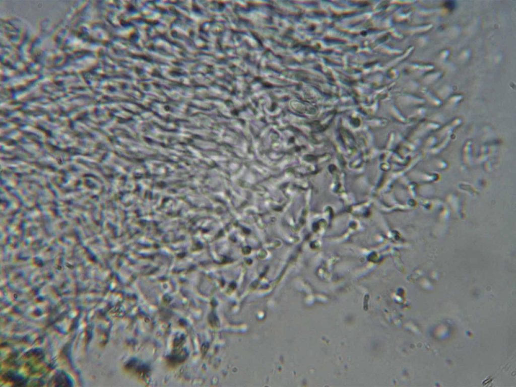
[attachment=3]Lecoros05.jpg[/attachment]
We can also look at the photobiont cells and find that they divide by producing a cluster of daughter cells before dividing, rather than the binary fission that would be seen in Trapelia. The effect here is a bit like those triangular cheeses wrapped in foil packed in their circular box.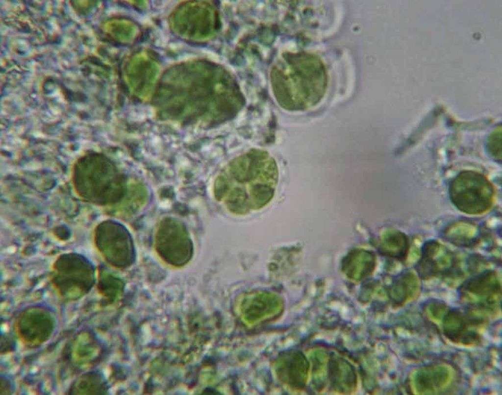
[attachment=2]Lecoros07.jpg[/attachment]
So far it hasn’t been very easy to make out individual asci and the nature of the paraphyses. It only takes a few moments to stain with ink-vinegar and now we see asci and that the paraphyses are somewhat branched and anastomosed, typical of many Lecanora species.
[attachment=1]Lecoros08.jpg[/attachment]
It doesn’t take long to stain the ascus tips (especially using my method) to get a good view of the Lecanora-type ascus tip (the non-staining apical cushion penetrating right to the top of the tholus).
[attachment=0]Lecoros09.jpg[/attachment]
Although I have shown the way I try to extract as much information from an apothecial section as possible, none of the characters (epithecial granules and their solubility, spore type and size, etc.) really provide any distinct differences from apothecial sections of related species such as L. sulphurea. We have proved that it is an usnic-acid containing Lecanora (seen more additional microscopic characters than we really needed) and have a good photo of its sorediate nature, allowing us to name it as L. orosthea.January 28, 2024 at 3:40 pm #45831Here is a section through one of the blackened apothecia. The apothecial tissues are much degraded and discoloured. They are heavily infected with a dematiaceous hyphomycete which is certainly a lichenicolous fungus but not one that I am confident to name. Such things are difficult, having few distinct characteristics and I remain to be convinced that this sort of thing can be properly named using simple inspection. 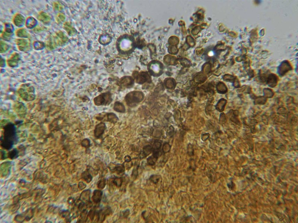
[attachment=0]Lecoros10.jpg[/attachment] January 28, 2024 at 7:40 pm #45832Thanks for the comprehensive and fascinating reply, Mark. Sylvia
January 30, 2024 at 3:20 pm #45833Anonymous
Here is a 2024 photograph taken by the sea of Lecanora sulphurea(probably parasitized) it looks a bit similar, but there are several taxa under the name of Lecanora sulphureawhich is a parasitic species which can also be parasitized which does not facilitate determinations. Alain
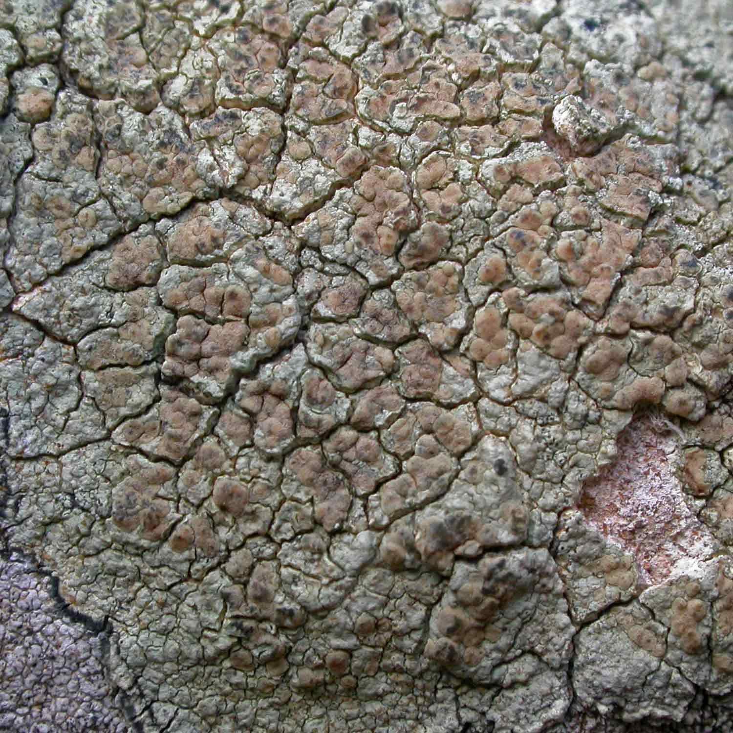
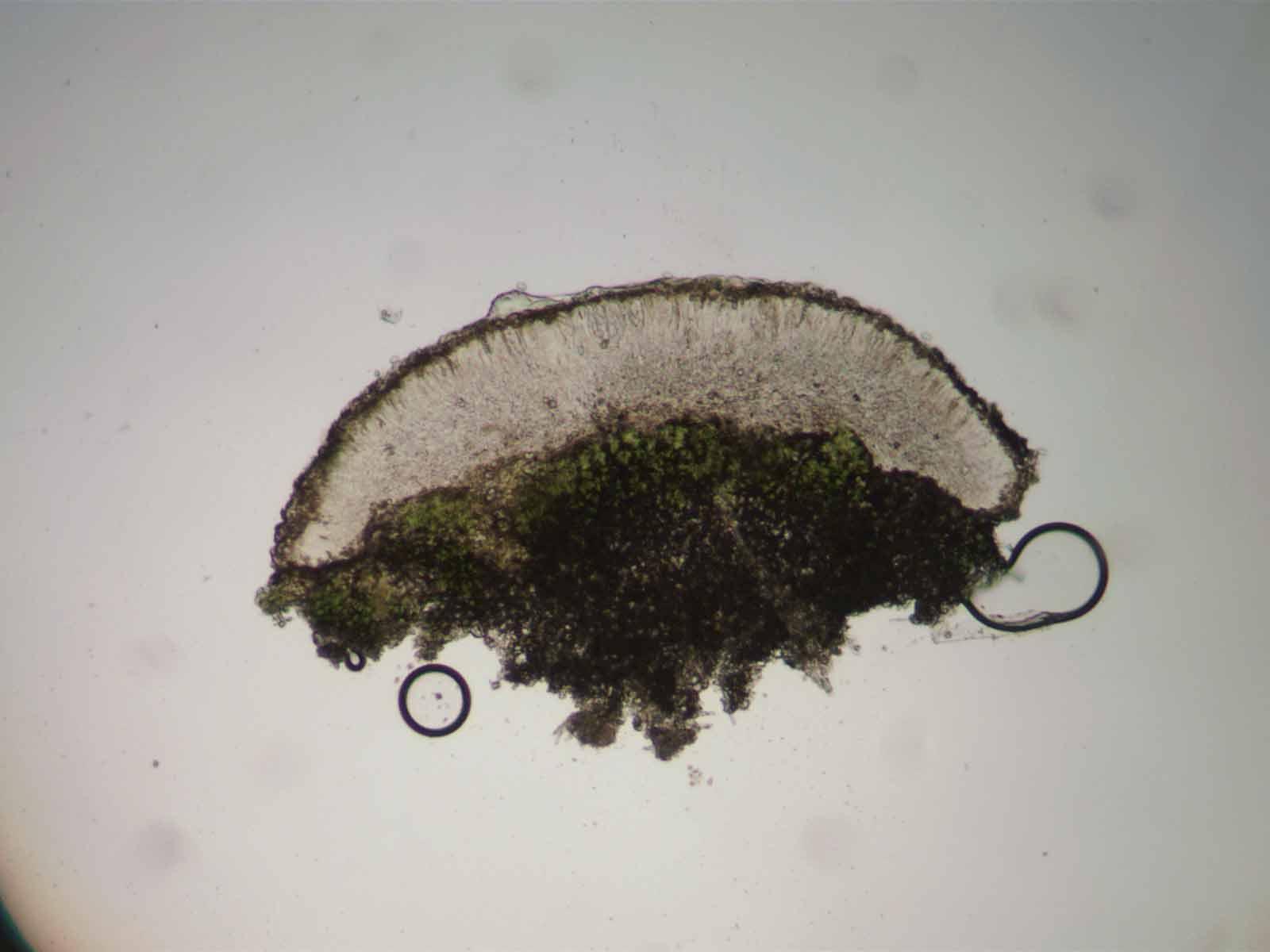
February 1, 2024 at 11:21 am #45834In my following comments I am going to describe the way these usnic-acid containing Lecanoraspecies are usually recorded by English churchyard recorders (I am not claiming any particular taxonomic insights). When growing on acidic/siliceous stone, and when containing usnic acid there are only five names we commonly play with. L. polytropaand L. stenotropaare almost invariably fertile, they are non-sorediate, and their apothecia have persistent margins. Many instances are recorded as L. polytropain the field even though microscopy (spore width) is required to be absolutely certain. L. polytropausually seems to contain more usnic acid making it appear more yellowish-green than L.stenotropawhich often has a bluish tint. L. stenotropawould be rare in my region except for its common occurrence on chemically treated fence rails. Lecanora soraliferahas a distinctive appearance when well-developed, with neatly delimited soralia. Lecanora orosthea(when fertile) and L. sulphureahave apothecia which lack distinct margins, the margins usually excluded at an early stage. We call the sorediate material L. orostheaand the non-sorediate material L. sulphurea(especially when apparently parasitic on Tephromela atra). -
AuthorPosts





- You must be logged in to reply to this topic.