
Melanomma pulvis-pyrius?
Forums › Forums › Fungi ID requests (post here if you aren’t sure what type of fungus you’ve found) › Melanomma pulvis-pyrius?
- This topic is empty.
-
AuthorPosts
-
March 3, 2017 at 6:27 pm #30590
Help please! Found something that has been tentatively id’d as M.pulvis-pyius by members of Glamorgan Fungus Group but question regarding very open pores in stromata. USBx20 photos attached (apologies not good quality). Found in dense masses stromata less than 1mm on well rotted log in damp woodland and my garden on old (20 plus years) Leylandii stump. Microscope work will be attempted next week (but not by me). Any thoughts would be appreciated March 3, 2017 at 10:03 pm #39824Hello, would be quite untypical for M. p., which is smaller, doesn’t grows in such dense communities and doesn’t open usually by half perithecia.
This looks more like Ruzenia spermoides, but should be verified by microscopy (what will not be possible in such overaged collections usually … )
best regards,
Andreas
March 4, 2017 at 8:11 am #39825Hi Andreas, Many thanks. Hopefully microscope work will be done next week. Mark
March 4, 2017 at 9:31 pm #39826Hi Mark, don’t spend to much efort in such an old collection – if you don’t see some spores in the first try, than rather throw it away than spoil more time for it ….
best regards,
Andreas
March 5, 2017 at 12:01 pm #39827Echoing Andreas’ comments, because many ‘pyrenomycetes’ have tough carbonaceous exteriors they last longer than their active life. Your example is clearly empty; here is an example where you can see the jelly-like contents of a ‘live’ specimen, in this instance of Diatrype bullataon Salix: 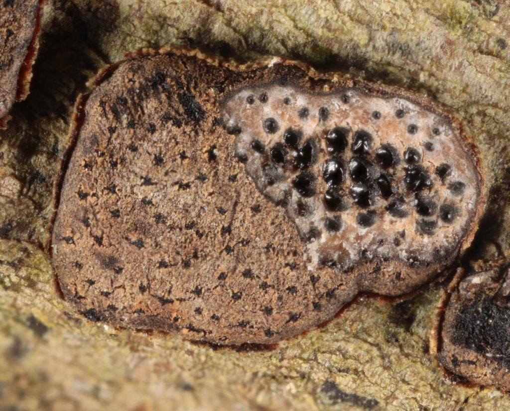
[attachment=0]Diabul03.jpg[/attachment]
I come across more dead perithecia than active ones. The best strategy is to cut transversely across any prospective collectionin the fieldwith a pen-knife or similar (often a finger nail can suffice). Then check with a handlens. This will save time, effort – and coverslips! Cheers
Chris
March 5, 2017 at 12:40 pm #39828Thanks Chris! Hopefully we will find live ones ‘in the field’ which is my garden! I’ll try and post again in the week. Mark
March 5, 2017 at 2:18 pm #39829Hello, definitely right was Chris says.
Sometimes when it is important to identify nevertheless, I try one (ONE!) preparation of the empty “crust” because you may find single spores that have been sticking to the interior wall – but that makes only sense when you have a species which can be identified by a singel spore, or (like in this case) when you just want to check wether your macroscopic determnation is correct and you know beforehand how the spores should look like.
best regards,
Andreas
March 13, 2017 at 5:46 pm #39830Hello Andreas and Chris, Ruzenia spermoides – thanks your suggestion Andreas!
Successfully found ‘live’ perithecia and able to extract ‘jelly’. Microscope work by Emma Williams Glamorgan Fungus Group revealed asci with ascospores – hyaline. When stained with Melzer’s showed up nicely. Ascospores allantoid musiform 20.6 -0 21.6 microns by 4.2 – 4.5 which corresponds to paper by R.Hilber and O.Hilber 2002. Moment of confusion when a very few ascospores similar to M.pulvis-pyrius appeared but put down to contamination from insects or other. Sorry no photos but I haven’t been able to resize.
Mark
-
AuthorPosts


- You must be logged in to reply to this topic.