
Forum Replies Created
-
AuthorPosts
-
June 11, 2021 at 7:07 pm in reply to: Please can I have some ID help with these white rhizomes #44161
Honey fungus rhizomorphs are black from a very early stage. I don’t think you need to worry about that particular group. If you want another opinion, though, you could tweet your photos and ask @Jassydrak. Interestingly, honey fungus rhizomorphs are bright white on the inside. If you split one with a finger nail it tears back like a cheese string and reveals the white central strand.
Great work Chris. How many volumes are you going to need?! I love seeing your compilations. Wish I could do as well with my photography.
Did you mean 1253, Chris? Didymosphaeria conoidea? That would be an interesting find. Have found both Leptosphaeria acuta and L. doliolum on nettle stems today. The later was only at the extreme base of the stem – that short section which is often more prostrate. Do you know if this a general trend?
Thanks for starting off this topic, Chris. I spent a while looking at nettle stems last year and will enjoy looking again. Isn’t there a second species of Leptosphaeria that can be found on nettle stems? How easy is it to tell the difference between them? Simon, when youre collecting Ramularia specimens, don’t forget that if put the sample in a plastic bag or box with some wet tissue for a couple of days, you’ll get more clumps of conidiophores forming and they’ll be easier to confirm. Have you seen Arthur Chater’s new Ramularia guide? Download from this page:
https://www.aber.ac.uk/waxcap/links/index.shtml Good to see posts on pathogens. Thank you.
It’s also possible that these brackets have a coating of algae growing on them rather than a moss. Sorry. I’ve just realised that I don’t have image permission yet for the images in the T. punctata description. I’ve taken it down and will seek permission before re-posting. I’d be happy to call this the first English record for Taeniolella pertusariicola, Chris. Taeniolella species have hyphae immersed within the host thallus and darker, multi-septate conidiophores which blend into chains of indistinctly separated conidia and emerge from the host thallus. It is often difficult to tell where each conidium ends and how many cells belong to each conidium. The literature claims that the conidial chains separate easily but this is not always true.
There are three species of Taeniolella that are not particularly fussy about their hosts. T. delicata has smooth cell walls. T. punctata has verriculaose walls but is observed under a dissecting scope to form tufts of dark conidiophores/conidial chains. T. verricosa (not yet recorded from the UK) has verriculose walls but each cell in the conidiophore bulges in the middle so that in extreme cases hyphae look like chains of beads. The material from Westmorland doesn’t match any of these.
T. pertusariicola has been recorded once in the UK from southern Scotland on Ophiopalma ventosa. It forms few conidial chains arising through the thallus surface but is always accompanied by dark material with no fixed structure. The coniophore and conidial cells are verriculose and only slightly constricted at the septa. The conidal chains should break mainly into two-celled conidia but these were not found loose on the material examined. The majority of conidial chains were intact with a paler, round-ended cell at the tip. The paler immersed hyphae could not be visualised, although the dark conidiophores were visible within the host thallus.
T.delicata
https://drive.google.com/file/d/1wH32Ol8mTxh4wGvkYQHwVhsqoJgvbiWZ/view?usp=sharing T. punctata
https://drive.google.com/file/d/1oPZ10RwPGAZwRA0x-6bYA8lL0vCgtZfV/view?usp=sharing To help the search for this very recognisable host and for Muellerella ventosicola, two species descriptions have been written. These can be accessed from Google Docs using the following links: Ophiopalma ventosa:
https://drive.google.com/file/d/1JpuSLJSVcUaOgiYtqeYjAIIMHrAVhbH-/view?usp=sharing Muellerella ventosicola:
https://drive.google.com/file/d/1oGASJGTCqhb8fT0vg8NcyD4nzoOtYh_R/view?usp=sharing I can only apologise for not putting the link here immediately. I don’t know why I didn’t think of it. I’m now working on a guide to LFs on Physcia species.
I’ll post anything else I release in this thread.
I hope the first guide is useful. Please send both positive and negative feedback through. And, if anyone has any better photos to share, I’m open to changing those too.
There are two other LFs on this lichen but neither has been confirmed as present in the UK. Taeniolella petusariicola is a hyphomycete. It doesn’t produce pycnidia or perithecia.
Polycoccum ventosicola does produce perithecia and also brown, one-septate spores. But there are only eight spores per ascus and the spores are thinner. The Muellerella has 64(ish) spores per ascus so that is the key thing to check.
There has been one report of a Taeniolella on Ophioparma ventosa in the UK but it was not identified to species. The Polycoccum is known in Spain. A careful search of this host may find either of these as new to Britain! Neither would be unlikely.
Many leaf spots caused by Ramalina spp do not have a pale neurotic centre. To confirm Ramularia you need to see a white felty layer on the underside of these spots. If not present, they may develop if a leaf is placed on some damp kitchen paper in a plastic box for a day or two. There are no downy mildew on this host to get confused with. Welcome Clare, I’ve found the book and will try to read it in time. Good to see you on the forum.
Thanks for looking into this Paul. My material all had white hairs early in development. Later the hairs close to the edge of the disc remained mainly white while a yellow blush, that deepened to a definite yellow, developed on the majority of the hairs. There was never the vivid bold yellow in many of the available photos online, though.
Perhaps that’s why synonymy was considered so early on in the history of these names.
Have now found one of the Periconia species on nettle. 
[attachment=2]Periconia sp 20-101 b – jpeg.jpg[/attachment]
There is definitely a septum at the base of the ‘head’ and possible signs of branching or spore chains at that point.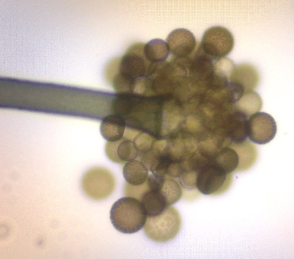
[attachment=1]Periconia sp 20-101 c – jpeg.jpg[/attachment] 
[attachment=0]Periconia sp 20-101 d – jpeg.jpg[/attachment]
Not sure whether this is P. byssoides or P. cookeri. Can anyone help, please? -
AuthorPosts

