- This topic is empty.
-
AuthorPosts
-
March 27, 2023 at 8:03 am #32588
Good morning, UK Fungi Lichen Forum — I’m a newly-minted lichen enthusiast living along the River Waveney on the border between Norfolk and Suffolk. I’ve recently been inspired by Neil Sanderson’s work on
Lecanora chlarotera s. lat.( https://www.fungi.org.uk/viewtopic.php?t=2404https://www.fungi.org.uk/viewtopic.php?t=2404” class=”bbcode_url”> L. chlarotera s. lat.material to confirm the extent of L. hybocarpavs the other Lecanorae of the subfuscagroup, particularly L. chlarotera s. str.( https://www.fungi.org.uk/viewtopic.php?t=3959https://www.fungi.org.uk/viewtopic.php?t=3959” class=”bbcode_url”> With this post I’m seeking advice on a first attempt at taking on Mark’s challenge. I collected this
L. chlarotera s. lat.material from a large twig that had fallen into a car-park on the Norwich Cathedral estate (TG 2369 0873) — BetulaI think. After a day in front of the microscope, I believe this material may be
L. hybocarpa. But I suspect that would be quite the result, and I’d be grateful for the views of UK Fungi Lichen Forum regulars. If it were L. hybocarpa, and if the BLS distribution map of January 2022 is still reasonably current, it might be a first for the hectad. A cautionary note before moving on to some photographic evidence — as mentioned, I’m only a newly-minted lichen enthusiast, and this specimen was just the second I’ve ever examined microscopically. Apologies for the inevitable deficiencies in technique!

[attachment=9]20230317-P1130726.jpg[/attachment]
The external morphology of the first specimen might suggestL. hybocarpa, showing the pink-brown apothecia mentioned in LGBI3 — but the discs aren’t entirely flat or concave, nor perhaps lightly pruinose. 
[attachment=8]20230326-P3260636.jpg[/attachment]
K+ yellow as expected.
[attachment=7]20230326-P3260644.jpg[/attachment]
An overview of an apothecial section in cross-polarised light. The polarisation was very much a DIY arrangement, with a Hoya 52mm linear polariser resting on the microscope stage atop the slide, and a Hoya 52mm circular polariser hand-held between the condenser and the illumination. Not the best section for the characteristic epithecial granules descending into the hamathecium — but it shows the expected large crystals in the thalline margin (and also the fact that I hadn’t really cut the section centrally to the apothecium).
[attachment=6]20230326-P3260676.jpg[/attachment]
Another peripheral section in cross-polarised light, showing what seem to be descending epithecial granules, yellowish in colour as expected.
[attachment=5]20230326-P3260674.jpg[/attachment]
The right extremity of the section above shown at higher magnification, to illustrate the descending epithecial granules more clearly.
[attachment=4]20230326-P3260677.jpg[/attachment]
An overview in cross-polarised light of another peripheral section with the putative descending epithecial granules.
[attachment=3]20230326-P3260682.jpg[/attachment]
The left extremity of the section above in brightfield, by way of an attempt to show the brownish epithecial granules descending between the paraphyses.
[attachment=2]20230326-P3260683.jpg[/attachment]
The same extremity in cross-polarised light, showing the descending epithecial granules more clearly.
[attachment=1]20230326-P3260684.jpg[/attachment]
The right side of the section above in brightfield.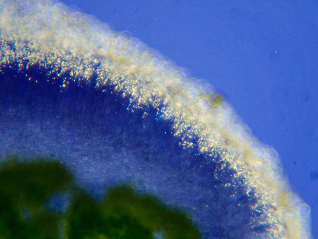
[attachment=0]20230326-P3260685.jpg[/attachment]
The right side of the section above in cross-polarised light.Based on this examination, I think there’s at least a decent argument that this specimen is
L. hybocarpa. But it’s worth mentioning that I don’t have any Pd, so I can’t rule out L. pulicarisby Pd+ red spot-test of the thalline margin. LGBI3 suggests that one could expect L. pulicaristo have darker apothecia with less crenulate margins — unless it’s that rare L. pulicarisform that LGBI3 says is virtually indistinguishable from L. hybocarpa. No doubt there are many other important identifying characteristics and possibilities that I’m simply missing as a beginner. I’d be chuffed of course if this specimen was indeed the first
L. hybocarpafrom TG20. But I’d also be chuffed with L. pulicaris, which I think from a look at the BLS distribution map might also be a first for the hectad, as well as a first in the immediate area since before 1960. But most of all I’d rather not get ahead of myself! It would be tremendous just to benefit from the guidance of the experienced lichenologists in this forum. I have other
L. chlarotera s. lat.material from the same hectad, as well as from another hectad in Suffolk — so it would be extremely helpful to know if this enthusiastic beginner is at least headed in the right direction! Many thanks,
Anthony
March 27, 2023 at 9:19 am #45379Following on the post above — on the same large twig there were many other similar Lecanora chlarotera-type thalli. 
[attachment=4]20230317-P1130729.jpg[/attachment]
I examined the thallus at the bottom of the photo here in an attempt to ascertain whether it might also beLecanora hybocarpa. It shows the flat-to-concave, more dispersed pink-brown apothecia mentioned in LGBI3 (as well as perhaps some apothecial initials characteristic of L. sinuosa? — which LGBI3 suggests might actually be the correct taxon for at least some European forms of L. hybocarpa). 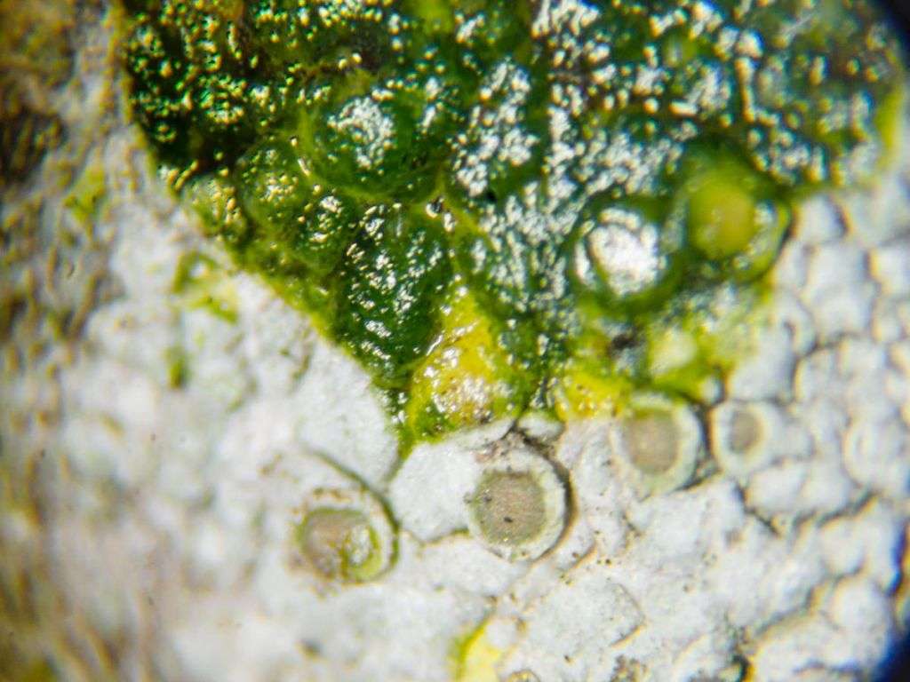
[attachment=3]20230326-P3260652.jpg[/attachment]
K+ yellow as expected, but less convincingly as the earlier thallus above (perhaps I hadn’t scratched the cortex properly, but for some reason I didn’t try again).
[attachment=2]20230326-P3260660.jpg[/attachment]
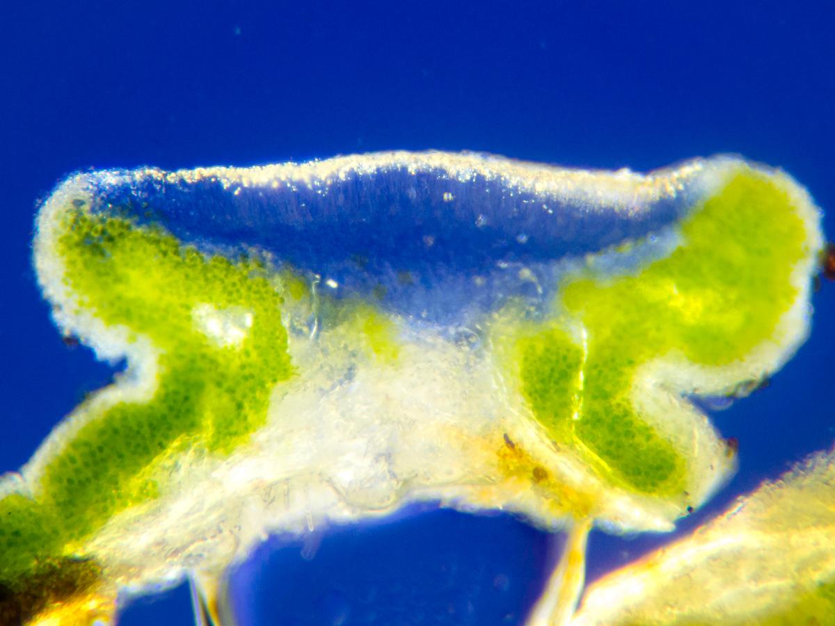
An overview of an apothecial section in cross-polarised light — and what seems to be a much more central section this time! The polarisation arrangement was the same as with the thallus above. This section shows the expected large crystals in the thalline margin, and perhaps some characteristic descending epithecial granules.
[attachment=1]20230326-P3260669.jpg[/attachment]
Another apothecial section (more or less from the centre) in brightfield.
[attachment=0]20230326-P3260668.jpg[/attachment]
The same section as above in cross-polarised light.Any advice welcome on this thallus, as well as the one above!
Thank you,
Anthony
March 27, 2023 at 12:56 pm #45380Anthony I am please to have inspired you!! You chose a very good clear
Lecanora hybocarpato start with: perfect specimen and clear features. Yes it would be ideal to check with Pd, but Lecanora pulicarisnever has the pale milky look to the disk: it is always chestnut brown (I will check, but I think LGBI 3 meant they were identical internally). Also on habitat grounds would be unlikely on a twig in Norwich; it is an acid loving lichen, if found at all in the area would most likely be on worked timber (it is on twigs in clean areas in acidic woods). The second is a less well developed Lecanora hybocarpa; it has small disks so that is expected. It is always good to go for the best developed material. Anyway
Lecanora hybocarpais now proving to be a very common species in the lowlands; finding Lecanora chlaroteras. str. is more of a challenge in some areas. Anyway very good work for the second specimen examined microscopically, your technique looks fine.
Neil
March 27, 2023 at 3:54 pm #45381I agree with Neil in his identifications of your material and all of his comments (including your clear micrographs and decent sections). The area that I know best is the Bedfordshire/Northamptonshire border and I would expect the species present in your area to be very similar. In my area
L. hybocarpaturns out to be extremely common, much more common than any of the other ‘subfusca’ group. It is frequently the only one of the group that I find on a tree. L. argentataturns up occasionally, including among colonies of L. hybocarpa. Candidates for L. argentatacan be picked out by the darker brown discs but, at a guestimate, one would have to look through several hundred thalli of L. hybocarpato find one of L.argentata. As Neil has suggested, L. pulicarisis almost entirely found on weathered wood in my area now. I have failed to find L. sinuosain the Midlands or East Anglia and I have also failed to find a single specimen of L.chlaroteras. str. in my region. Bear in mind that you might encounter corticolous occurrences of L. campestris(and not just on dusty bases of tree trunks). Here is
L. pulicariswith its Pd reaction (if you ever get to the stage of needing to test things with Pd, then private message or email me as I have a large stock of it). 
[attachment=1]E101BT-WQAIpew1.jpeg[/attachment] The discs of your
L. hybocarpaare the typical pale colour but you will occasionally find darker individuals such as this: 
[attachment=0]FICb43QWYAQzzM0.jpeg[/attachment]
https://twitter.com/obfuscans3/status/1477352704439635977 In case you start to get bored in your search for species other than
L. hybocarpain your subfuscastudies, perhaps some other challenges would interest you? Fifteen years ago X. polycarpawas abundant in my area but now much declined: https://twitter.com/obfuscans3/status/1494392417579057162 Similarly
Amandinea punctatahas much declined, but a near look-alike Catillaria nigroclavatahas appeared and become common. https://twitter.com/obfuscans3/status/1206182408875319296 Similarly (but more difficult to identify)
Scoliciosporum chlorococcumhas much declined and a near look-alike Halecania viridescenshas become common. https://twitter.com/obfuscans3/status/1208423621296840705 These are examples of the sort of dramatic changes that can be witnessed by examining the lichens growing on twigs.
March 27, 2023 at 7:41 pm #45382Dear Neil and Mark — Many thanks for your time — and for your votes of confidence on both the
Lecanora hybocarpaidentification and my microscope technique. Unless either of you object, I’ll submit this to the BLS as a record for TG20, which isn’t shown as hosting
L. hybocarpaon the January 2022 BLS distribution map. Or would a true record demand a Pd- spot-test to be absolutely certain? If so, would you have any advice for using and storing Pd safely considering the toxicity? It’s especially helpful to know that
L. pulicarisdoesn’t ever show pale discs like these, and that I shouldn’t generally expect to find it in urban settings. It’s also useful to know that the second L. hybocarpaspecimen is simply less well-developed — which I’ll use as a guide when picking over my other collected L. chlarotera s. lat. material for the best apothecia to examine. I appreciate Mark’s very helpful pointers about dealing with the
subfuscagroup — and the offer of Pd if necessary. I intend to continue to examine local L. chlarotera-type material, including on twigs I’ve collected from a couple of adjoining hectads in north Suffolk not shown on the January 2022 BLS distribution map as hosting L. hybocarpa. Based on all the advice, I won’t be surprised to find that this material is indeed L. hybocarpa. At any rate, the thalli look similar to the ones I posted above, at least at first glance. Mark’s additional challenges are welcome! I’m particularly curious about the
Amandina punctatachallenge. Before tackling the L. chlarotera-type material, I practised sectioning apothecia from a Lecidella elaeochromathallus on the same twig, which I aimed to prove wasn’t A. punctataby examining the spores. It wasn’t — and perhaps no surprise if A. punctatais in decline. Thanks again, and hope to stay in touch from time to time.
Anthony
-
AuthorPosts




- You must be logged in to reply to this topic.