Forums › Forums › Fungi on miscellaneous substrates (not soil or wood) › Tremella versicolor?
- This topic is empty.
-
AuthorPosts
-
June 20, 2016 at 1:39 pm #30218
Hi, I noted this tiny jelly fungus only after repeated but unsuccessful (so far) attempts to identify a Peniophora crust fungus collected from Longshaw, Derbyshire. In fact I didn’t spot the jelly on the crust for a fortnight – it’s less than a millimetre across and easy to miss when dry. A squash preparation didn’t show the “tuning” fork structures or allantoid basidiospores of a typical Tremella – instead it provided clusters of conidia-like bodies. Buckzacki gives the following: “SPORES: basidiospores are absent but clusters of ellip CONIDIA 3-5 x 1.5-5 um” – this doesn’t look a million miles away from my specimen. According to Buckzacki this tiny Jelly was regularly recorded in the 19th C but “forgotten about” till the middle of the 20th C.
Steve
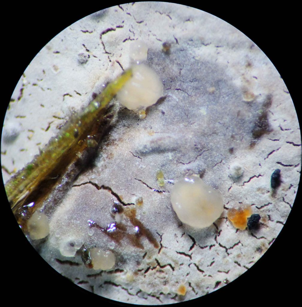
[attachment=5]1 Stereo image x45 wet.JPG[/attachment] 
[attachment=4]2 Stereo image x45 dry.JPG[/attachment] 
[attachment=3]3 Stereo image.JPG[/attachment] 
[attachment=2]4 Squash x 600 congo red.jpg[/attachment] 
[attachment=1]5 Squash x 1000 in Congo Red.jpg[/attachment] 
[attachment=0]6 x1000 in Met Blue – stack.jpg[/attachment] June 20, 2016 at 10:33 pm #38397Steve wrote:Hi,I noted this tiny jelly fungus only after
repeated but unsuccessful (so far) attempts to identify a Peniophora crust fungus . . . .
Hi Stevedon’t have any thoughts at the moment re the jelly. But forget the
Peniophora– in my experience unless you get them in prime condition you’ll get nowhere (except perhaps another order for coverslips 
cheers
Chris
June 21, 2016 at 12:41 pm #38398Hi Chris, Those are wise words. The key in CNE starts off with dendrohyphidia and then sulfocystidia, moving on to subiculum properties. Very scary. And
just21 species 😆 The cystidia can be beautiful though – this one especially so,
Steve

[attachment=1]16 oile.jpg[/attachment] 
[attachment=0]2016-06-21 13-35-26 (B,Radius8,Smoothing4).jpg[/attachment] June 21, 2016 at 11:32 pm #38399Steve wrote:Hi Chris,
. . . . The cystidia can be beautiful though – this one especially so,
Steve
Agreed Steve, but those cystidia are clearly dead – and I’d be surprised if you got a spore-dropbest wishes
Chris
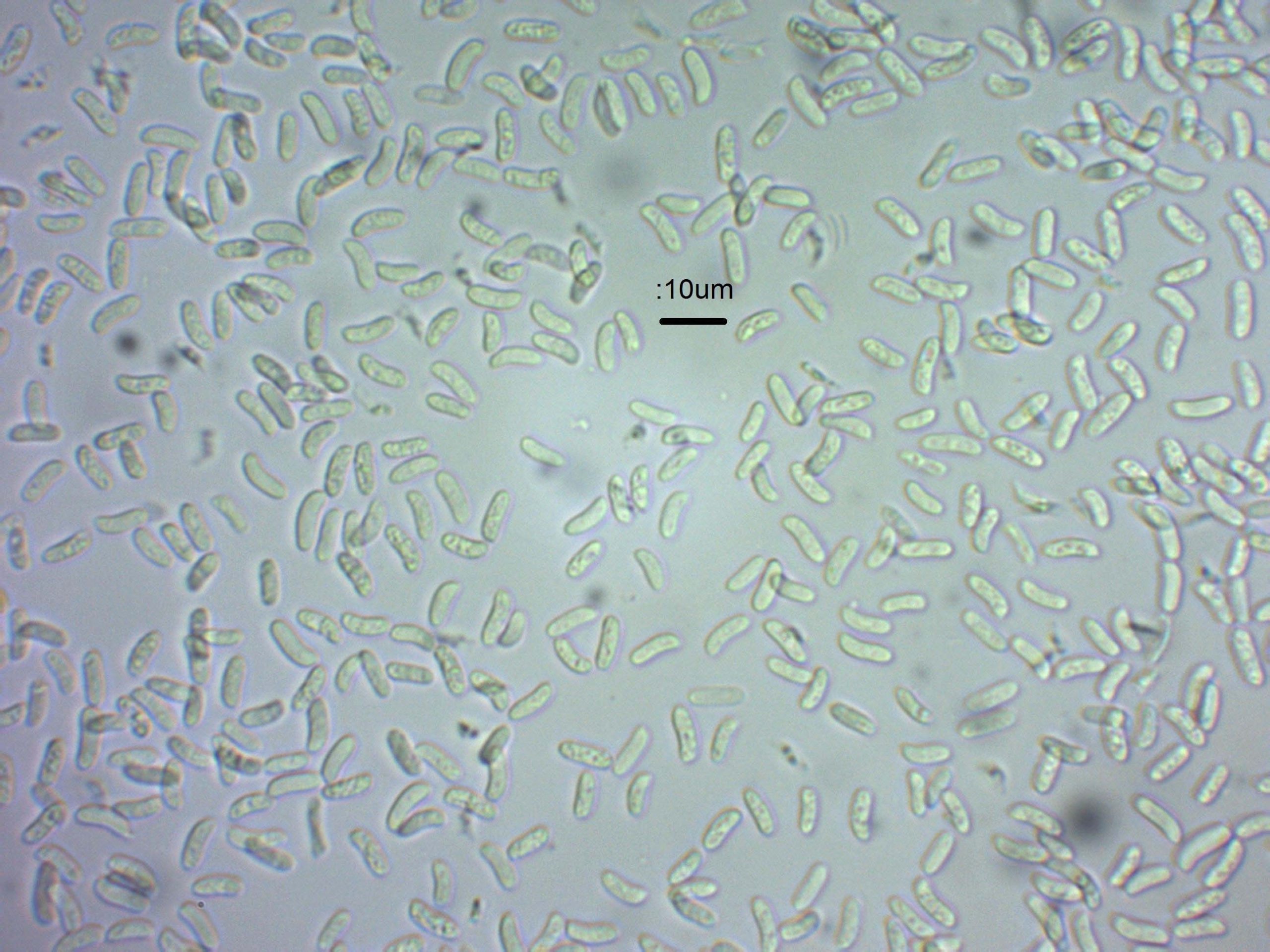
June 28, 2016 at 10:43 pm #38400Hi Chris, In fact I got a lovely spore deposit of pale pinkish spores! The Congo Red slides were in water and ammonia, and also in KOH and NH3 (the denuded cystidia) so were going to be dead. NH3 is nasty stuff and so is KOH. So I always look at fungi slides in water first, as I am noticing a drastic effect of KOH solution on crystalline covering of cystidia (it gets dissolved). With all kinds of cystidia.
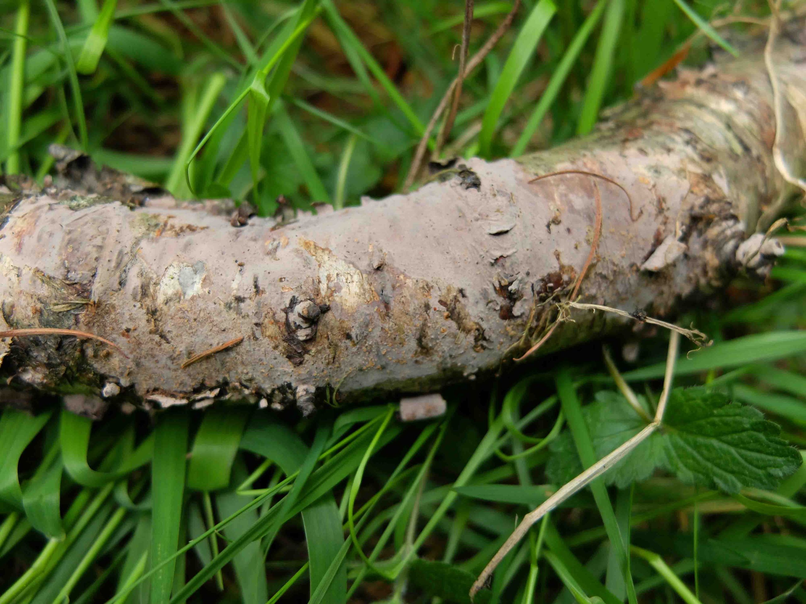
Here are some of the photographic “evidence base” for this specimen of Peniophora (which I collect for each of the fungi I collect – before I go near a book). To me that’s the only way to try to get an objective identification. It’s hard work and I get 3-400 photos/micrographs for each trip to Longshaw, for about 20-30 specimens.
I am stuck on this Peniophora as I don’t keep the nasty chemicals required for ID in the house – I have an inquisitive granddaughter.
But I agree that it’s likely a lost cause to try to ID some Peniophoras, as they are hard and unfriendly to say the least. I couldn’t find basidia on this one, perhaps because I didn’t spend enough time searching.
Cheers,
Steve

[attachment=3]Field DSCF7582.JPG[/attachment] 
[attachment=2]Stereo DSCF7648.JPG[/attachment] 
[attachment=1]Spores 16 600.jpg[/attachment] 
[attachment=0]Water 16 oil wat.jpg[/attachment] July 3, 2016 at 1:00 pm #38401Well it’s clearly not one of the orange ones, nor is it P. limitataor P. quercina; looks very like P. cinerea– I’d start from there . . . cheers
Chris
July 4, 2016 at 9:48 am #38402Agreed! -
AuthorPosts



- You must be logged in to reply to this topic.