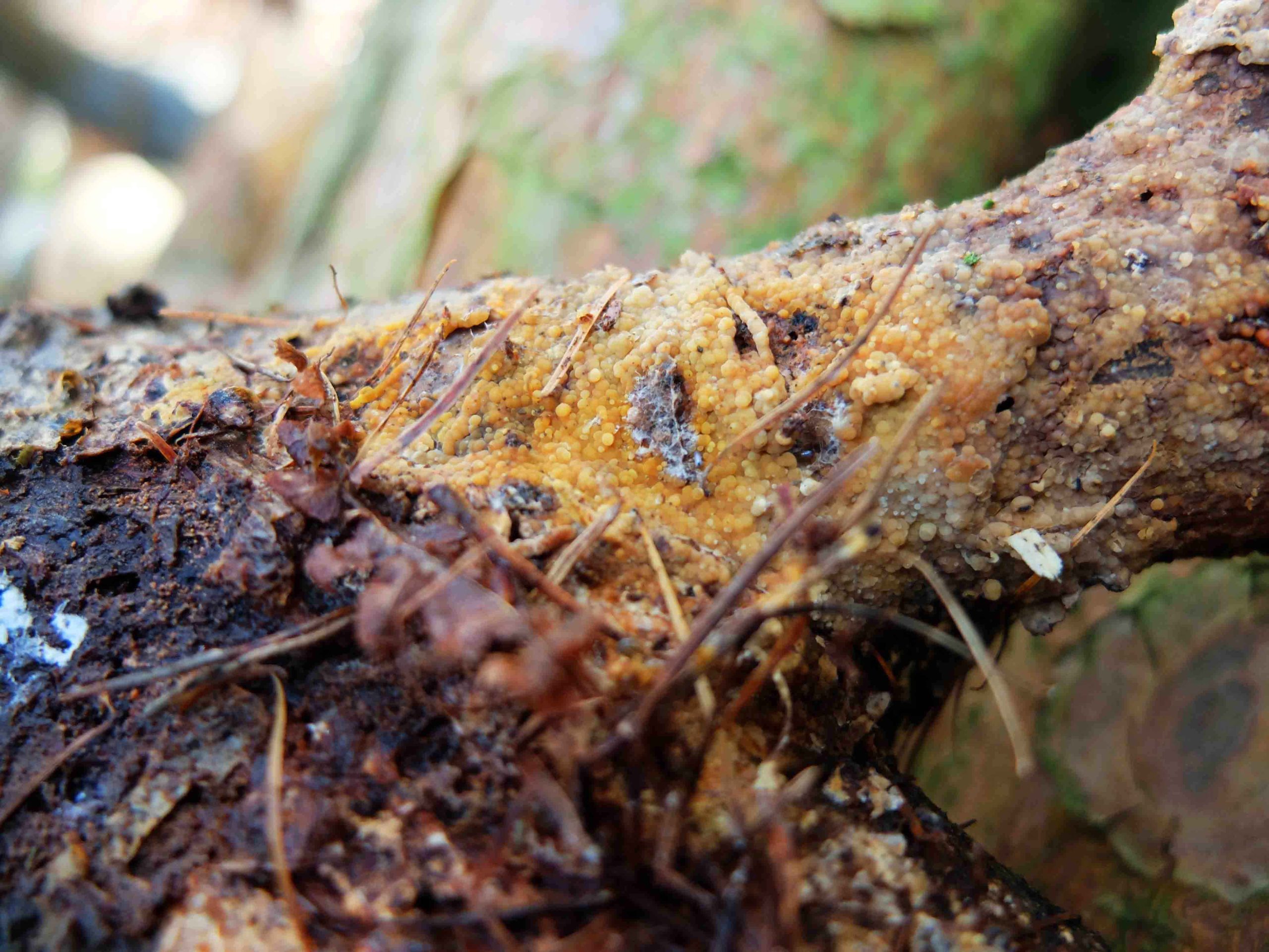
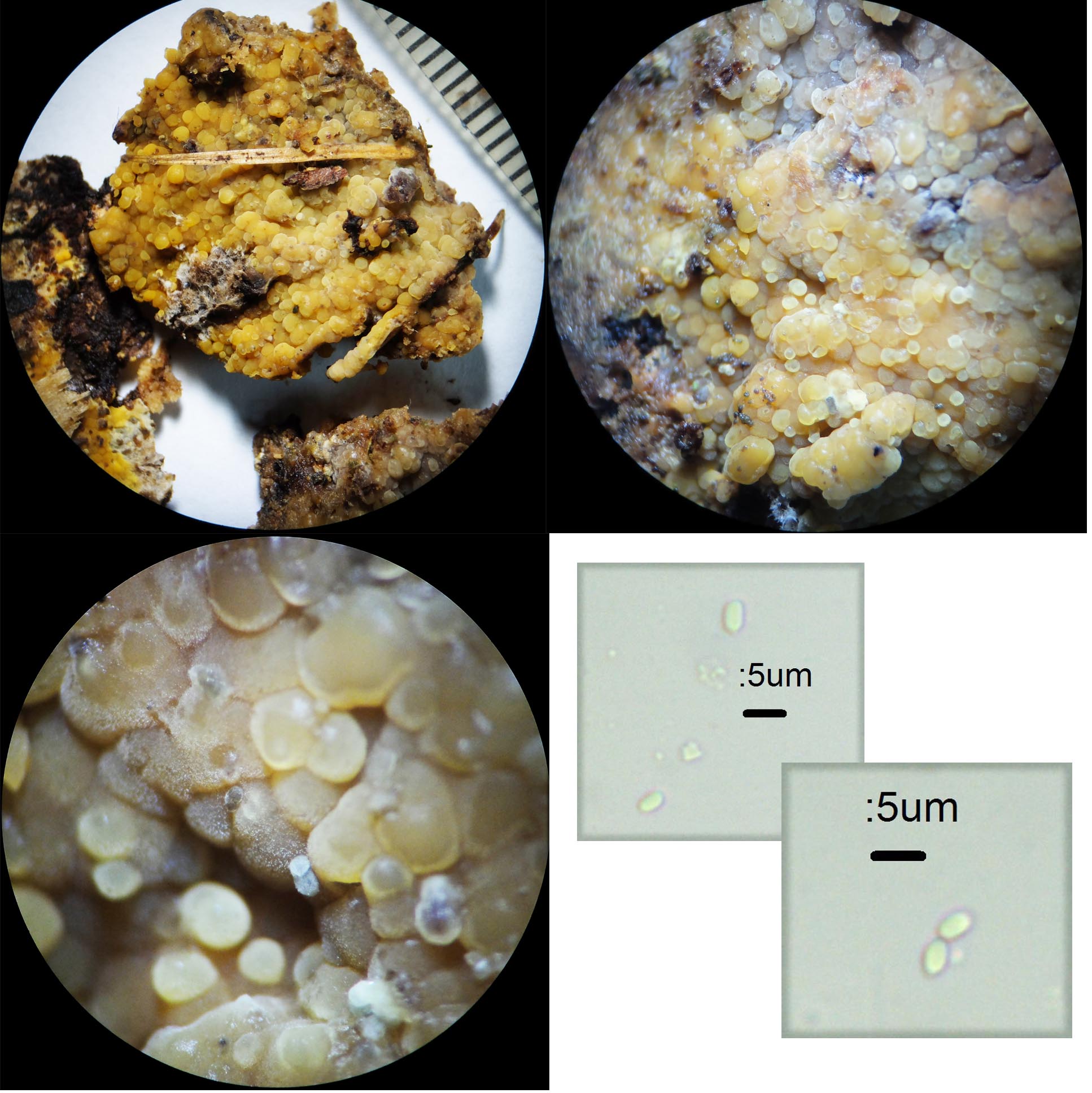
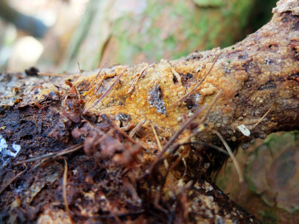
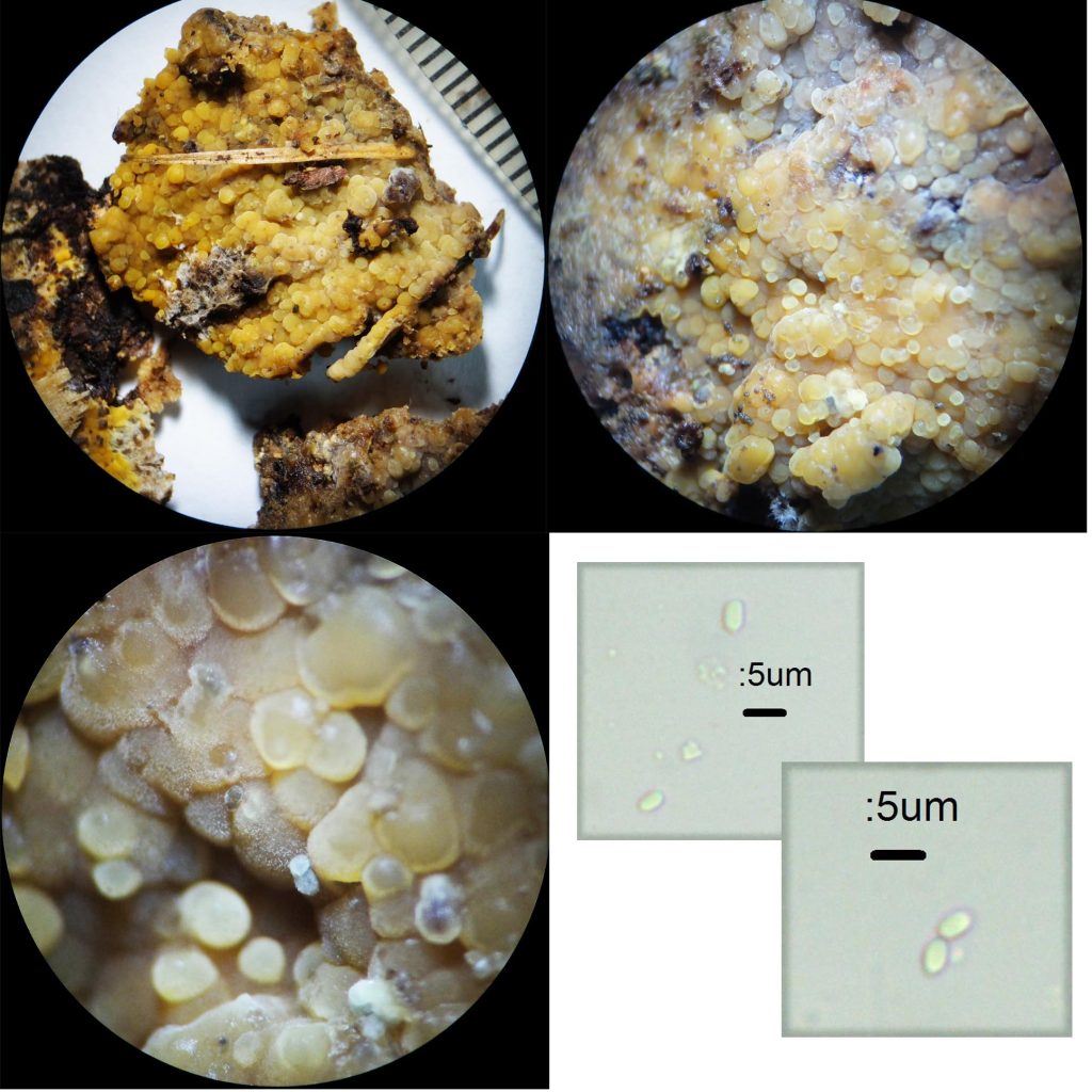
This yellowish “pustular” crust was found last week at Longshaw in a conifer plantation – mostly Scots Pine, which included some birch and other broadleaf.
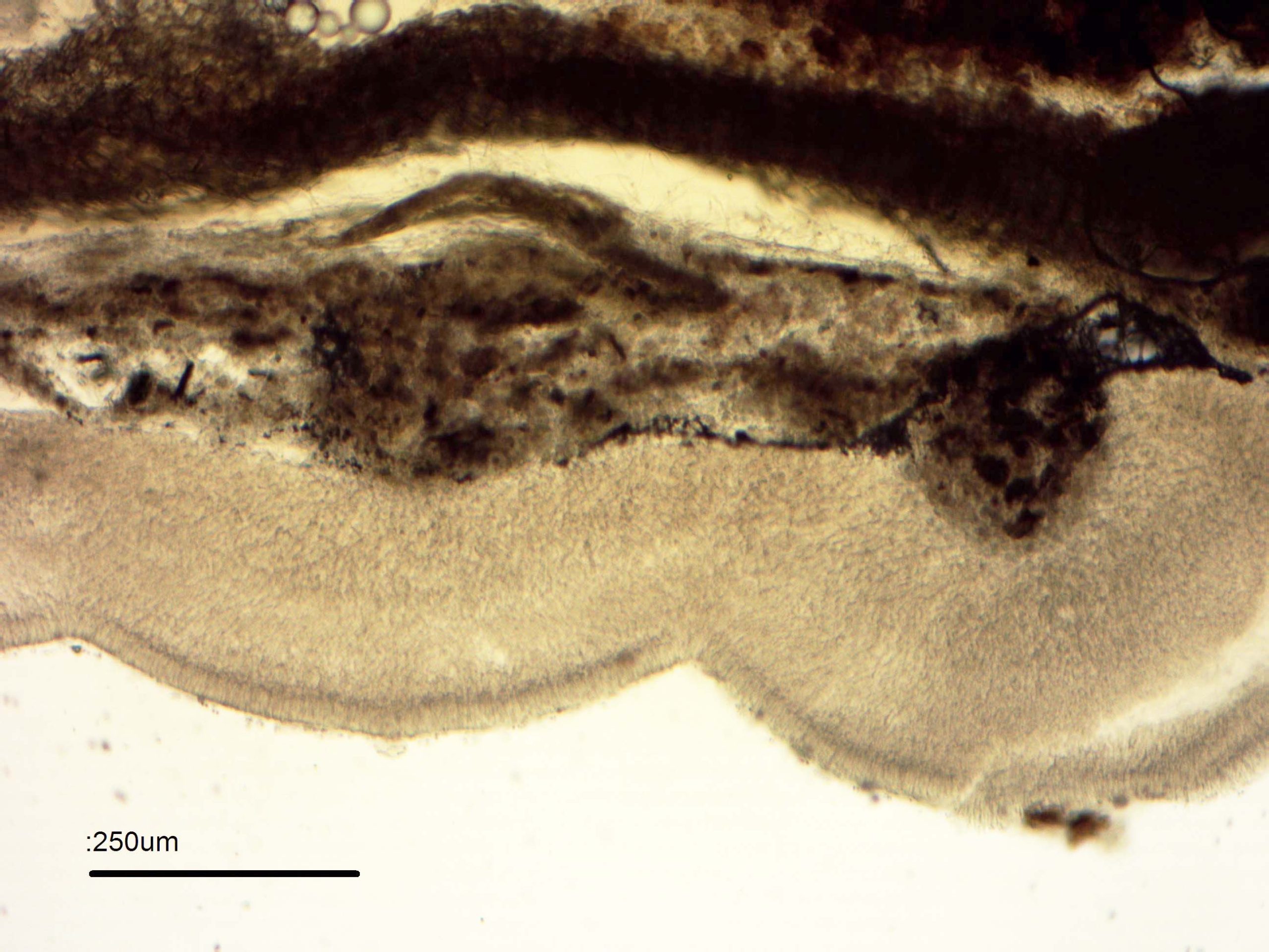
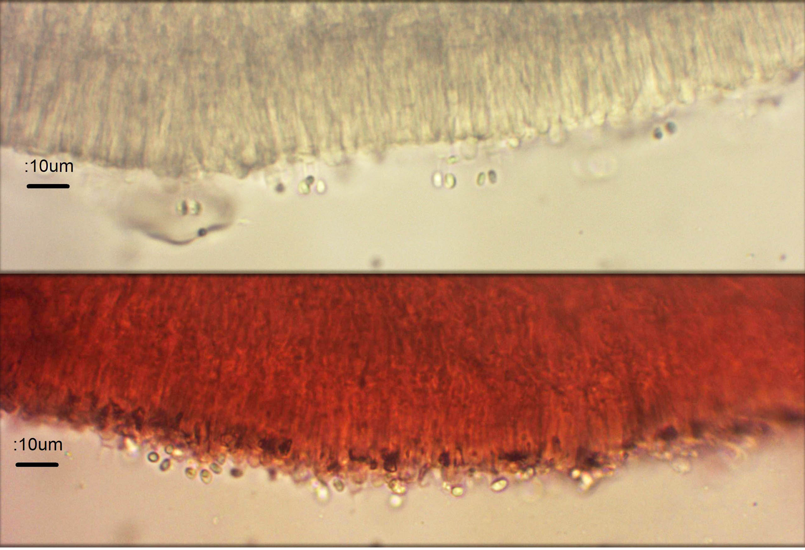
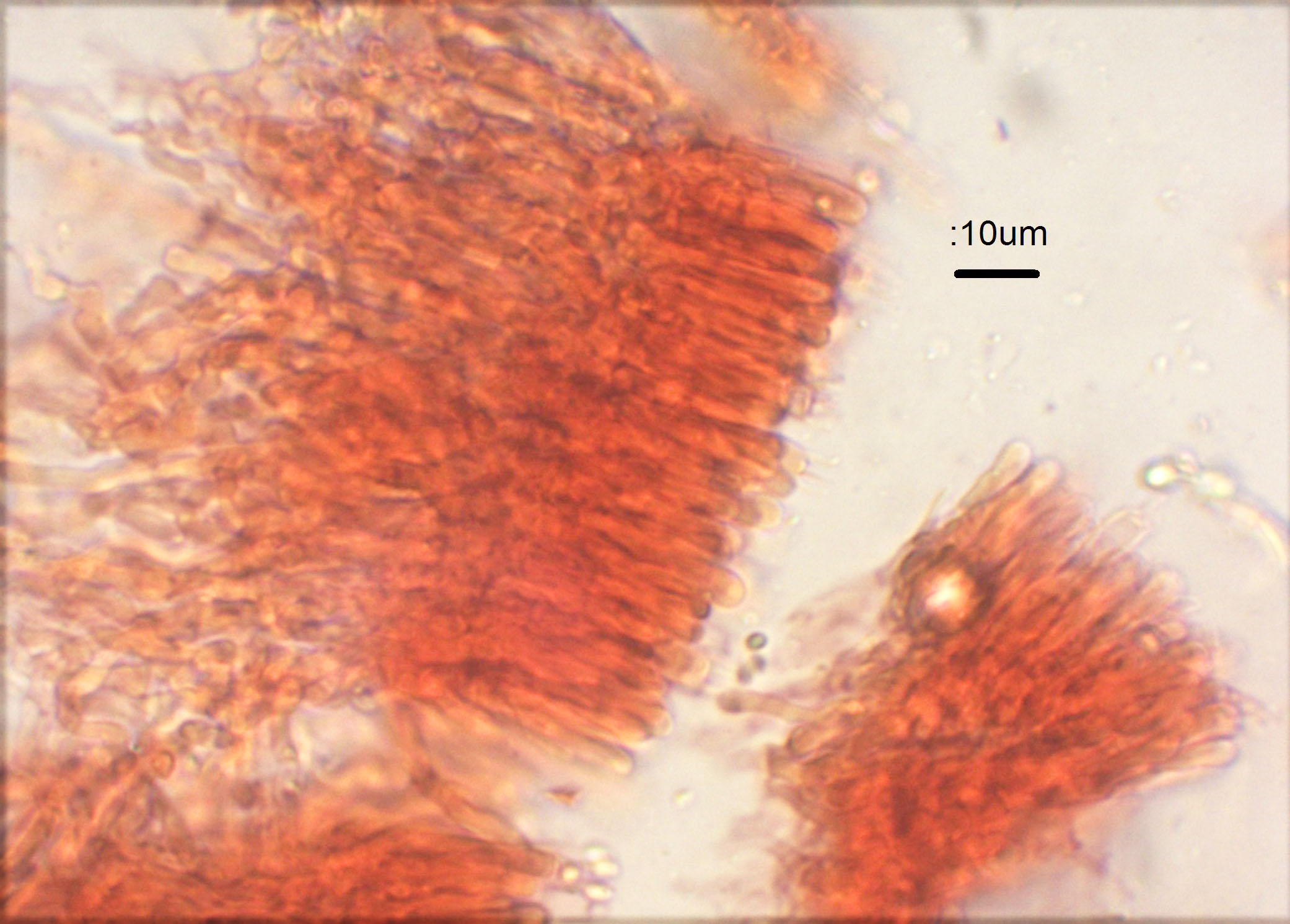
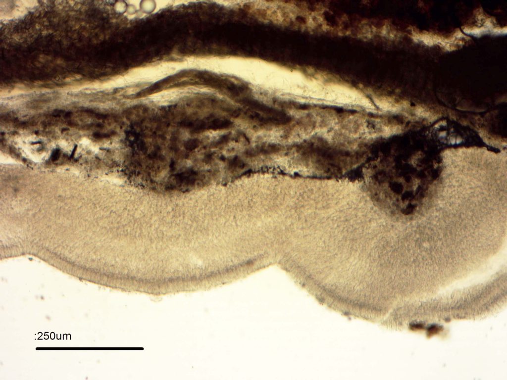
At first sight it looks like Megalocystidium leucoxanthum cf. in FOS. The microscopy soon dispelled this, and suggested Phlebia. Very few spores were shed overnight, so the second specimen (immediately dried) was checked – the same kind of spores were attached to basidia. They were 3.5-4 x 2-2.5 …ish. No obvious cystidia were noted on the surface. Hyphae were very twisted and irregular, about 3-5 wide bit with swellings along the hyphae up to 8. A few clamps were seen. Large terminal swellings up to 10 wide (lenghth not discernable) on some hyphae might conceivably be interpreted as leptocystidia, embedded in the crust fungus. This would fit Phlebia rufa as shown in FOS. The hyphae were gelatinised and hard to separate on squashing even in KOH. I concluded that this was most likely Phlebia rufa, which I think is quite variable in colour as is Plebia radiata.
Steve
[attachment=5]UK1 field.JPG[/attachment]
[attachment=4]UK2 STEREO SPORES1.jpg[/attachment]
[attachment=3]uk3 11 100 koh.jpg[/attachment]
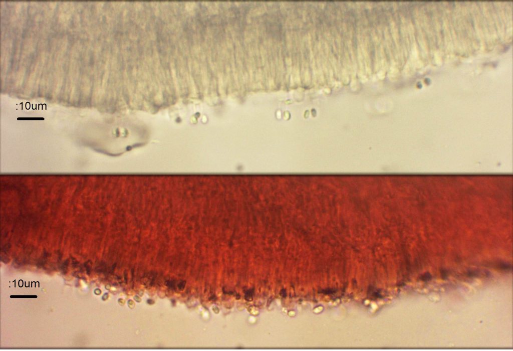
[attachment=2]UK4 SURFACE1.jpg[/attachment]
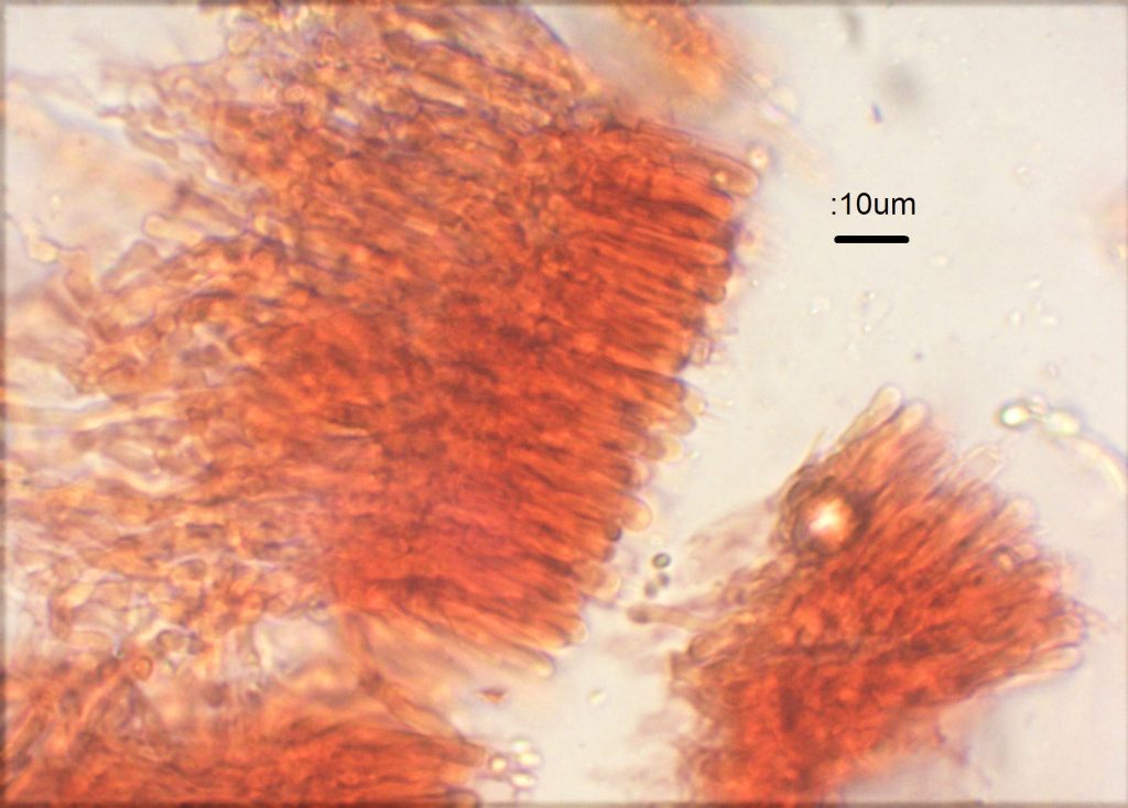
[attachment=1]UK5 Gelat kohcrc.jpg[/attachment]
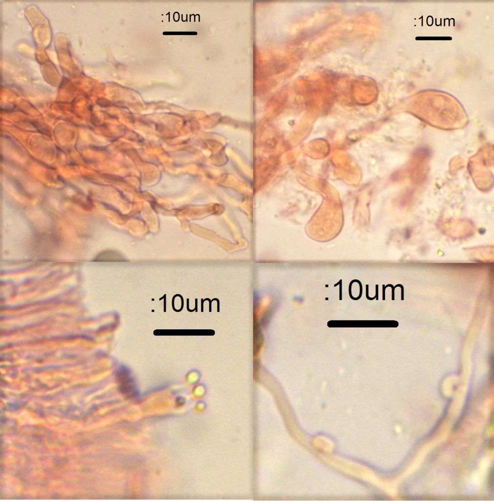
[attachment=0]UK6 micro.jpg[/attachment]