Forums › Forums › Fungi on miscellaneous substrates (not soil or wood) › Myxo on dung?
- This topic is empty.
-
AuthorPosts
-
September 26, 2015 at 12:57 pm #29773
Found on horse dung near a Pine stand on Ashdown Forest. I would love to do microscopy but I’m not sure how. 
[attachment=1]RSCN9167.JPG[/attachment] 
[attachment=0]DSCN9168r.jpg[/attachment] September 26, 2015 at 4:23 pm #36463Hi, not a Myxomycete but, with that granular outer surface, possibly Cheilymenia granulata, a member of the Ascomycota. You can suspend it over a slide overnight to collect some spores (if you are lucky) see if you can see the striations under oil X100. To see the asci and paraphyses you need to take a very thin section of the hymenial surface and gently squash it on the slide.
http://www.newforest.com/page_2.asp?epivalue=Cheilymenia+granulata&x=70&y=56 ” class=”bbcode_url”> http://www.newforest.com/page_2.asp?epivalue=Cheilymenia+granulata&x=70&y=56 Dave
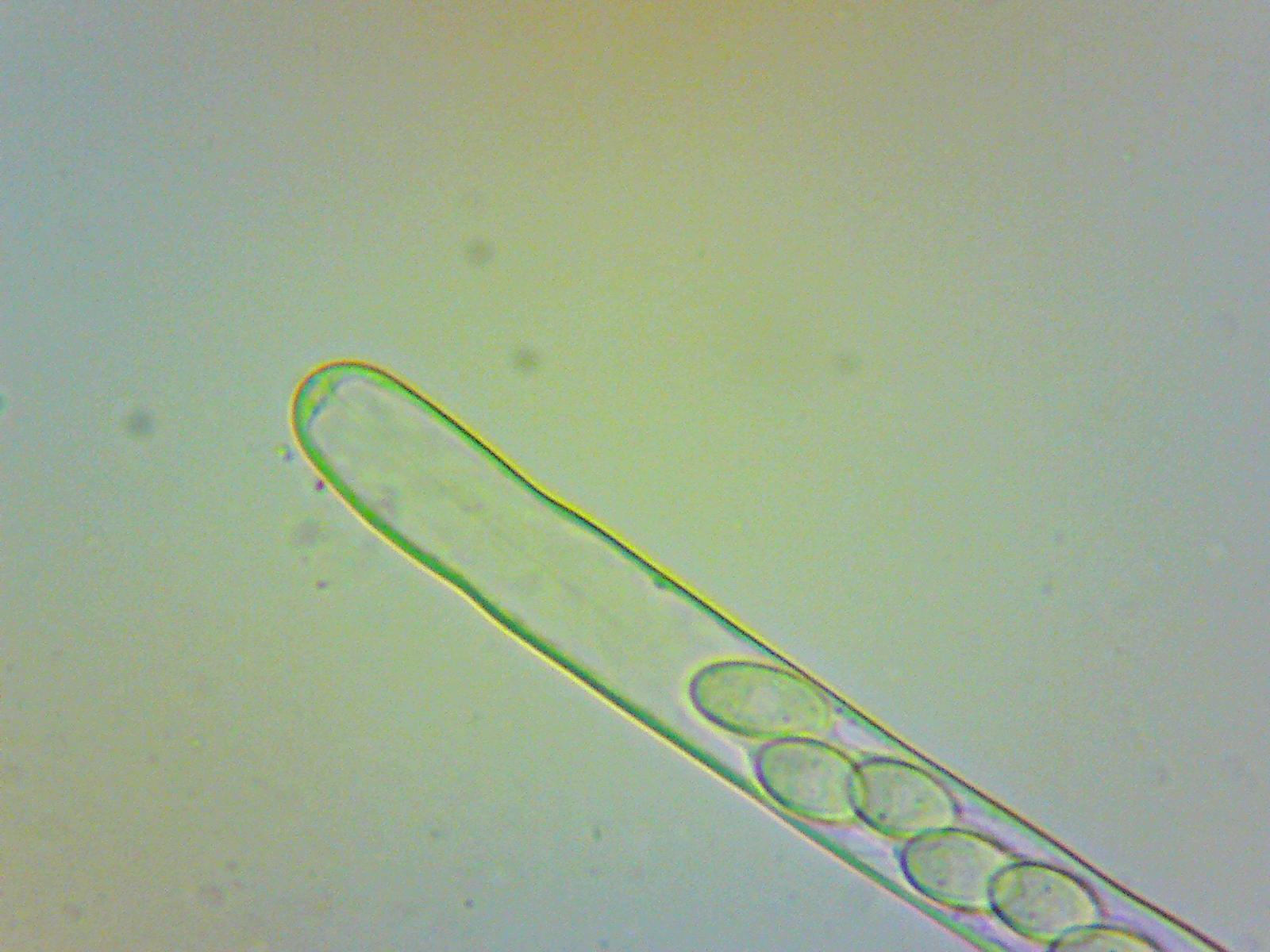
September 26, 2015 at 5:14 pm #36464Thanks Waxcap Are these OK? The spores, if they drop, will be coming soon.

[attachment=3]Ashdown Forest – Dung-dwelling Asco – Asci001 – Unmeasured.jpg[/attachment] 
[attachment=2]Ashdown Forest – Dung-dwelling Asco – Asci002.jpg[/attachment] 
[attachment=1]Ashdown Forest – Dung-dwelling Asco – Asci003 – Apical Pore.jpg[/attachment] 
[attachment=0]Ashdown Forest – Dung-dwelling Asco – Asci003 – Apical Pore002.jpg[/attachment] September 26, 2015 at 6:18 pm #36465An excellent start. Which objective were you using? I’d suggest asci at 40X and spores at 100X in oil. Dave
September 26, 2015 at 6:45 pm #36466That’s good to hear! I’m labelling the photos with the objectives used.
September 27, 2015 at 9:18 am #36467With Asco’s the shape of the paraphyses is important and you can see the inflated ends nicely in your 10x photo. Most asco’s have 8 spores in each ascus but there are exceptions. Seeing the spores in the ascus is helpful as you get an idea of the size and shape but to get an accurate size and shape – and see the all important surface ornamentation (or lack of it) – you must look at naturally released spores in something like Cotton Blue stain at 100x, under oil. Dave
-
AuthorPosts


- You must be logged in to reply to this topic.