
Possible Melaspilea amota?
- This topic is empty.
-
AuthorPosts
-
February 2, 2025 at 12:48 pm #32992
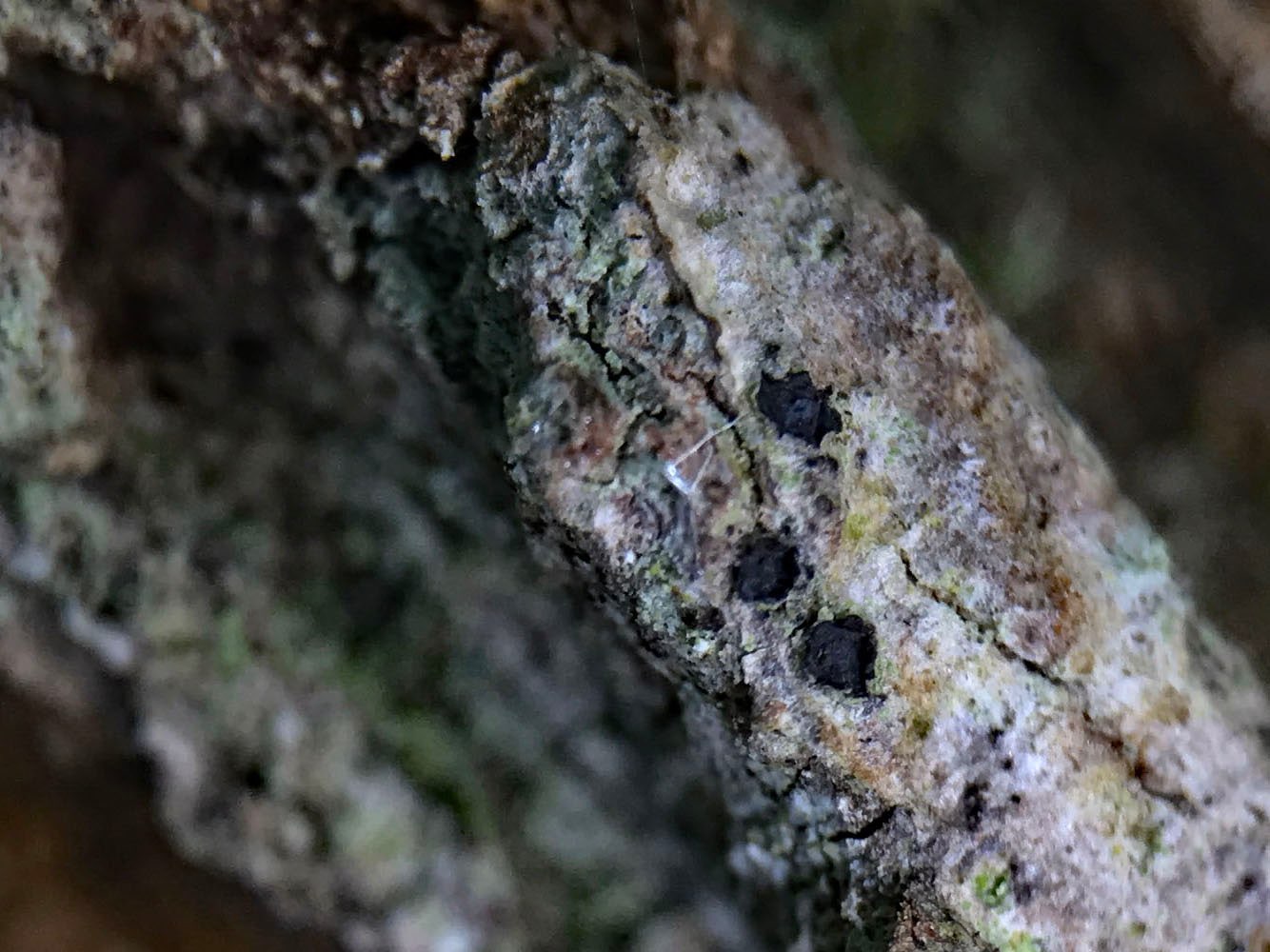
Going through various fallen twigs and small branches from a Dorset roadside and I’m wondering if I might have come across a possible Melaspilea amota. It was (probably) on Holly with erumpent lecedeine apothecia partially covered by bark cells. The asci appeared to be (4-)8 spored, spores 1-septate, green, 17-20 x 7-8 µm and there was no K reaction.
Comments appreciated as I can find no images online to compare with.

[attachment=6]z Melaspilea Q 250130 005.jpg[/attachment] 
[attachment=5]z Melaspilea Q 250130 007.jpg[/attachment] 
[attachment=4]z Melaspilea Q 250130-4.jpg[/attachment] 
[attachment=3]z Melaspilea Q 250130-5.jpg[/attachment] 
[attachment=2]z Melaspilea Q 250130-7.jpg[/attachment] 
[attachment=1]z Melaspilea Q 250130-7b.jpg[/attachment] 
[attachment=0]z Melaspilea Q 250130-1.jpg[/attachment] February 2, 2025 at 6:25 pm #46481Looking at last nights sections (all in K) the spores were now pale brown 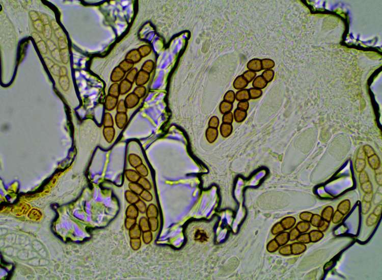
[attachment=1]z Melaspilea Q 250130-11.jpg[/attachment] but the asci tips weren’t K/I+ blue when I removed the cover slip and added a drop of Melzers
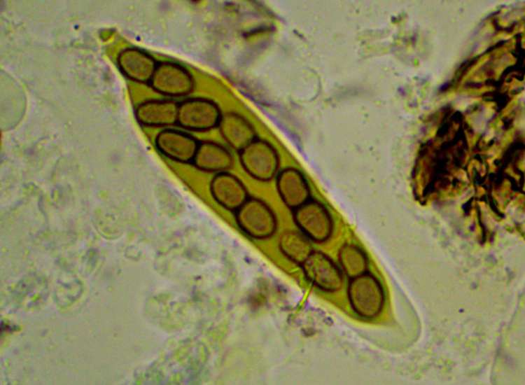
[attachment=0]z Melaspilea Q 250130-13 Melzers.jpg[/attachment] In this case, and despite the erumpent apothecia, is it just Melaspilea ochrothalamia?
February 2, 2025 at 7:04 pm #46482Jenny It is certainly not
Melaspilea amota, that looks very different and also has distinctly different spores, they are strongly sole shaped and shorter and wider than your spores and are colourless or pale brown. I thought I would have more pictures than I have but this one of typical apothecia (now up on the BLS website too). 
[attachment=0]2019-05-21-20.jpg[/attachment]
Melaspilea amota, old Oak, Coed Glan-yr-afon, Coedydd Maentwrog NNR Melaspilea amotais a bark fungus that erupts from some depth in rough thick bark and I have only seen it on old trees in ancient woodlands. It never grows on smooth thin bark. Your apothecia looks to be growing under the outer epidermis of the Holly bark, and looks very similar to
Arthothelium ruanumapothecia. The spores are not muriform, so it can not be that, but I am not sure what that is at all. Do you have a photo of an apothecia cross section? Neil
February 2, 2025 at 9:18 pm #46483Thanks for your reply Neil – and I obviously went very wrong with this one! Unfortunately these images are all that I have and I didn’t take any of an apothecia in cross section.
Ah well, another for the ‘unknown folder’.
February 3, 2025 at 12:27 pm #46484Sure it’s not a Mycomicrothelia? Paul
February 3, 2025 at 3:27 pm #46485Thank you Paul! Not sure how I missed that as the green > brown spores are virtually identical to those in a post of mine towards the end of last year that you confirmed as Mycomicrothelia confusa.
But ……. this specimen appears to have an obvious halo of bark cells?
-
AuthorPosts



- You must be logged in to reply to this topic.