
Anisomeridium viridescens?
- This topic is empty.
-
AuthorPosts
-
July 29, 2024 at 1:38 pm #32849
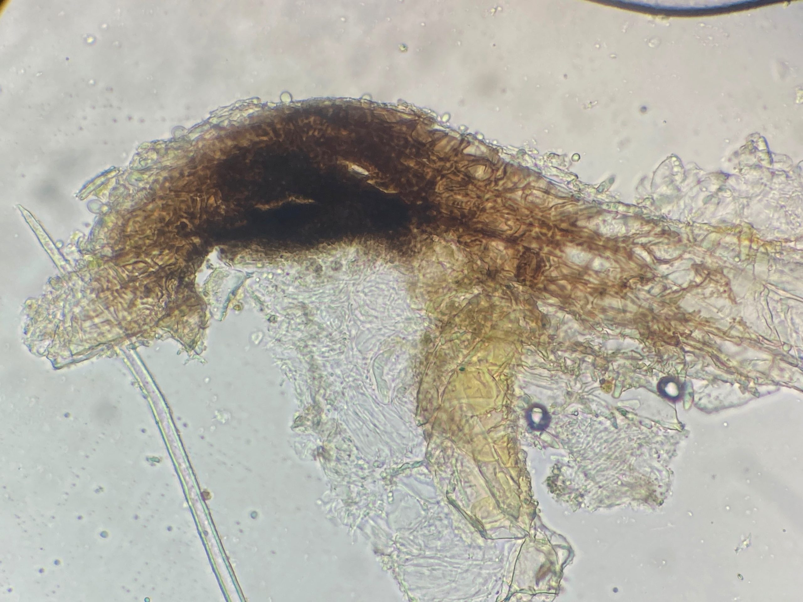
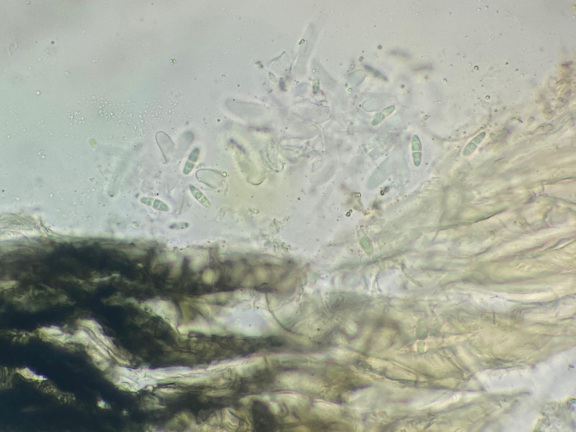
SW Ireland on a dead Hazel twig – is this Anisomeridium viridescens rather than Arthopyrenia carneobrunneola? Perithecia 0.1-0.2mm diameter, spores becoming brown with age 1-septate 15-17(-20) x (3.5-)4-4.5(-5) µm. Pseudoparaphyses (?) visible left side of first two micrographs.

[attachment=4]Anisomeridium viridescens Q 240719 008.jpg[/attachment] 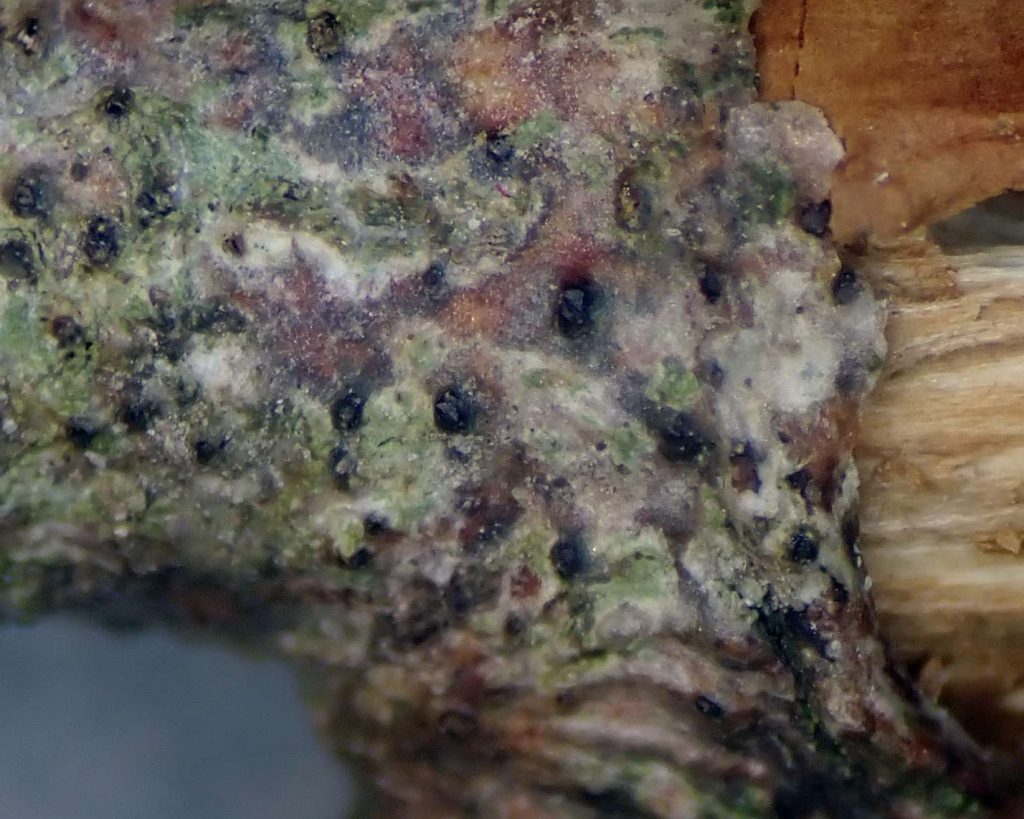
[attachment=3]Anisomeridium viridescens Q 240719 009.jpg[/attachment] 
[attachment=2]Anisomeridium viridescens Q 240719-1.jpg[/attachment] 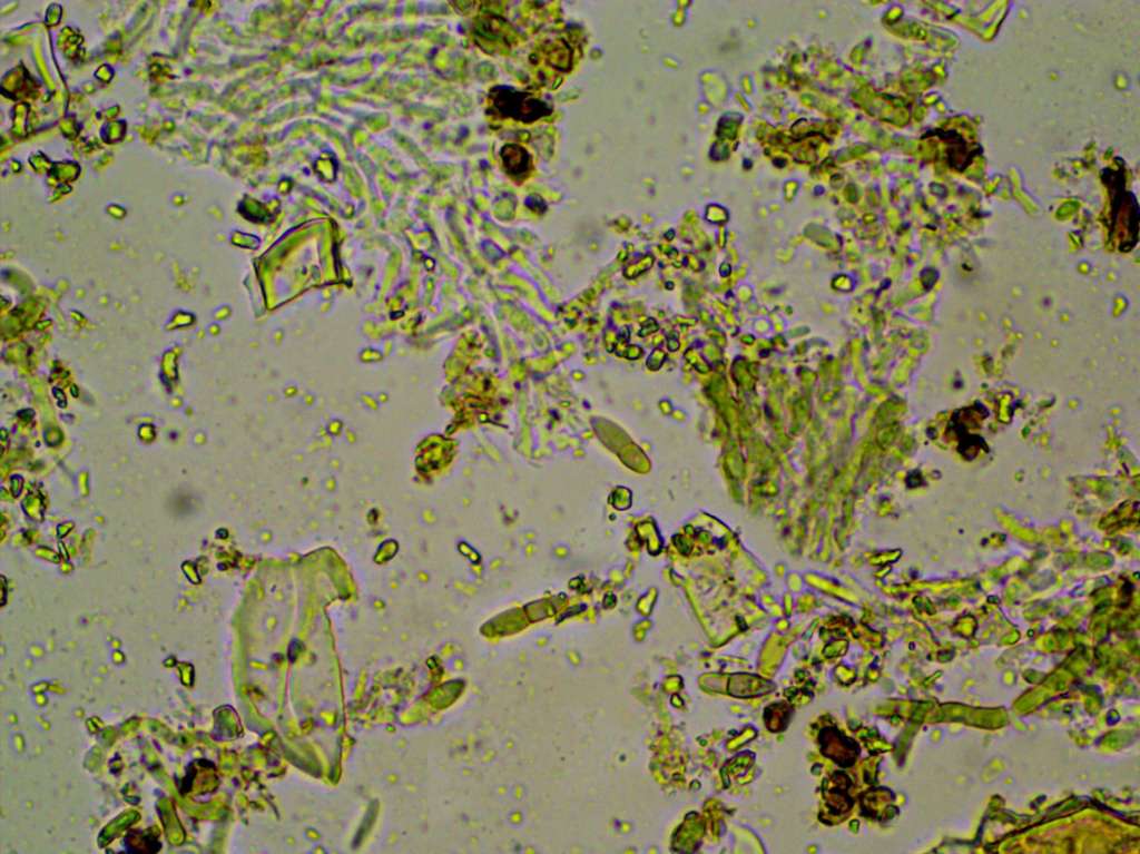
[attachment=1]Anisomeridium viridescens Q 240719-5 pseudoparaphyses q.jpg[/attachment] 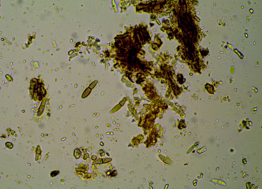
[attachment=0]Anisomeridium viridescens Q 240719-9.jpg[/attachment] July 29, 2024 at 11:51 pm #46129And from a bit further along the same twig both perithecia and pycnidia present, spores 13-16 x 3.5-4.5 µm, macroconidia 10-12x 2-2.5 µm. 
[attachment=2]w Anisomeridium viridescens 2 240719-10b.jpg[/attachment] Macroconidia

[attachment=1]w Anisomeridium viridescens 2 240719-4 con.jpg[/attachment] 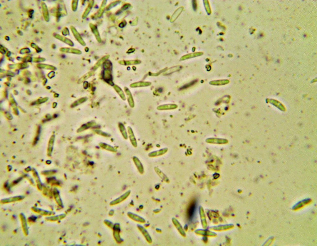
[attachment=0]w Anisomeridium viridescens 2 240719-3 con.jpg[/attachment] July 31, 2024 at 1:52 pm #46130Dear Jenny — In his monograph on pyrenocarps, Alan Orange wrote that the clearest character distinguishing between
Anisomeridium viridescensand Arthopyrenia carneobrunneolais that the involucrellum of the former is K+ green, whereas that of the latter remains brown. Not sure what mount you used for your slides, but perhaps mounting in K might help if you haven’t yet tried it? I think the thallus of
A. carneobrunneolashould also be browner — yours seems to be white in places, which suggests A. viridescensas you’ve already suspected. Best regards,
Anthony
July 31, 2024 at 3:31 pm #46131Thanks for the reply Anthony, and the slides were mounted in K. The lack of an obvious K+ green reaction in my first section concerned me so I was relieved to find both perithecia and pycnidia in the second section as macroconidia aren’t mentioned for Arthopyrenia carneobrunneola.
I checked the LGBI3 descriptions for both Anisomeridium viridescens and Arthopyrenia carneobrunneola and though the K reaction and presence/absence of pycnidia are the most obvious differences there is also a slight difference in the spore shape:
A. viridescens “usually constricted at the septum; the upper cell usually distinctly wider than the lower”
A. carneobrunneola “not or only slightly constricted at the median or somewhat supramedian septum”
Though some of the spores in my sections are a bit intermediate they do compare better with Mark Powells’s illustration of Anisomeridium viridescens here:
https://fungi.myspecies.info/sites/fungi.myspecies.info/files/Anisomerid%20viridescens.jpg ” class=”bbcode_url”> https://fungi.myspecies.info/sites/fungi.myspecies.info/files/Anisomerid%20viridescens.jpg than with the Asco France photographs of Arthopyrenia carneobrunneola here:
http://www.ascofrance.com/recolte/3729/dothideomycetes-pleosporales-arthopyreniaceae-arthopyrenia-carneobrunneola ” class=”bbcode_url”> http://www.ascofrance.com/recolte/3729/dothideomycetes-pleosporales-arthopyreniaceae-arthopyrenia-carneobrunneola Jenny
August 1, 2024 at 2:10 pm #46132Agree this is most like Anisomeridium viridescens, Naetrocymbe carneobrunneolahas quite a distinct dark brown thallus. The K+ green involucrellum is not that obvious, a dull green rather than anything very distinctive. Neil
August 1, 2024 at 2:45 pm #46133Thanks for the confirmation Neil – and very reassuring that the K+ isn’t obvious! (Any chance of adding that to LGBI3?) August 3, 2024 at 8:58 pm #46134Got some Anisomeridium viridescenstoday on a Hampshire Flora Group meeting in N. Hants, new to the vicecounty. So I took some pictures: 
[attachment=2]2024-08-03-05.jpg[/attachment]
Cross section of perithecia in water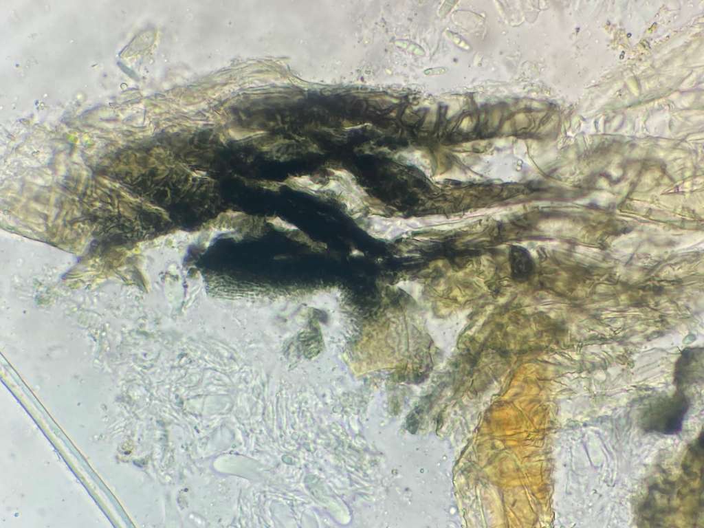
[attachment=1]2024-08-03-06.jpg[/attachment]
Cross section of perithecia in K
[attachment=0]2024-08-03-07.jpg[/attachment]
SporesSeeing it to day reminded me that the thallus on your specimen looks far to well developed for
Anisomeridium viridescens, so still could be something else, possibly. Neil
August 3, 2024 at 9:53 pm #46135Thanks for posting the micrographs from your specimen Neil, and might you have any suggestion of what mine might be if its not Anisomeridium viridescens? Photograph below shows where both the first and second group of micrographs were from (the second with both spores and macroconidia).

[attachment=0]zz Anisomeridium viridescens Q 240719 008.jpg[/attachment] August 4, 2024 at 7:40 pm #46136Is the thallus something else over growing it? In which case, with the conidia, it is fine (i.e. it can it be Anisomeridium polypori). Not always clear what one is looking at on pictures. All the best
Neil
August 4, 2024 at 10:41 pm #46137Hi Neil,there seems to be a green alga over-growing the area round the perithecia and pycnidia, and the same on other parts of the twig too 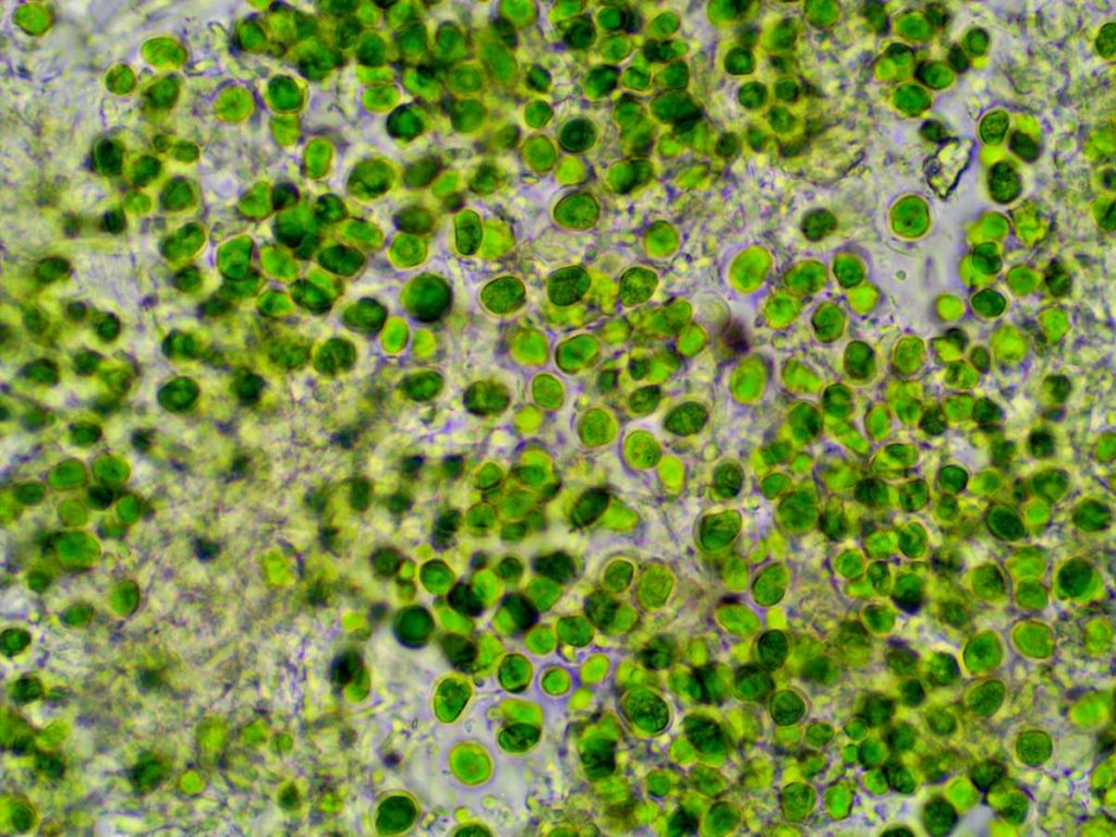
[attachment=0]zz algae An vir thall 240719-1.jpg[/attachment] -
AuthorPosts




- You must be logged in to reply to this topic.