
Sterile crust with K+ violet soredia
- This topic is empty.
-
AuthorPosts
-
March 16, 2023 at 5:58 pm #32582
Hello all, Is this sterile crust with granular K+ violet soredia possible to ID? I am lingering around C. chlorina or R. pityrea, but I am unsure if I should be considering other possibilities as well (such as C. turkuensis, C. virescens or other Rinodina spp.).
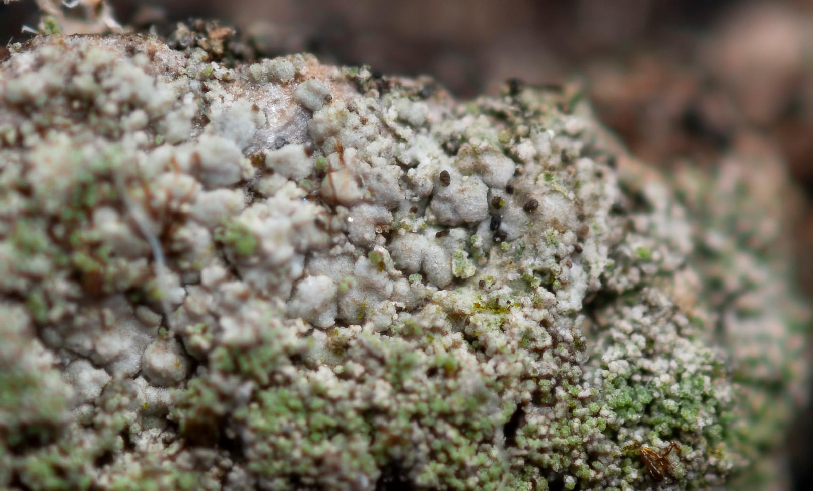
The soredia appear quite coarse (~50 µm diameter) but as far as I can see those are consoredia consisting of smaller soredia (~20-30 µm). The soredia are confluent and form a crust of coralloid isidioid consoredia (if I am interpreting it correctly). Most, but not all soredia have a distinct purple reaction with K (sedifolia grey?). The soredia are formed along the margins of the areoles, but seemingly also from more crater-like soralia.
The thallus was wide spreading (~15 x 15 cm) and appears to be overgrowing moribund mosses quite willingly. It is rather thick in places and areolate to almost small squamulose.
Found near the base of a park tree (likely Crataegus sp.) in Aarhus, Denmark.

[attachment=5]cf. Caloplaca sp. (Uniparken, basis, cf. hvidtjørn, aarhus) 1-1.jpg[/attachment] 
[attachment=3]cf. Caloplaca sp. (Uniparken, basis, cf. hvidtjørn, aarhus) 1-4.jpg[/attachment] 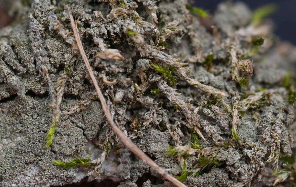
[attachment=2]cf. Caloplaca sp. (Uniparken, basis, cf. hvidtjørn, aarhus) 1-2.jpg[/attachment] 
[attachment=4]cf. Caloplaca sp. (Uniparken, basis, cf. hvidtjørn, aarhus) 1-3.jpg[/attachment] 
[attachment=6]cf. Caloplaca sp. (Uniparken, basis, cf. hvidtjørn, aarhus) 1-7.jpg[/attachment] 
[attachment=0]cf. Caloplaca sp. (Uniparken, basis, cf. hvidtjørn, aarhus) 1-5.jpg[/attachment] 
[attachment=1]cf. Caloplaca sp. (Uniparken, basis, cf. hvidtjørn, aarhus) 1-6.jpg[/attachment] March 17, 2023 at 9:37 am #45369Unfortunately, I think it is going to be difficult to be certain about your lichen without apothecia present. When I first came across Rinodina pityreait was tentatively misidentified for me as Caloplaca chlorinaand it was only after I found fertile colonies that I realised it was actually R. pityrea. The published descriptions perhaps give a slightly misleading impression of R. pityreaand certainly don’t give enough detail and discussion to guide someone whether or not to record sterile colonies. Here are some of my own notes: 
[attachment=5]Screenshot 2023-03-17 9.02.40 AM.png[/attachment] 
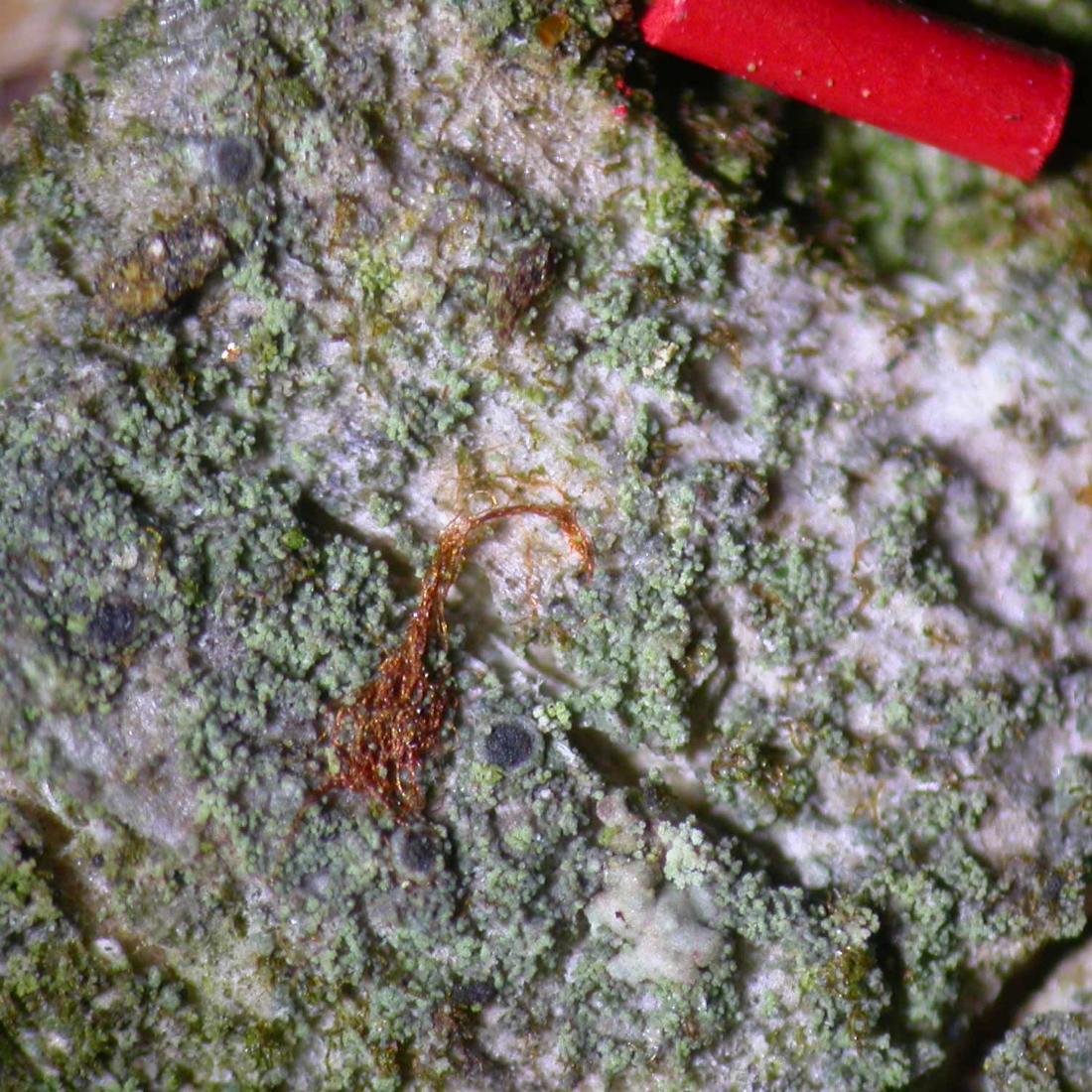
[attachment=4]Screenshot 2023-03-17 9.04.08 AM.png[/attachment] Below is an image of
R. pityreawith 0.5 mm red pencil lead for scale. 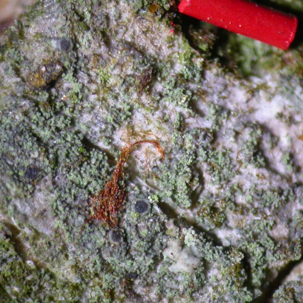
[attachment=3]FozCxk-XoAAXQWu.jpeg[/attachment] Interpreting the morphology of your lichen may be complicated by the fact that it was growing near the base of a tree and so might be subject to mollusc browsing. Browsing sometimes planes off areas of soredia which then regrow patchily and give an atypical appearance.
You are right in thinking that the K+ violet pigment is mainly found in some ‘
Caloplaca‘ species and in a few Rinodinaspecies. However, I have also found a similar pigment in Fellhanera viridisorediatawhich I illustrate and discuss in the following twitter thread: https://twitter.com/obfuscans3/status/1244236290901651457 My gut reaction is that your lichen is most likely to be
R. pityreabut I’m afraid I don’t have confidence to confirm it as such and don’t know quite how you can prove it without apothecia. In case you do find apothecia, I have written notes about these and ways of observing the ‘extra layer’ present in
Tunicata-type’ spores. 
[attachment=2]Screenshot 2023-03-17 9.05.00 AM.png[/attachment] 
[attachment=1]Screenshot 2023-03-17 9.05.23 AM.png[/attachment] 
[attachment=0]Screenshot 2023-03-17 9.06.02 AM.png[/attachment] March 17, 2023 at 11:25 am #45370Thank you for your detailed reply! After reading your notes I am both relieved that I am not the only one struggling with this specimen and I am now leaning heavily towards R. pityrea as the most likely ID. I have seen that species in fertile condition and much to my delight, the spores are very characteristic and makes for an easy identification in an otherwise quite difficult genus.
My main concern with identifying the current specimen as R. pityrea lies in the quite well developed (and in places sub-squamulose) thallus. But that could be a variation of the species that has yet to make it into the various descriptions – I would not be surprised if this is the case.
I will visit the site again in the hope of finding an apothecium or two. If I do I will update the thread here.
All the best,
Jonas
-
AuthorPosts




- You must be logged in to reply to this topic.