
Lichen on sandstone churchyard wall
- This topic is empty.
-
AuthorPosts
-
August 28, 2022 at 7:44 pm #32382
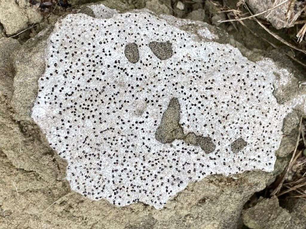
[attachment=5]IMG_5350.jpeg[/attachment] Some help with this would be appreciated. On a sandstone wall around a churchyard in the midlands. On a flake of loose stone. I think the apothecia are lecanorine. The discs are black and seem to pull away from the surrounding very white thallus, which is thin – sand grains easily visible. Spores are around 17 um long. A cross-section of the apothecium looked distinctly pink initially (and even pinker when one dried out), but khaki-brown in water. K- C-. Tried staining the asci, but don’t think I’ve got the hang of that yet. Paraphyses maybe anastomising. Probably missing something really obvious.August 28, 2022 at 9:30 pm #44851Great photos! Trapelia coarctatagroup, possibly T. elacista. August 29, 2022 at 8:33 am #44852I agree with Alan, that this is Trapelia coarctata group. The combination of sandstone substratum and the halo-like thalline rim around the apothecia would lead my thoughts straight towards T. ‘coarctata’. You also report that the apothecia appear distinctly pink and this is a Trapelia trait (either in section or when wet). You probably made a better job of staining the asci than you realised as Trapelia has a tholus which stains only weakly (if at all) in iodine. You report that your lichen is C-. You might like to try again with some strong, fresh bleach as I think you will find it will be C+ red. If you do it on your collected fragment, use the tip of a cocktail stick or similar to apply a tiny drop so you don’t ruin your specimen by flooding it with bleach. The difficulty of demonstrating and interpreting C reactions is a frequent problem in lichen recording. Here are a couple of my own images of T. coarctata s. lat., one dry and one wet.

[attachment=4]Trapelia coarctata 2.jpg[/attachment] 
[attachment=3]Trapelia coarctata 4.jpg[/attachment] You will see from the following maps that records for T. coarctata are abundant and widespread, whereas T. elacista has very few records.

[attachment=2]Trapelia coarctata.jpg[/attachment] 
[attachment=1]Trapelia elacista.jpg[/attachment] This extract from the key to Trapelia in LGBI3 indicates the difficulty of separating these two species.

[attachment=0]Screenshot 2022-08-29 9.03.02 AM.png[/attachment] I don’t feel confident in separating the two myself. One problem is that we don’t have a recording unit for T. coarctata s.lat. so the records of T. coarctata will probably continue to be a mix of the very few identifications which are reliably T. coarctata s. str. and the vast majority of records which don’t have the benefit of well-developed material and/or molecular confirmation. In summary, for the purposes of your own interest, consider this to be ‘T. coarctata group’ and if submitting the record to the database, probably best to call it ‘T. coarctata’ (even though I agree with Alan that it *could* be T. elacista). Records of T. coarctata in the database must already be considered to be a mixture of T. coarctata and T. elacista while it would be best to restrict records of T. elacista to those that have been confirmed (e.g. by sequencing or examination of good material by a referee).
August 29, 2022 at 11:06 am #44853Thank you so much for the detailed response. I have tried C again, but cannot get any trace of red reaction with bleach, neither on the thallus nor apothecia (using cocktail stick) nor on apothecial sections (they start reddish, dry or in water, but just bleach out with C). I have tried it four times, and haven’t seen even a fleeting reaction (watching under dissecting microscope). The same bleach gives a strong red reaction with Punctelia this morning, so I don’t think it’s the bleach. No red with KC either. Puzzling. Apologies for photo quality – some are through the dissecting microscope. 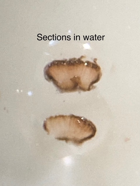
[attachment=6]IMG_5413.jpeg[/attachment] Thick section under x100 compound microscope – it was wetted, and harder to cut.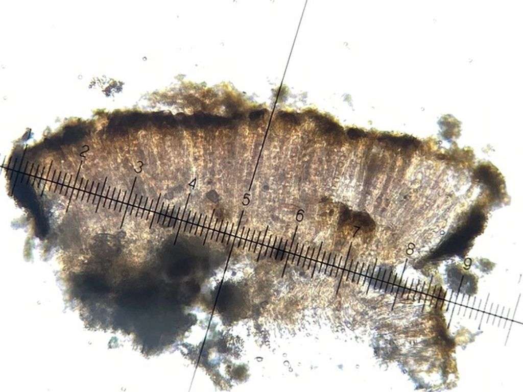
[attachment=2]IMG_5448.jpeg[/attachment] 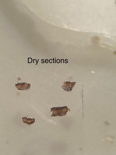
[attachment=5]IMG_5414.jpeg[/attachment] 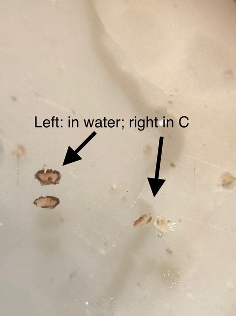
[attachment=4]IMG_5415.jpeg[/attachment] This photo is of an algal blob found in the thallus – it isn’t a discrete algal layer, there just seem to be odd pockets of algae mixed up in the cotton-wool like thallus.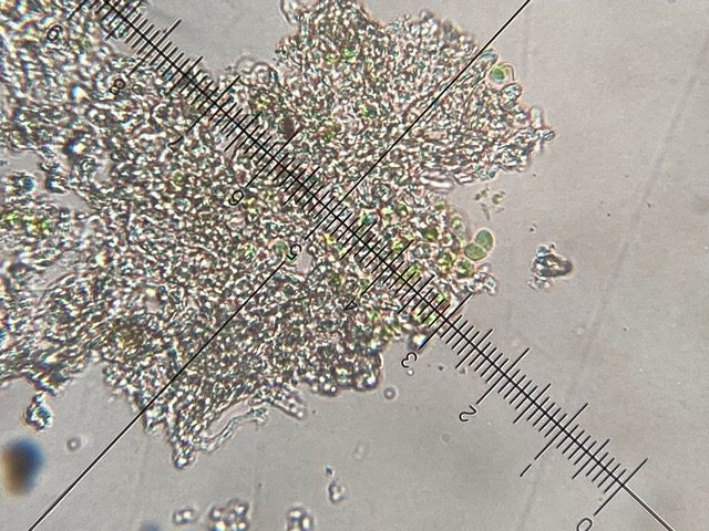
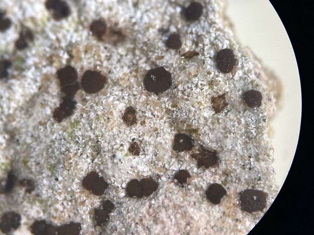
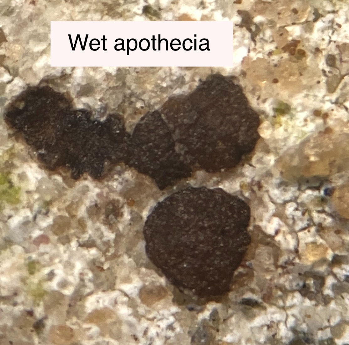
[attachment=3]IMG_5417.jpeg[/attachment] Last two photos are after spraying it with water, and then after leaving a droplet of water on the surface for about ten minutes. Spraying didn’t seem to do much, but after about ten minutes of being submerged, the apothecia have swollen up quite a bit, and look much more reddish-brown rather than black. It is very fragile – the sandstone keeps just rubbing off the back, and the main piece has fractured into several bits. I’m keeping it for future reference. I’ve looked up Trapelia in several places and can see it’s a tricky genus. Happy to leave it at this now.
[attachment=1]IMG_5449.jpeg[/attachment] 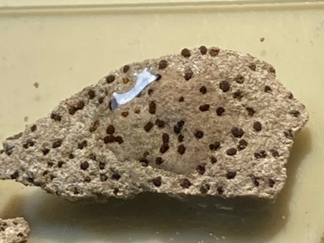
[attachment=0]IMG_5450 2.jpeg[/attachment] August 29, 2022 at 7:29 pm #44854Mark is correct. The distinction between T. coarctataand T. elacistaare subtle, and I went for T. elacistasolely on the presence of the crack between thallus and apothecia. The T. coarctatamap should really be renamed T. coarctatas. lat. as all pre-2028 collections of T. elacistawould have been called T. coarctata. Also agree about your ascus staining. The
Trapelia-type ascus is described as “tholus I–, with I+ blue flanks, which is exactly what you photo shows! The C– reaction is puzzling. I always check for C+ reaction under a compound scope, so you should try that, but without tlc there’s not much more to be done – except to note on the packet that it is C–.
Cheers – Alan
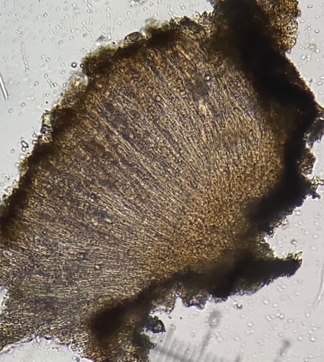
August 29, 2022 at 9:08 pm #44855Thank you for the tip about watching it under higher magnification. After considerable battling with very awkward samples and many tiny grains of sand, I saw a definite C+ red under the compound microscope in this rather thick and messy section. I videoed it with my phone (in a clamp, which meant I could concentrate on putting the C on the slide), and then took screenshot stills. It took about 6-10 seconds before it started reacting, and lasted another 10 or so seconds before fading. Very satisfying. The only ‘fleeting’ C+ red that I had come across before was with foliose lichens, where I could easily see the reaction under the dissecting microscope, even though it was so quick to disappear. Section in water:
[attachment=2]IMG_5477.jpg[/attachment] Section about 10 seconds after adding a drop of C to the slide: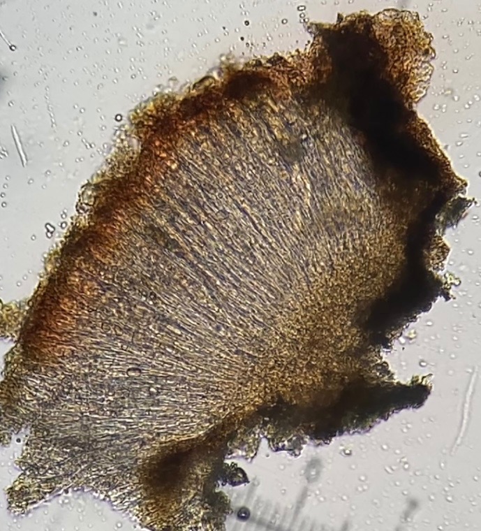
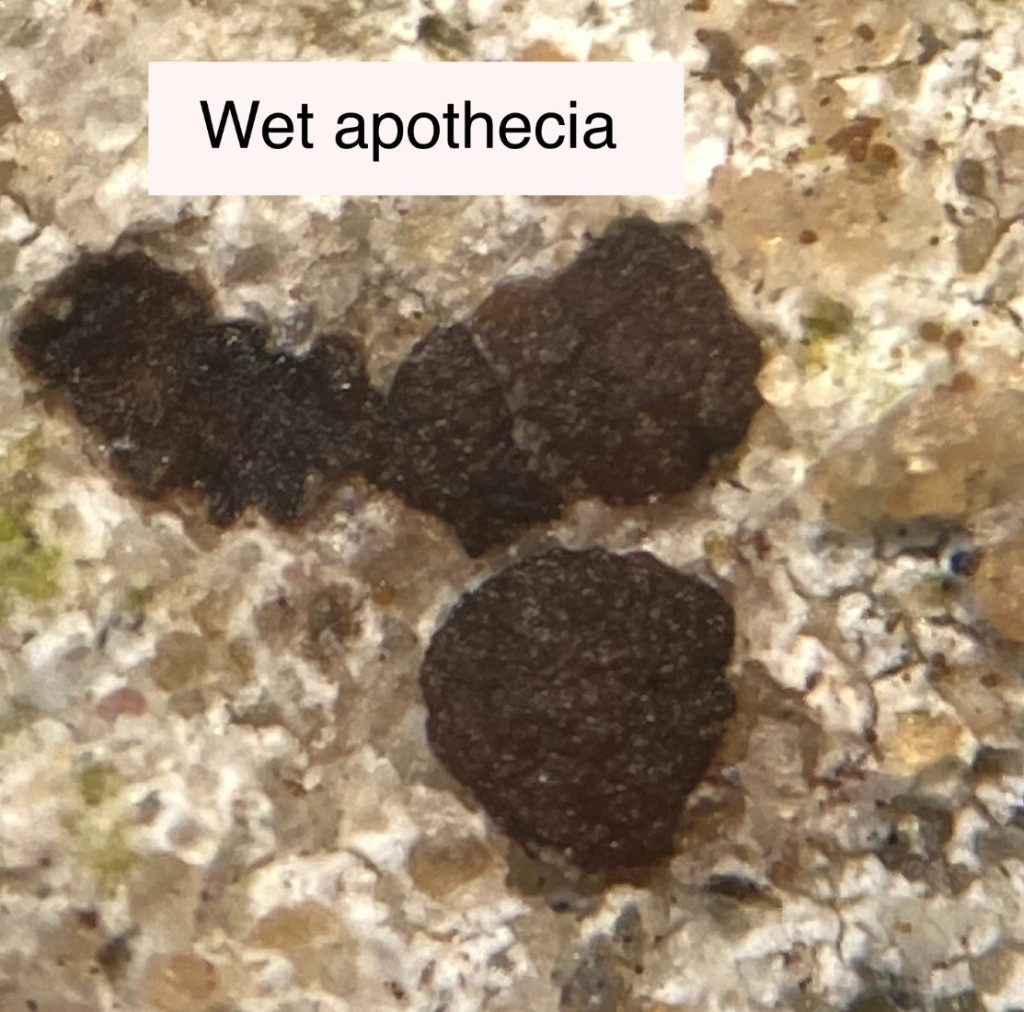
[attachment=1]IMG_5478.jpg[/attachment] After maybe half an hour in water, the apothecia swelled up considerably and changed from black to brown.
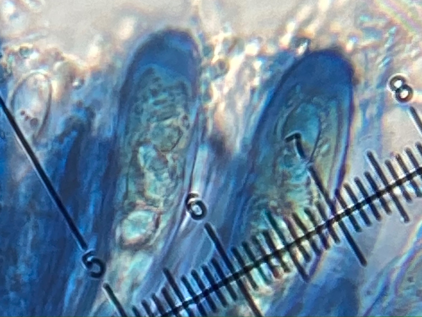
[attachment=0]IMG_5464.jpg[/attachment] October 13, 2022 at 3:06 pm #44856Just adding some more ascus photos after a bit more practising. I think they look right for Trapelia. The only thing that really bothers me with this is that the thallus resolutely refuses to react to C. 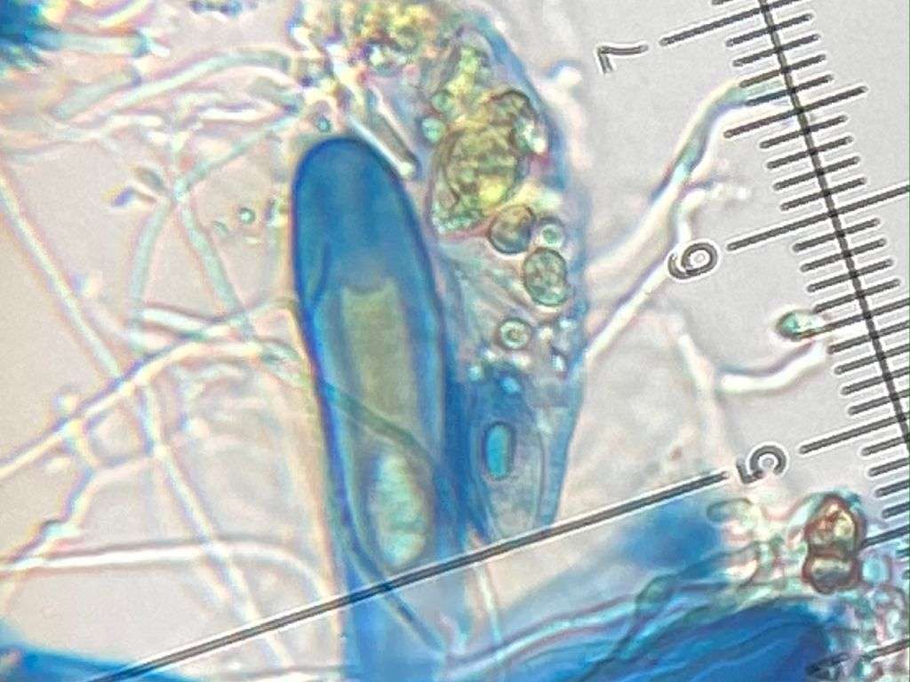
[attachment=1]IMG_8709.JPG[/attachment] 
[attachment=0]IMG_8720.JPG[/attachment] -
AuthorPosts


- You must be logged in to reply to this topic.