
Possible commensal association between a cyanobacterium and Lecidella elaeochroma
Forums › Forums › Lichens › Possible commensal association between a cyanobacterium and Lecidella elaeochroma
- This topic is empty.
-
AuthorPosts
-
August 15, 2022 at 7:31 pm #32375
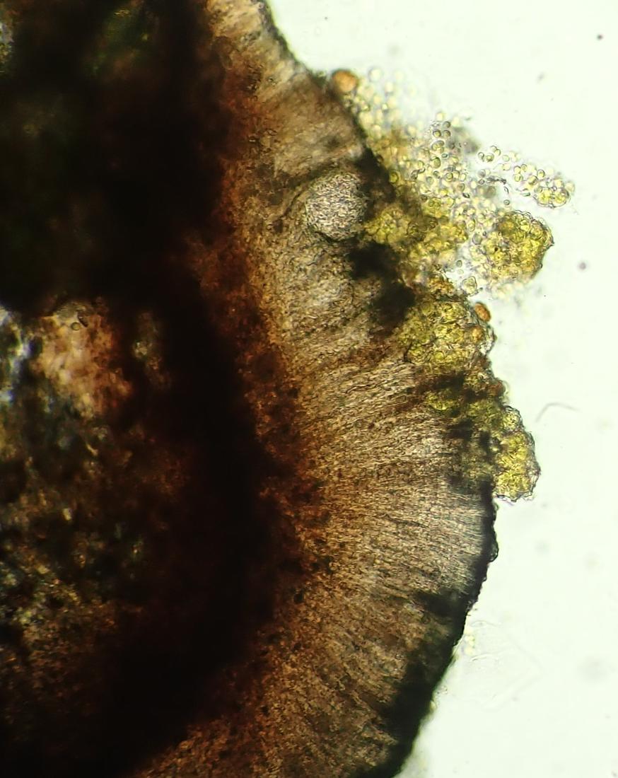
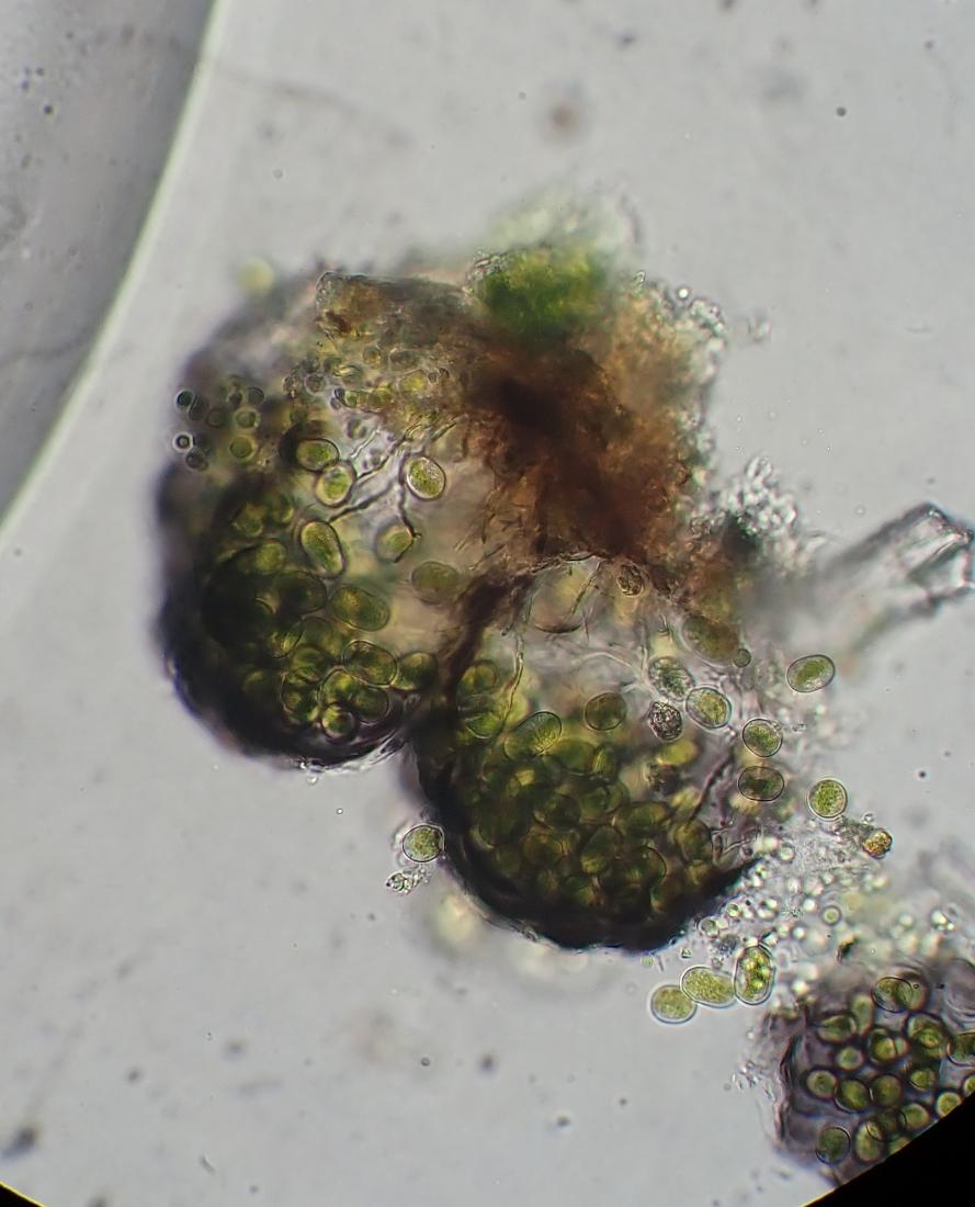
A black crust on a timber bench slat appears to be an extensive thallus of Lecidella elaeochroma in close association with a cyanobacterium. The lichen thallus is poorly developed and the cyanobacterium is present as numerous small, shiny black clusters on the thallus as well as around the edge of the apothecial discs.It’s commonplace to find cyanobacteria growing freely among lichens but in ths case sections of the lichen apothecia show invasion of the hymenium by the cyanobacterial cells. 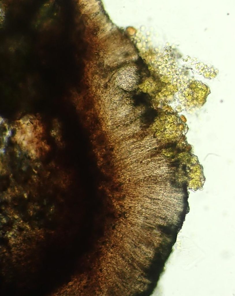
[attachment=6]cyano on elaeo (2).JPG[/attachment] 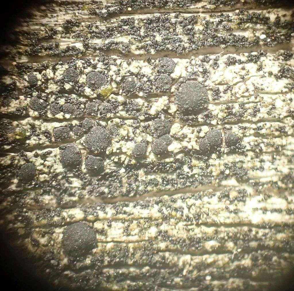
[attachment=0]cyano on elaeo.JPG[/attachment]
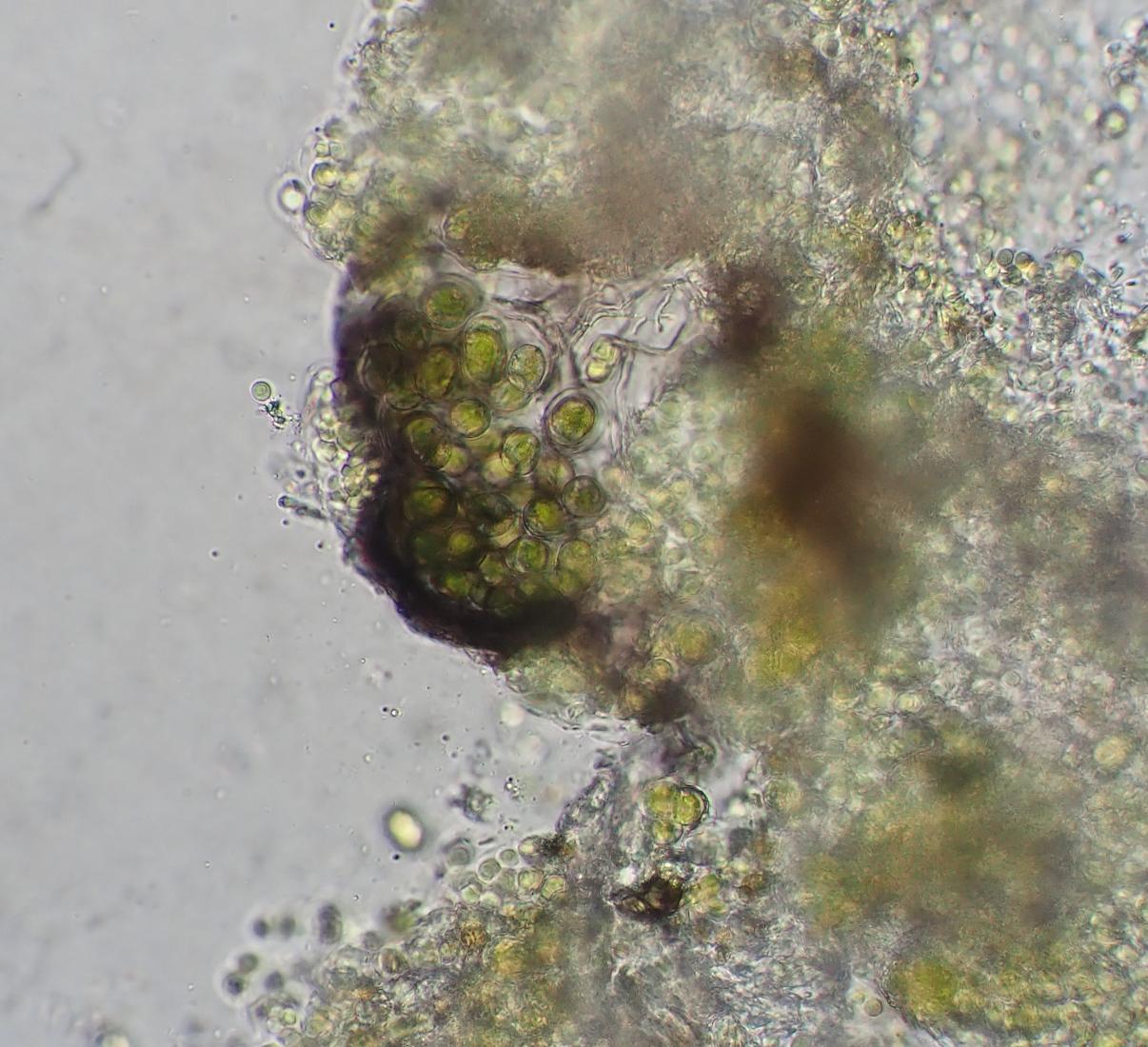
Distinct globose structures with dark walls can be seen and the lichen epithecium is thickened and darkened in these areas.Additionally some areas of the hymenium are filled with green cyanobacterial cells. There are also clouds of algal[attachment=6]cyano on elaeo (2).JPG[/attachment] cells and dark globose structures freely occurring all over the whole area.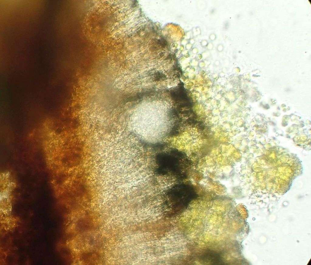
[attachment=5]cyano on elaeo (3).JPG[/attachment] 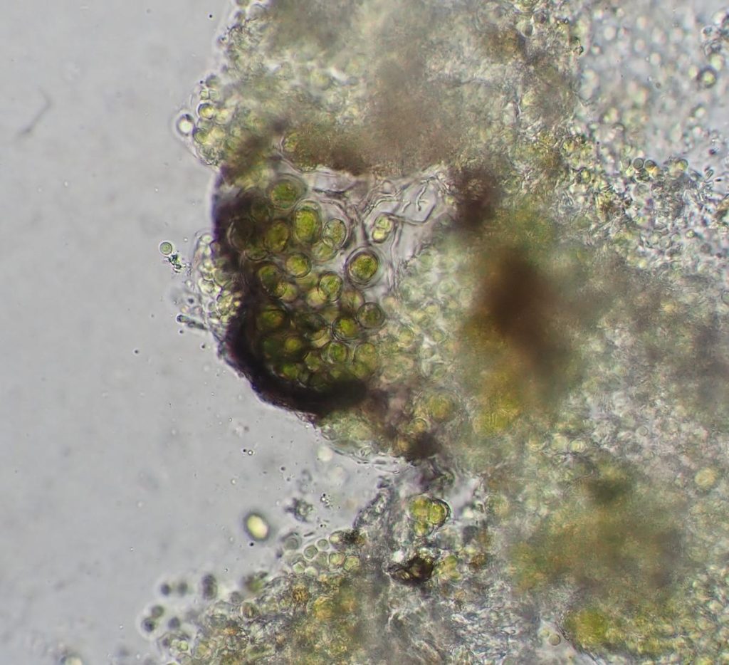
[attachment=4]cyano on elaeo (4).JPG[/attachment] [attachment=6]cyano on elaeo (2).JPG[/attachment] [attachment=6]cyano on elaeo (2).JPG[/attachment] 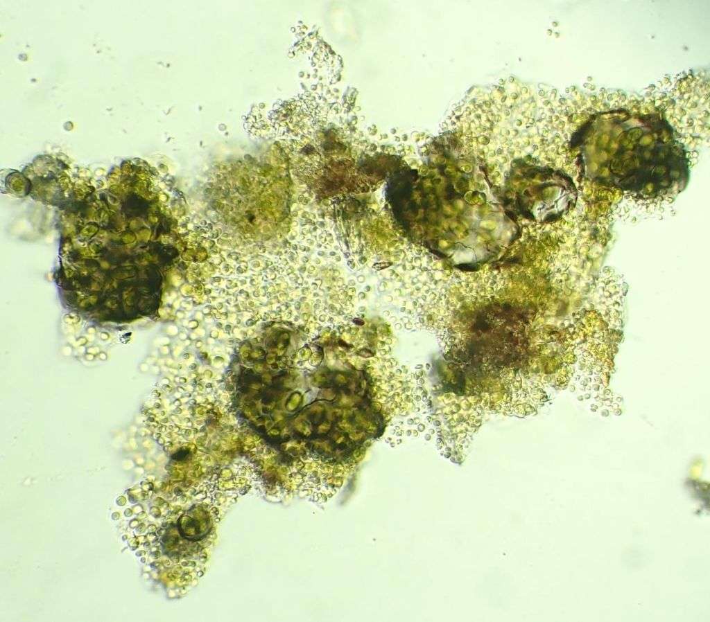
[attachment=1]cyano on elaeo (7).JPG[/attachment] August 16, 2022 at 1:14 am #44838Hi: It’s not clear from your photos, but are you sure these are cyanobacteria and not green algae – they look very green! If they are green algae then you probably have a species of
Moriola. This is a genus of fungi that forms goniocysts consisting of a spherical shell of fungal cells up to 100 µm diam filled with cells of green algae. They are not rare in alpine habitats growing on damp soil and among bryophytes and over crustose lichens, but they also occur in lowland habitats, usually on old wood or lignum. They are interesting because the exhibit a primitive form of lichenization but haven’t been studied for c.100 years – although I have a colleague in Sweden who is trying to extract DNA from them…. They are rarely fertile so an ID to species is not possible but M. pseudomycesis a name given to some specimens. Here is a plate I made for a recent publication, but was edited out of the final paper.
Alan
August 19, 2022 at 10:57 am #44839You’re probably right in thinking the alga involved in this relationship is a green alga rather than a cyanobacterium. I was interested to learn about the goniocysts of Moriolaso looked more closely at the structure of the clumps of algal cells I have. I could find no fungal hyphae and the tissue containing the algae consists of blue/black parenchymatous cells (ca. 25 microns diameter). 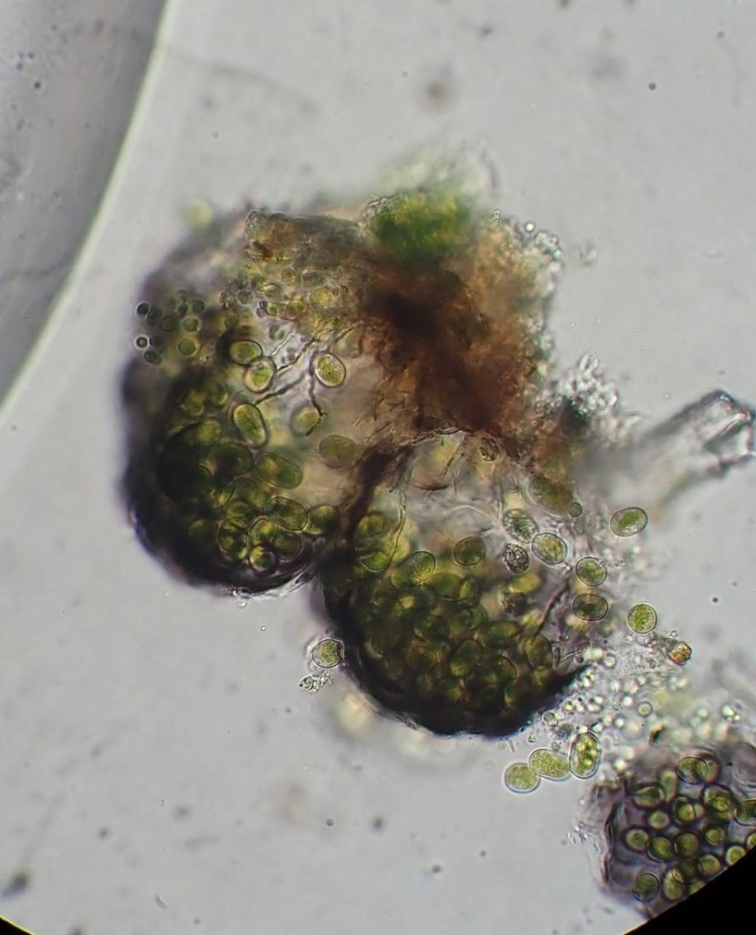
[attachment=0]Alga elaeo (2).JPG[/attachment]
This is probably going to be a case in which we see algae living closely alongside lichens and more intimately involved with them than we know. We have growing knowledge concerning lichenicolous fungi and some cases of lichenicolous lichens but the situation regarding algae and lichens is more likely to be one of the lichens benefiting from the photosynthetic powers of the algae rather than harming them.I did however observe that the hymenium of the lichen was shrunken and discoloured and that asci and spores were not produced where the alga had invaded the hymenium.
I shall keep an eye out for
Moriolaon wood and lignum and thank you for your interest. August 24, 2022 at 8:49 pm #44840Maxine: I can see hyaline tissue and what appears to be hyaline hyphae, so I suspect there is a fungus involved here as well. Your collection is clearly, morphologically similar to Moriola and it would be interesting to know if the fungus was related. If two unrelated lineages both produce these goniocysts it could prove, either that they are possibly widespread in the ascomycetes, or, if it is the same or related alga, that goniocyst formation is initiated by the alga – in which case it could have fundamental implications for how lichens in general are formed. Perhaps it’s the alga calling the shots, not the fungus…
All very interesting and lots of scope for speculation…
Alan
-
AuthorPosts


- You must be logged in to reply to this topic.