- This topic is empty.
-
AuthorPosts
-
October 2, 2020 at 5:29 pm #31812
Can anyone help with this ID? I collected this sterile crust at Postbridge, Dartmoor, Devon, growing on a heather stem in moorland. It has no reactions with C,K and Pd but shows a brilliant white fluorescence under UV. The whole thallus is approximately 25mm across its longest length and the individual soralia are about 0.4 to 0.5mm across, becoming confluent. The soredia are pale green, surprisingly looking quite brown under the microscope and measuring about 25u across. The photobiont cells are about 10-12.5u diameter.
According to LGBI, the possibilities are Fuscidea pusilla and Ropalospora viridis, the only two corticolous sterile crusts with negative reactions and white fluorescence. I have seen F, pusilla (I think), like a diminutive F. lightfootii, and it didn’t look quite like this. On the other hand, I get the impression from the literature that R. viridis is a lichen of old woodland.

[attachment=4]Ropalospora Dartmoor a small.jpg[/attachment] 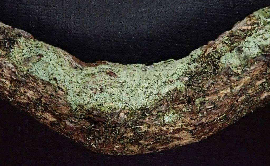
[attachment=3]Ropalospora Dartmoor b small.jpg[/attachment] 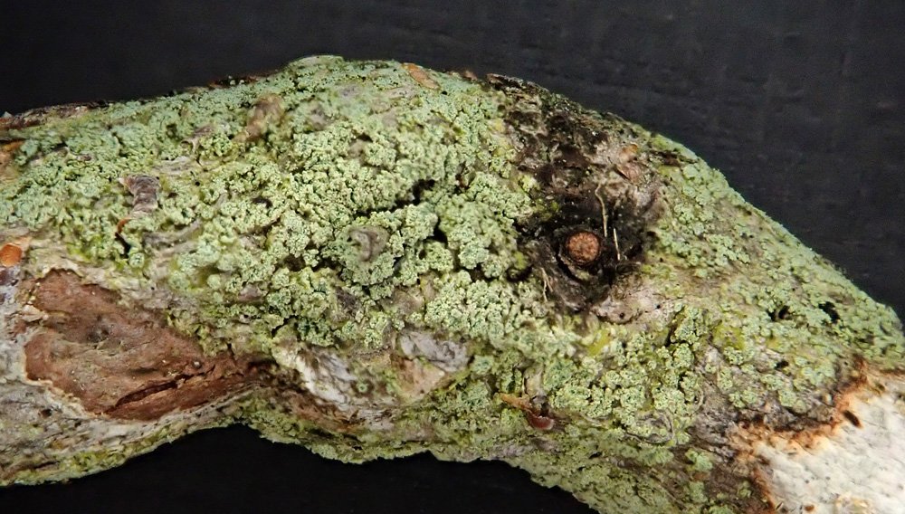
[attachment=2]Ropalospora Dartmoor c small.jpg[/attachment] 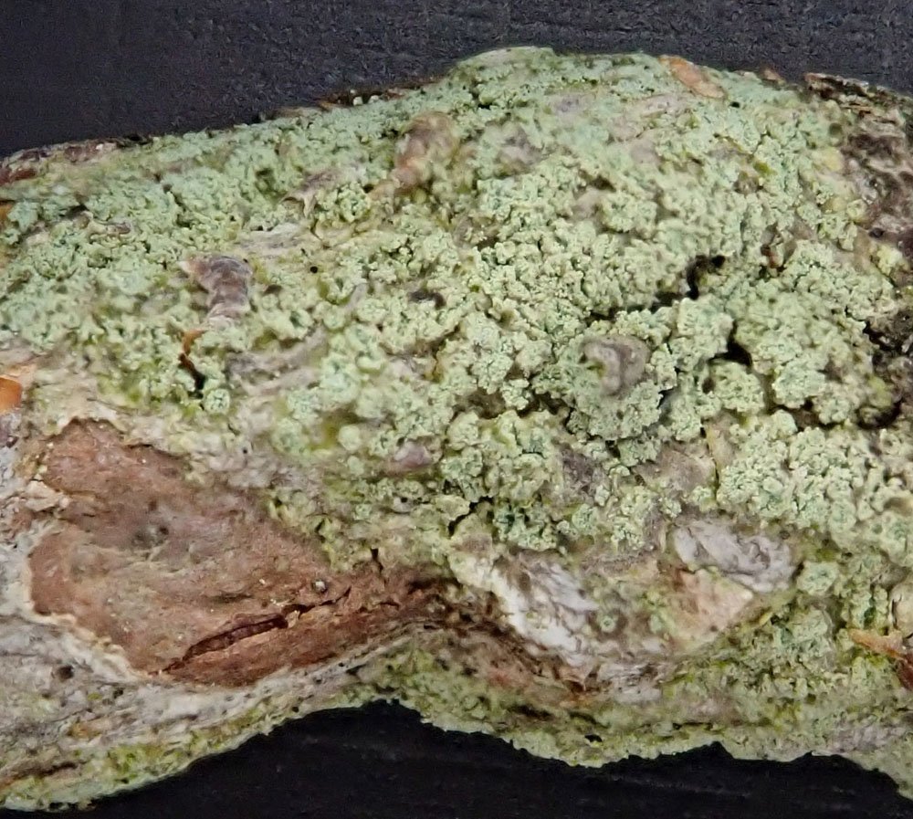
[attachment=1]Ropalospora Dartmoor d small.jpg[/attachment] 
[attachment=0]Ropalospora Dartmoor in UV small.jpg[/attachment] October 3, 2020 at 1:15 pm #43348Hi John: These two are difficult to separate morphologically – you really need tlc. However this publication from James Lendemer, which is freely available on the web, may help.
Lendemer, J. C. 2011: A review of the morphologically similar species Fuscidea pusilla and Ropalospora viridis in eastern North America. Opuscula Philolichenum 9: 11-20.
Alan
October 3, 2020 at 1:21 pm #43349The UV flourescence of Ropalospora viridisis quite week, and is mainly the thallus with the soralia dull in UV. The soralia are also green not yellowish as this material. This looks like sterile Fuscidea lightfootiito me, which has very bright UV flourescence. It is often sterile and frequently occurs on Heather stems. Neil
October 3, 2020 at 2:00 pm #43350Looking up Fuscidea pusillayou could call it that, but not sure how you would demarcate from sterile small Fuscidea lightfootii. It certain has not been recorded much in Britain. The Fuscideaaccount in LBGB2 did not seem to rate Fuscidea pusillaas much more than a morph of Fuscidea lightfootiibut opinions may have firmed up since then if someone has sequenced them. The picture of typical F. pusillain Lendemer Fig 2 F is different from Fuscidea lightfootiihow? Neil
October 3, 2020 at 2:25 pm #43351Neil: Good question. Fuscidea lightfootii is not known to occur in North America – which is to say, no fertile material has been collected. I collected some good F. lightfootii for James one time when I was back in the UK hoping he would sequence it, but I don’t think he has…
Alan
October 4, 2020 at 7:57 am #43352Alan Yes sounds like we definitely need sequencing here to be sure what is going on. I can see that the small, sterile and strongly sorediate morph found in boreal Europe and N. America could be phyologentically separate from a more oceanic
Fuscidea lightfootii, but this would still leave us in oceanic Europe with the problem that I am fairly sure that Fuscidea lightfootiiis capable of growing in the Fuscidea pusillamorph when stressed, as on a heather stem. So we need to find out if the two species are separate and, if they are, also sequence potential Fuscidea pusillamorphs within the distribution Fuscidea lightfootiito see if they can be separated if they grow together. Aaagggghhhhhh
Neil
October 4, 2020 at 12:09 pm #43353In Lichenologist50(4);425-438 (2018), Zahradnikova et al address this question, using DNA sequencing on many examples of F.pusilla and F. lightfootii. They concluded that ‘even though some morphotypes appeared distinct and could be assigned to one of the two species with some degree of certainty, the use of DNA sequencing is recommended for their identification’. If only there were ways of getting work like this done without having to ask favours of the small number of people who have access to the facilities. Thank you both for your advice, it really has helped.
October 4, 2020 at 12:31 pm #43354Neil: Couldn’t agree more with your final comment – “Aaagggghhhhhh” just about sums it up!
It seems to me that BLS/ABLS/IAL needs to fund a sequencing facility that can help resolve problems like this. How about an annual contribution to RBGE to cover staff and material costs?
Alan
October 4, 2020 at 7:40 pm #43355Alan Quote:It seems to me that BLS/ABLS/IAL needs to fund a sequencing facility that can help resolve problems like this. How about an annual contribution to RBGE to cover staff and material costs?
Yes this sort of idea has been mulled over before and Becky, the current BLS president, is keen to get more money into small scale lichen research, definitely worth pushing for.
Neil
October 5, 2020 at 7:48 pm #43356John Thanks, I had forgotten that paper, so they are different, but impossible to tell apart morphologically. So until we have hand held DNA scanners, it does not seem realistic to record them
Neil
-
AuthorPosts


- You must be logged in to reply to this topic.