aspeca

Forum Replies Created
-
AuthorPosts
-
Following up on Paul’s suggestion of Acremonium/Pronectria, I’ve found that there has indeed been at least one Pronectriaspecies very recently described on Lecanora. Valerii Darmostuk (2021) describes Pronectria gromakovaeas new to science from an occurrence on Lecanora populicolain the region of Kharkiv, Ukraine. I’m not sure how strongly this bears on the LF described here, though. The details are available open-access from Lindbergia— https://doi.org/10.25227/linbg.01141https://doi.org/10.25227/linbg.01141” class=”bbcode_url”> Many thanks for starting this forum topic, Mark. Last month I found ostensibly the same mould-like anamorphic conidial fungus in a specimen of
Lecanora hybocarpafrom near Lavenham, Suffolk (VC26/TL921498). The thallus is on I think a Corylustwig — the twig was broken and could have come from another tree, but it was well caught in a hazel at the edge of an arable field, and most of the surrounding trees seemed to be hazel. As with Mark’s specimen, the LF shows up as a red staining and ‘rotting’ in the apothecia. It doesn’t seem to affect the thalline margins or the thallus more generally.

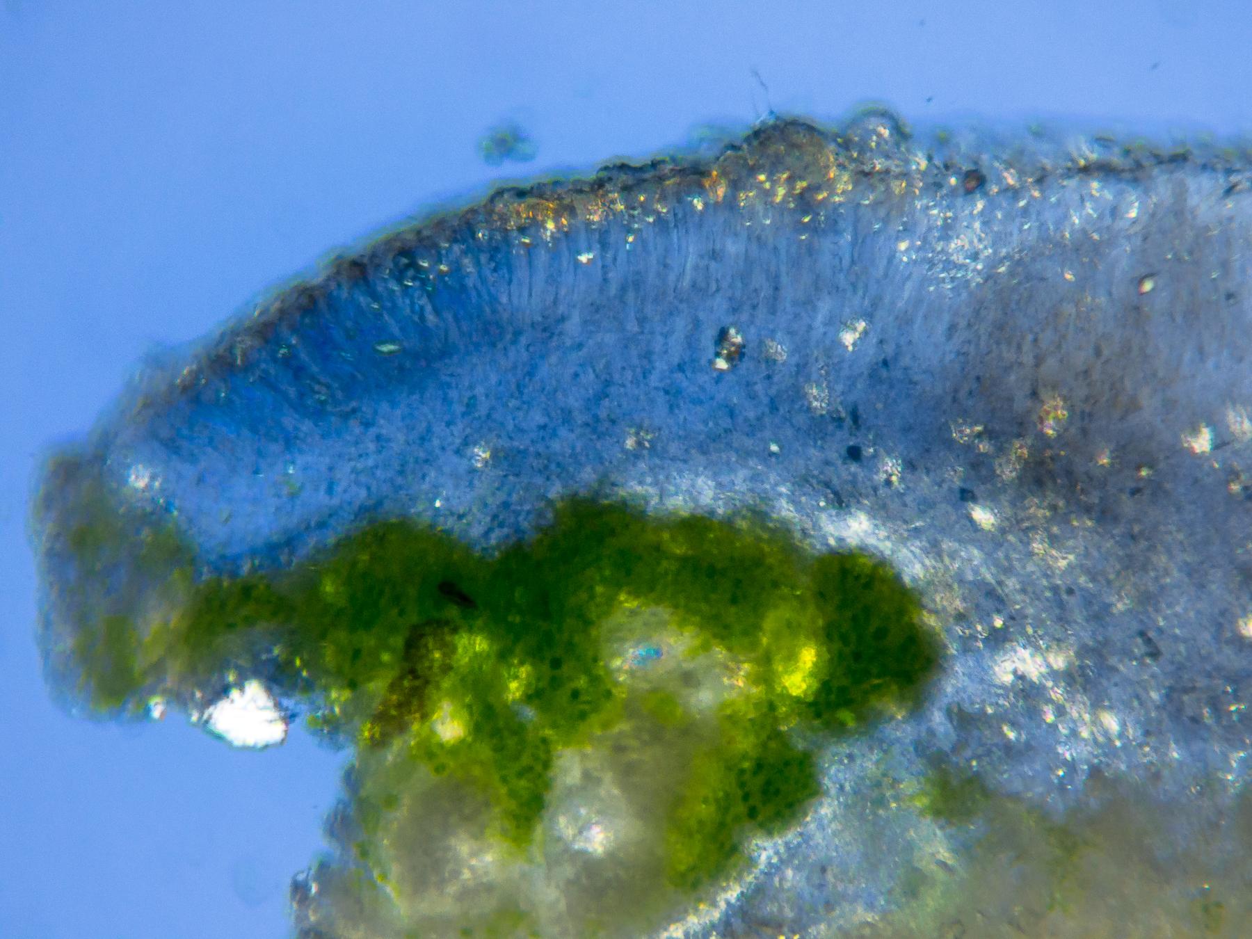
[attachment=9]20231005-PA050262.jpg[/attachment] Even apothecia that seem relatively unblemished are affected. In this section, mounted in water in cross-polarised light, a reddish stain appears within the hymenium. The LF seems to have inhibited or degraded the normal development of the apothecium. The descending POL+ epithecial granules characteristic of
L. hybocarpaare missing, except in the ‘healthy’ part of the apothecium. Asci with spores are visible, but again only in the healthier part. Where the reddish stain is present, there seem to be no asci at all. The LF seems to have inhibited or destroyed them as well. 
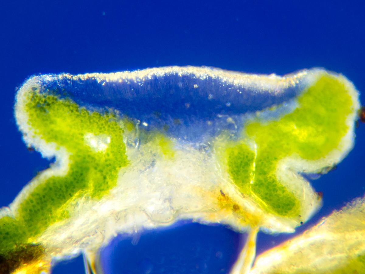
[attachment=8]20231005-PA050265.jpg[/attachment] An overview section of a visibly fully parasitised apothecium, again mounted in water, shows that the reddish stain of the LF dominates the fruiting body. No asci are seen at all (nor POL+ epithecial granules, not shown here due to the limit on the number of photos per post).
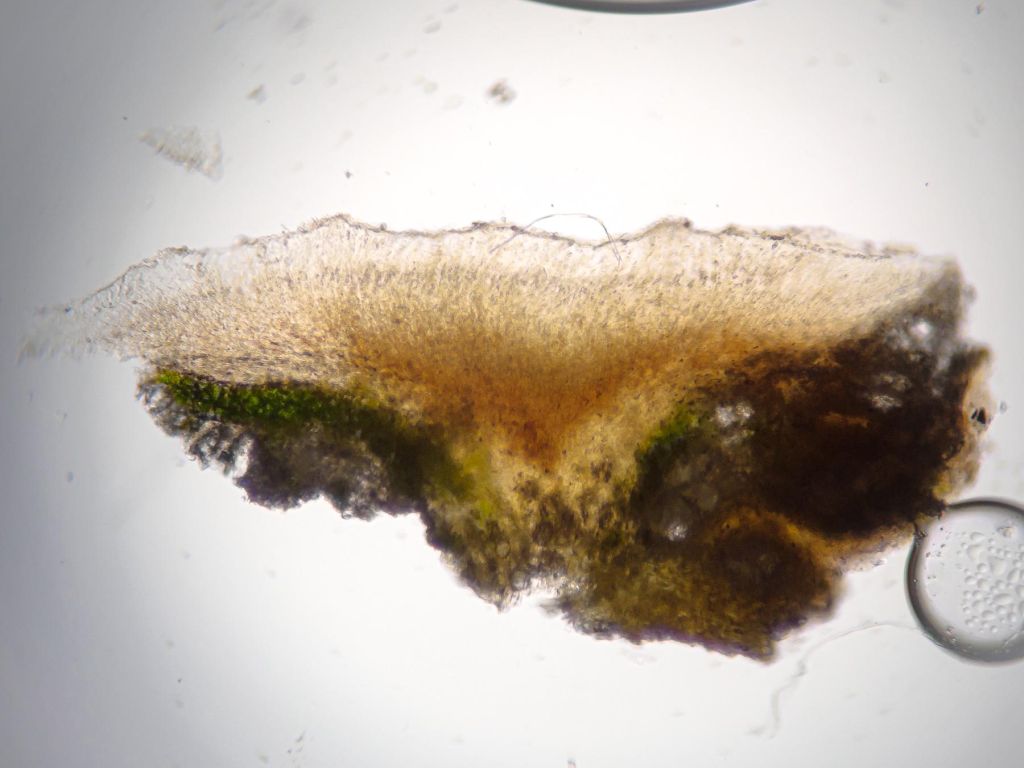
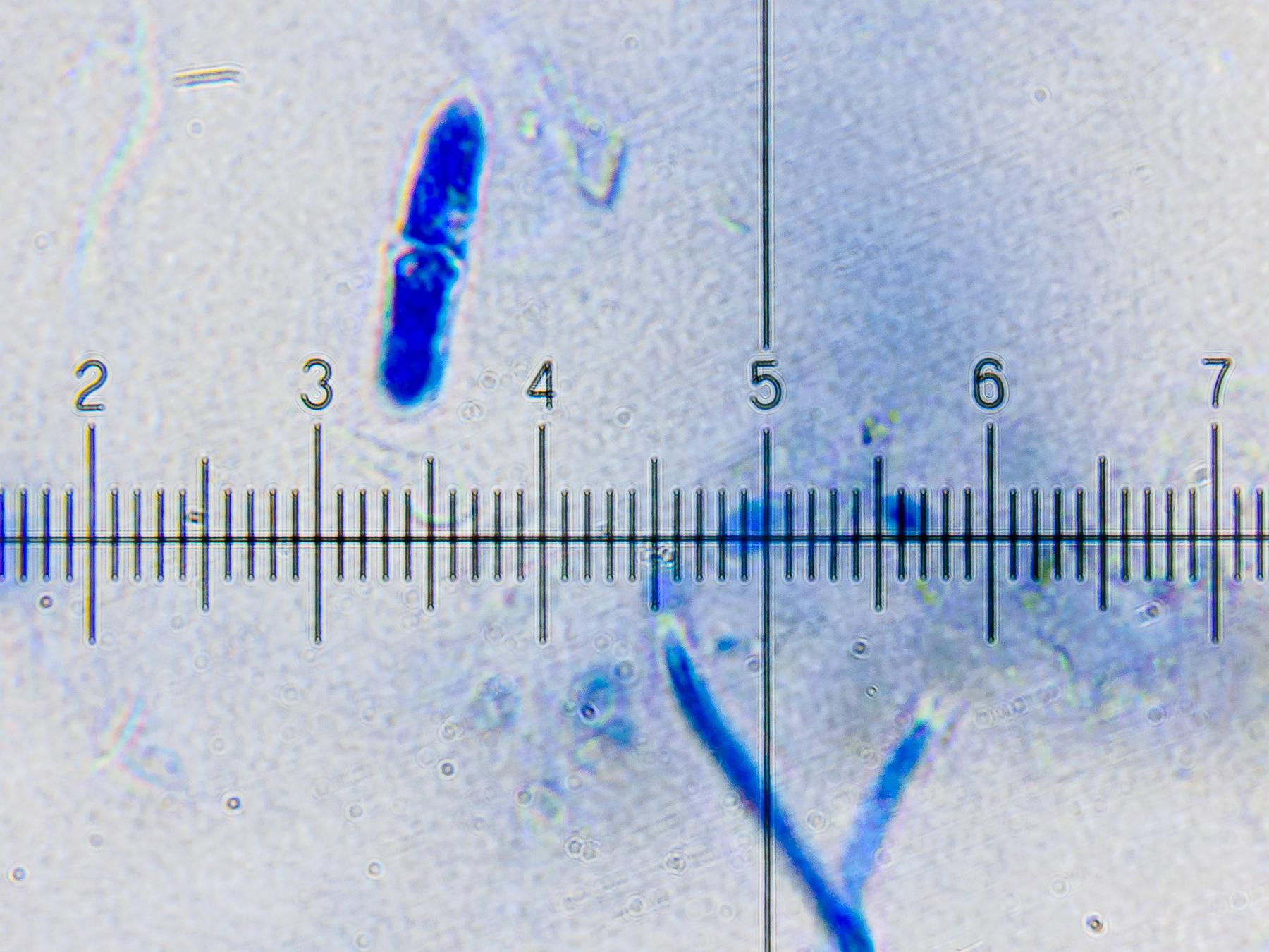
[attachment=7]20231005-PA050268.jpg[/attachment] However, the LF’s hyaline conidia can be made out peeping about the surface of the apothecium (a different apothecium than the one above). As with Mark’s specimen, the conidia are 1-septate (1 div = 1 µm).

[attachment=6]20231110-PB100790.jpg[/attachment] Clearing and staining in K-ink-vinegar brings out the LF’s hyphal network. Again, the LF appears confined to the hymenium of host apothecia, seemingly drawing its nutrients from the hymenial tissue, not the photobiont. Clearing and staining also make obvious that the host’s asci and paraphyses are nearly entirely inhibited or degraded or destroyed. Essentially only the LF’s own hyphae remain (1 div = 10 µm).

[attachment=5]20231110-PB100793.jpg[/attachment] The LF’s hyphae appear mostly to be trending towards the surface of the host apothecium to sporulate. Developing conidia and perhaps also phialides are visible near the 5 to 8 marks on the scale (1 div = 2.5 µm).

[attachment=4]20231110-PB100797.jpg[/attachment] The hyphae themselves are hyaline as the unstained images above show. They seem to be roughly 3-4 µm wide, including a roughly 0.5 µm cell wall. It’s hard to get a sense of the length of each hyphal cell, as the hyphae seem to twist. They don’t seem to branch too much, though (1 div = 1 µm).

[attachment=3]20231110-PB100800.jpg[/attachment] A close look at free and developing conidia shows that fully mature spores are roughly 13-16 µm in length and 4-5 µm in width, including a roughly 0.5 µm cell wall — in line with the size of the conidia in Mark’s specimen (1 div = 1 µm).

[attachment=2]20231110-PB100803.jpg[/attachment] 
[attachment=1]20231110-PB100806.jpg[/attachment] 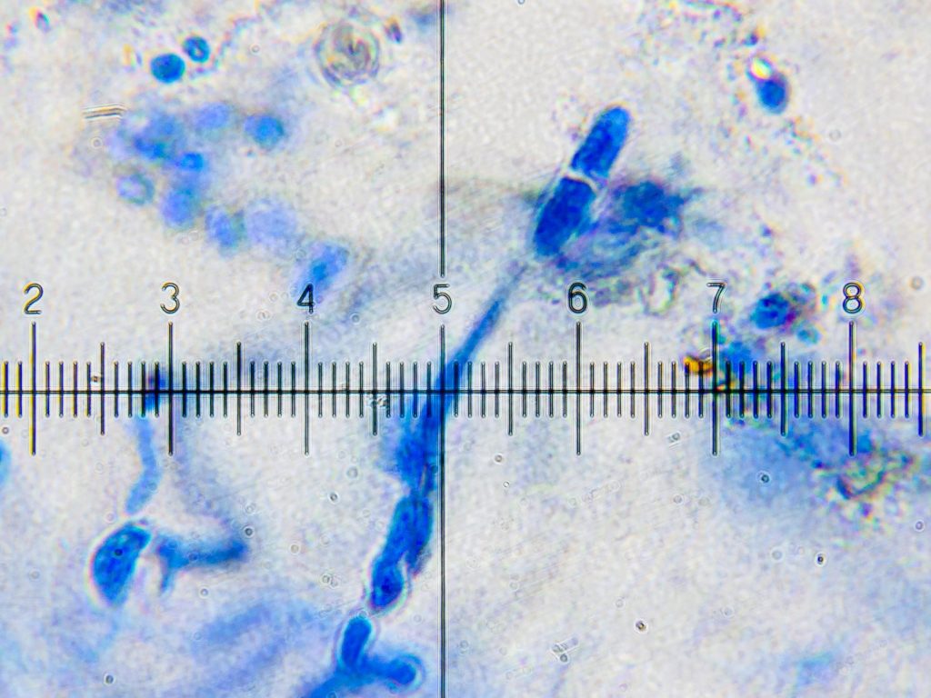
[attachment=0]20231110-PB100809.jpg[/attachment] Apart from this thallus, there are other
L. hybocarpathalli from the same twig that appear to be similarly infected. I also have a more recently collected L. hybocarpaspecimen from near Glandford, Norfolk (VC27/TG037408) that I think might also host this LF. Many thanks, Paul, for your suggestion of
Acremonium pedatum/ Pronectria anisospora. Acremoniumhas come up in the Twitter discussion, which has also brought forward additional finds that others may wish to post here. Grateful for any further advice about next steps towards identifying or describing this LF.
Many thanks, Paul. My oversight not to have checked your FGBI website before posting. The images of H. aractinaand H. oraethere don’t match my specimen as well as I thought they might from the descriptions and photos in Alan Orange’s monograph. As you point out, that tends to confirm Alain’s view that my specimen is likely an atypical form of H. maura — or as he suggested perhaps an as-yet unclear cryptic taxon. All very useful — I’ll be referring to
H. maura s. lat.from now on, and no longer without that explicit qualification! It will be interesting to hear if future study of Hydropunctariaturns up any morphological characteristics that could be relied upon. Many thanks, Alain — that’s very helpful. It’s interesting that you think the lichen may be a cryptic taxon within the broader conception of H. maura s. lat.I hope you’re able to recommence your study of Hydropunctaria. I wonder if
H. oraecan be completely discounted? [list][*]The image is from the interior of the thallus, so the prothallus wouldn’t be visible. I don’t particularly remember a white prothallus, but as I was unaware ofH. oraeat the time, I wouldn’t have looked for it specifically. [*]Alan Orange does indeed mention that H. orae likes freshwater, but he says it can be found ‘especially where slightly irrigated by rainwater’. The Scottish coast on which I found this specimen is extremely rainy, and that location might have been ‘slightly irrigated’ by rainwater fairly regularly. I don’t really remember it as a rainwater channel — but again I wasn’t looking specifically.[*]The colour in the image could easily be an artefact of the camera. When I reduce the colour temperature even slightly, it looks yet greener to me (see below). But I suppose it’s hard to say.[*]The thin patches in the thallus, and the limited cracking even in the thicker parts, seems more like Alan Orange’s description ofH. oraethan anything else — especially considering that he says of H. maurathat ‘The thallus is always well-developed and cracked into mostly discrete areoles’. But that doesn’t rule out another cryptic taxon apart from H. orae. [/list] 
[attachment=0]20230807-P8070802-2.jpg[/attachment] As for
H. oceanica— yes, the reason I downplayed that possibility below was that the perithecia didn’t seem pointed in the way you suggest, and as shown in Alan Orange’s pictures. In any case, it seems perhaps that the image (regrettably the only one, as I took it uncontroversially to be
H. maura) doesn’t suggest H. oraeor another known Hydropunctaria taxon clearly enough to follow up. Perhaps I’ll return there next summer! Thanks again for your help.
Yes, indeed — many thanks, Paul. I’ll inform Martin at Graz and others via a tweet. Best regards,
Anthony
Dear Paul — Many thanks for your reply — and for your encouragement to promote LGBI3.
My apologies — but the correction doesn’t yet seem to have appeared on the BLS site. I’ve cleared my browser cache to ensure I was downloading the latest PDF, but couplet 19 remains missing from the genera key.
I’ll be sure to post a Tweet about the correction once its available, so that Martin Grube and others are aware!
Thanks again,
Anthony
Dear Paul — Many thanks for this exciting announcement. As a lichen beginner with a serious interest, I await these additions to LGBI3 with some anticipation! Most grateful for all the work you and your collaborators are putting in on this project.
I see that you’re not on Twitter often @fgbi_pfc, so I thought I’d let you know that I added your announcement on my Twitter feed (with some very minor comments of my own) —
https://twitter.com/specanatura/status/1674512677211602946 Quite quickly, Martin Grube of the University of Graz replied to the tweet, having spotted that the key to genera seems to be missing couplet 19 —
https://twitter.com/symbiodeme/status/1674535813399339012 Thanks again — and best regards,
Anthony
PS — Looking very much forward to the volume on Teloschistaceae. Any expected date?
Dear Neil and Mark — Many thanks for your time — and for your votes of confidence on both the
Lecanora hybocarpaidentification and my microscope technique. Unless either of you object, I’ll submit this to the BLS as a record for TG20, which isn’t shown as hosting
L. hybocarpaon the January 2022 BLS distribution map. Or would a true record demand a Pd- spot-test to be absolutely certain? If so, would you have any advice for using and storing Pd safely considering the toxicity? It’s especially helpful to know that
L. pulicarisdoesn’t ever show pale discs like these, and that I shouldn’t generally expect to find it in urban settings. It’s also useful to know that the second L. hybocarpaspecimen is simply less well-developed — which I’ll use as a guide when picking over my other collected L. chlarotera s. lat. material for the best apothecia to examine. I appreciate Mark’s very helpful pointers about dealing with the
subfuscagroup — and the offer of Pd if necessary. I intend to continue to examine local L. chlarotera-type material, including on twigs I’ve collected from a couple of adjoining hectads in north Suffolk not shown on the January 2022 BLS distribution map as hosting L. hybocarpa. Based on all the advice, I won’t be surprised to find that this material is indeed L. hybocarpa. At any rate, the thalli look similar to the ones I posted above, at least at first glance. Mark’s additional challenges are welcome! I’m particularly curious about the
Amandina punctatachallenge. Before tackling the L. chlarotera-type material, I practised sectioning apothecia from a Lecidella elaeochromathallus on the same twig, which I aimed to prove wasn’t A. punctataby examining the spores. It wasn’t — and perhaps no surprise if A. punctatais in decline. Thanks again, and hope to stay in touch from time to time.
Anthony
Following on the post above — on the same large twig there were many other similar Lecanora chlarotera-type thalli. 
[attachment=4]20230317-P1130729.jpg[/attachment]
I examined the thallus at the bottom of the photo here in an attempt to ascertain whether it might also beLecanora hybocarpa. It shows the flat-to-concave, more dispersed pink-brown apothecia mentioned in LGBI3 (as well as perhaps some apothecial initials characteristic of L. sinuosa? — which LGBI3 suggests might actually be the correct taxon for at least some European forms of L. hybocarpa). 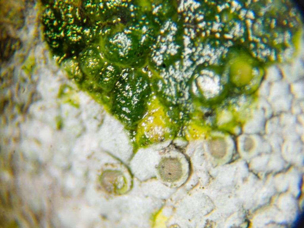
[attachment=3]20230326-P3260652.jpg[/attachment]
K+ yellow as expected, but less convincingly as the earlier thallus above (perhaps I hadn’t scratched the cortex properly, but for some reason I didn’t try again).
[attachment=2]20230326-P3260660.jpg[/attachment]
An overview of an apothecial section in cross-polarised light — and what seems to be a much more central section this time! The polarisation arrangement was the same as with the thallus above. This section shows the expected large crystals in the thalline margin, and perhaps some characteristic descending epithecial granules.
[attachment=1]20230326-P3260669.jpg[/attachment]
Another apothecial section (more or less from the centre) in brightfield.
[attachment=0]20230326-P3260668.jpg[/attachment]
The same section as above in cross-polarised light.Any advice welcome on this thallus, as well as the one above!
Thank you,
Anthony
-
AuthorPosts